Short Communication / Kısa Bilimsel Çalışma
Multi-organ damage in a cow with diabetes mellitus
Şima ŞAHİNDURAN
1, Özlem ÖZMEN
2, Necmettin Sarp SEVGİSUNAR
1University of Mehmet Akif Ersoy, Faculty of Veterinary Medicine, Departments of Internal Medicine1 and Departments of Pathology2, Burdur, Turkey.
Summary: A clinical, hematological and pathological study was carried out on a bovine case of diabetes mellitus (DM)
presenting clinical sings of polydipsia, polyuria, glycosuria, hyperglycemia, progressive emaciation and decreased milk yield. The body temperature, heart and respiratory rates were in normal range. Also there was no significant change in hematological values. Prior to slaughter; blood and urine samples were taken twice 48 hour interval from the cow. At necropsy, liver was pale and enlarged. Multiple small white foci were observed on the cortical area of the kidneys. Lung edema and foamy exudates in the lumen of the trachea and bronchus was present. At the gross examination pancreas was in normal appearance. Histopathologically reduced number and size were observed at the pancreatic islet localized in all pancreatic lobes. Decreased cell number observed in the Langerhans islets. Vacuolization and degenerations at the beta cells were commonly seen. Immunohistochemical study of pancreatic tissue revealed a marked reduction in the concentration and distribution of insulin, proinsulin, and amylin, but an increase in glucagon secreted cells. According to the clinical, histopathological and immunohistochemical findings, a combination of diabetes mellitus and multiorgan damage were diagnosed in a cow.
Key words: Cow, diabetes mellitus, immunohistochemistry, pathology.
Diabetes mellituslu bir inekte çoklu organ hasarı
Özet: Çok su içme, çok idrar yapma, ilerleyen zayıflama ve süt veriminde azalma şikayetleri ile kliniğe getirilen diyabetli bir
inekte klinik, hematolojik ve patolojik incelemeler yapıldı. Hayvanın vücut ısısı, kalp ve solunum sayıları normal sınırlar içerisindeydi. Ayrıca hemogram muayenede de önemli bir bulguya rastlanmadı. İnek kesime gitmeden önce 48 saat arayla 2 defa kan ve idrar numunesi toplandı. Nekropside, karaciğer büyümüş ve soluk renkliydi. Böbreklerin kortikal bölgelerinde çok sayıda beyaz renkli ve küçük odak saptandı. Akciğerlerde ödem, trahea ve bronş lumenlerinde köpüklü eksudat vardı. Dış bakıda pankreas normal görünümdeydi. Histopatolojik incelemede bütün pankreatik loplarda pankreatik adacık sayısı ve boyutunda azalma gözlendi. Langerhans adacıklarında hücre sayısının azaldığı saptandı. Genel olarak beta hücrelerinde vakuolizasyon ve dejenerasyonlar sıklıkla dikkati çekti. Pankreatik dokunun immunohistokimyasal muayenesinde insülin, proinsülin ve amilin sentezleyen hücrelerde belirgin bir azalma olduğu, buna karşın glukagon sentezleyen hücrelerde bir artış olduğu gözlendi. Klinik, histopatolojik ve immunohistokimyasal bulgulara dayanarak diabetes mellitus ve çoklu organ hasarı teşhisi konuldu.
Anahtar sözcükler: Diabetes mellitus, immunhistokimya, inek, patoloji.
Diabetes mellitus (DM) is a state of chronic hyperglycemia usually accompanied by glycosuria. Primary DM is a disorder involving the beta cells that result in decreased insulin levels and hyperglycemia (Koneko and Rhode, 1964; Van Metre, 2009; Ciobotaru, 2013). DM in domestic animals has been most commonly reported in small animals but rarely reported in other species such as cattle, horse, goat and sheep (Gold, 1981; Elliott et al., 1991; Charles, 2007; Braun et al., 2008; Giri et al., 2011). It is a quite a rare condition in cattle (Mostaghni et al., 1977; Radostits et al., 2008; Ciobotaru, 2013).
DM is a complex syndrome affecting humans and animals; the disease comprises a heterogeneous group of
hyperglycemic disorders. If insulin deficiency is severe, the hormonal abnormalities result in ketoacidosis and other manifestations of accelerated catabolism and diseases characterized by glycosuria, osmotic polyuria,
polydipsia, dehydration, and increased appetite.
Susceptibility to secondary infections increases in cases with DM (Unger et al., 1998; Jones et al., 1997; Van Metre, 2009). The main laboratory and clinical findings in cattle include emaciation, polydipsia, polyuria, glycosuria and hyperglycemia (Radostits et al., 2008; Ciobotaru, 2013). Histopathologically the most common findings are reduction in the number and size of pancreatic islet and vacuolization in beta cells (Koneko and Rhode, 1964; Mostaghni et al., 1977).
DM is generally accompanied with multiorgan damage and dysfunction in human. Only fatty liver and DM combination reported in cattle. The aim of this study is to report clinical, pathological and immunohistochemical findings of fatty liver, nephritis and pulmonary edema possibly related in a cow with DM.
A five year-old Holstein cow was brought to the Veterinary Medical Teaching Hospital from a farm from Burdur with complain of progressive emaciation, polyuria, polydipsia and decreased milk yield. According to the owner, the cow was healthy but in a period of 1 month lost weight and frequent urination started and then progressed. The cow avoided eating last 3 days. Previously a private veterinarian had suspected from nephritis and had treated her by cephalosporin (third generation) and vitamin B complex. The cow was vaccinated against bovine viral diarrhea (BVD) and infectious bovine rhinotracheitis (IBR) one year ago. On physical examination, the cow was depressed and
temperature, respiratory rate, heart rate were 38.4◦C, 40
breaths/min, 90 beats/min respectively. Moderately dehydration, polyuria and polydipsia were also observed at the clinical examination.
Urine and blood samples were collected for laboratory analyses. Serum biochemical analyses were performed with IDEXX Vet-Test equipment and reagents. MS9 blood counting equipment was used for hematological analysis of the blood drawn in EDTA tubes. For urine analysis IDEXX VetLAB UA equipment and URIPSIN 10 urine test strips were used. The serum was analyzed for glucose, alanine aminotransferase (ALT), aspartate aminotransferase (AST), alkaline phosphatase (ALP), creatinine, urea, amylase, cholesterol, triglyceride and total protein. Because of the poor prognosis the cow was sent to slaughter. Prior to slaughtering, blood and urine samples were taken twice with 48 hour interval. Anorexia, depression and coma occurred during the last 48 hours.
After slaughter tissue samples were collected from all visceral organs and the head, body and tail region of the pancreas and fixed in 10% buffered formalin. After routine procedure, tissues were blocked in paraffin and cut to 5-µm thickness. Tissue sections were stained with hematoxylin-eosin (HE) and examined microscopically. Oil Red O staining was used on frozen sections to demonstrate fat in the liver cells. Afterward, pancreas samples were immunostained with insulin antiserum (insulin mouse monoclonal antibody, NCL-INSULIN, 1/100 dilution; Novocastra), proinsulin antiserum (proinsulin mouse monoclonal antibody, NCL-PROIN-1G4, 1/200 dilution; Novocastra), glucagon antiserum (glucagon rabbit polyclonal antibody, NCLGLUCp, 1/25 dilution; Novocastra), and amylin peptide antiserum (amylin peptide mouse monoclonal antibody, NCL-AMYLIN, 1/100 dilution; Novocastra). The sections
were incubated with the primary antibodies for a period of 60 minutes, and immunohistochemistry was carried
out using biotinylated secondary antibody and
streptavidin-alkaline phosphatase conjugate. The
antigens were demonstrated by using diaminobenzidine (DAB) as the chromogen.
The body temperature, heart and respiratory rates were in normal range. There was no significant change regarding hematological values. The cow was depressed, severely emaciated, had polyuria, polydipsia, and loss of appetite. Because of the hemogram was normal and histopathological findings supported the cow has no other disease except DM; these clinical symptoms attributed the DM.
Results of a urinalysis were within normal limits,
except for glucose, ketone, and pH values in first and
second samples (Table 1).
On serum analysis, glucose levels were elevated. Results of serum samples analyses are shown in table 2.
Table 1: The biochemical analysis results of the first and second urine samples.
Tablo 1: İlk ve ikinci idrar örneğinin biyokimyasal analiz sonuçları.
Urinalysis First Results Second Results Reference levels (Radostits et al., 2008)
pH 5.5 5 7-9
Leucocytes Negative Negative Negative Protein Negative Negative Negative Glucose 1000 mg/dL 1050 mg/dL Negative Ketone 16 mmol/L (+++) 17 mmol/L (+++) Negative Urobilinogen Negative Negative Negative Bilirubin Negative Negative Negative Blood Negative Negative Negative
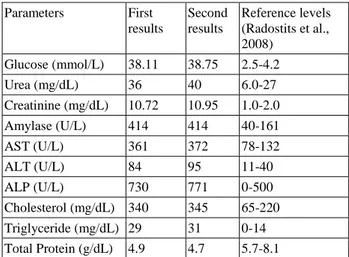
Table 2: Serum biochemical analysis results of the first and second samples.
Tablo 2: İlk ve ikinci serum örneğinin biyokimyasal analiz sonuçları. Parameters First results Second results Reference levels (Radostits et al., 2008) Glucose (mmol/L) 38.11 38.75 2.5-4.2 Urea (mg/dL) 36 40 6.0-27 Creatinine (mg/dL) 10.72 10.95 1.0-2.0 Amylase (U/L) 414 414 40-161 AST (U/L) 361 372 78-132 ALT (U/L) 84 95 11-40 ALP (U/L) 730 771 0-500 Cholesterol (mg/dL) 340 345 65-220 Triglyceride (mg/dL) 29 31 0-14 Total Protein (g/dL) 4.9 4.7 5.7-8.1
On gross examination, liver was the pale and enlarged. Multiple small white foci were observed on the cortex of the kidneys. Edema in lungs and foamy exudates in the lumen of the trachea and bronchus were noticed. At macroscopically examination, the pancreas was in normal appearance.
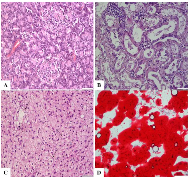
At the microscopical examination, fatty degeneration on hepatocytes, lymphocyte infiltrations at the cortical area of the kidney and pulmonary edema were diagnosed. Histopathologically number and size were decreased at the pancreatic islets localized in all pancreatic lobes. Reduced cell number observed in the central area of Langerhans islets (Figure 1A-C). Vacuolization at the beta cells were commonly seen. But the vacuoles were negative for lipid at the frozen section stained with Oil Red O. Frozen tissue from the liver stained positively with Oil Red O (Figure 1D).
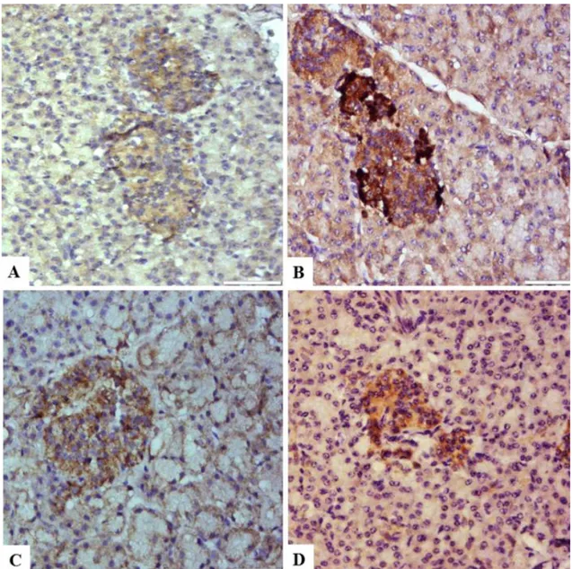
Immunohistochemical examination of all the pancreatic tissue revealed a marked reduction in the concentration and distribution of insulin, proinsulin, and amylin, but an increase in glucagon secreting cells (Figure 2A-D). Only a few islet cells that located centrally were slight reactive for insulin, proinsulin, and amylin. Glucagon positive cells were observed at the periphery of the islets.
DM is a syndrome complex affecting humans and animals; the unifying phenomenon is abnormal or inappropriate metabolism of glucose due to an absolute or relative deficiency of insulin. DM is characterized by fasting hyperglycemia, glycosuria, osmotic polyuria, polydipsia, dehydration, increased appetite an often ketoacidosis (Koneko and Rhode, 1964; Van Metre, 2009; Gold, 1981; Elliott et al., 1991; Jones et al., 1997; Charles, 2007; Braun et al., 2008; Giri et al., 2011;
Figure 1: (A) Decreased cells in the central areas of the Langerhans islet (arrows); (B) Non-supurative interstitial nephritis, lymphocyte infiltration (arrows) and severe degenerative changes in kidneys; (C) Severe lipoid degeneration of the liver (arrows), HE, Bars= 50µm (D) Positive staining for oil in liver, Oil Red O, Bar=30µm.
Şekil 1: (A) Langerhans adacıklarının merkezindeki hücrelerde azalma (oklar); (B) Böbrekte non-suppuratif intertisyel nefritis, lenfosit infiltrasyonu (oklar) ve şiddetli dejeneratif değişiklikler; (C) Karaciğerde şiddetli yağ dejenerasyonu (oklar), HE, Barlar=50 µm; (D) Karaciğerde pozitif yağ boyanma, Oil Red O, Bar=30µm.
Ciobotaru, 2013). In this case, glucose and ketone levels were very high in urine. Serum biochemical analysis results revealed very high glucose value which is consistent with DM.
At necropsy, the pancreas presents normal volume or various degree of atrophy, granular surface due to interstitial fibrosis and yellowish-brown color of the parenchyma in diabetic subjects. The liver is generally friable and pale. The kidneys present the same discoloration, observed mainly in the cortex (Jones et al., 1997; Clark, 2003; Van Metre, 2009). There were no macroscopical changes in pancreas in our case. Fatty liver and nephritis were observed at gross and histopathological examination. The etiology of fatty liver and nephritis was unknown in this case but because of the common association of DM and liver or kidney damage, the possible cause of these lesions may be
related with DM. The normal hemogram findings were supported our idea because no indication observed for any infectious disease.
At the histopathological examinations decrease in number and size of the pancreatic islets, vacuolar degeneration of residual cells, and a lymphocytic islet adenitis were commonly reported in DM cases. Marked decreased in the cells of Langerhans islets have been recognized in bovine DM (Jones et al., 1997; Van Metre, 2009). In this study, although decreased in number in islet cells and vacuolization were observed in pancreatic islet, inflammatory reactions were not seen. Taniyama et al., reported that almost complete absence in atrophied islet cells by immunohistochemical techniques (Taniyama et al., 1993). Similarly, in present study, very slight staining was observed in the pancreatic islet cells with, insulin, proinsulin and amilin by immunohistochemical
Figure 2: Decreased insulin (A), increased glucagon (B), decreased proinsulin (C) and decreased amyline (D) immuoreactivity in Langerhans islet, Streptoavidine Biotin Peroksidaz Method, Bars=50µm.
Şekil 2. Langerhans adacıklarında azalmış insülin (A), artmış glukagon (B), azalmış proinsulin (C) ve azalmış amylin (D) immunoreaktivitesi, Streptoavidine Biotin Peroxidase Method, Barlar=50µm.
method. Diagnosis of DM was supported by immunohistochemical findings.
In human and experimental animals DM is a serious disease with high morbidity and an early mortality rate, causing vascular, renal, retinal, or neuropathic disorders in the long term as well as acute metabolic complications. Much research shows that long-term hyperglycemia causes adverse changes in blood vessels and different organs. Hyperglycemia, which occurs long before diagnosis, may cause organ damage (Molitch, 1990; Van Metre, 2009). In addition to this, susceptibility to infections increases in DM. Diffuse thickening of the glomerular mesangium can be seen. Hepatic lipidosis and hepatomegaly are also seen (Stogdale, 1986; Jones et al., 1997; Charles, 2007; Van Metre, 2009). Fatty liver in a diabetic cow previously reported (Nazifi et al., 2004). In this case, liver lipidosis, non-suppurative interstitial nephritis and lung edema may be related to vascular damage because of the hyperglycemia. High urea and creatinine levels together with high blood glucose supported possible relation with kidney damage and DM. Similarly high AST, ALP, cholesterol and triglyceride levels accompanying to elevated blood glucose level indicated liver damage and DM relation. Possible reason for decreased total protein level may be attributed to inappetence and liver damage.
Numerous possible mechanisms of
insulin-dependent DM onset in cattle are reported, such as viral infection, metabolic disorders such as fatty liver syndrome, parturition and chronic insulitis (Stogdale, 1986; Charles, 2007). Hepatomegaly occurs as a result of fatty degeneration. Lipids accumulate the liver as the result of increased fat mobilization and hepatocytes injured by ketonemia, have decreased utilization of fats. Most of the extra hepatic lesions of DM such as chronic
renal disease or blindness are the result of
microangiopathy characterized by thickening of the capillary basement membrane (Capen, 2001; Jones et al., 1997). In this study, fatty liver, nephritis, pulmonary edema and enteritis were diagnosed in the cow. Because of the laboratory findings were occurred together with organ damages the lesions attributed to hyperglycemia. No partition or viral infection history in this case.
DM is considered as a common metabolic disease diagnosed frequently in canine and feline pathology. Clinical syndrome of diabetes is described rarely in cattle, small ruminants, swine and horses. Pathology and immunohistochemistry findings rarely reported in these
species. This study reported clinical, histopathological
and immunohistochemical findings in a diabetic cow with multiorgan damage.
References
1. Braun U, Gansohr B, Seidel M, Dumelin J, Wenger B, Schade B, Pospischil A (2008): Diabetes mellitus in a goat. SAT, 150, 608-612.
2. Capen CC (2001): Endocrine System. 279-323. In: Thomson’s Special Veterinary Pathology. Third Edition, Mosby, Inc., Missouri.
3. Charles J (2007): Pancreas. 389-424. In: MG Maxie (Ed), Jubb, Kennedy and Palmer's Pathology of Domestic Animals, Elsevier Limited, Philadeplphia.
4. Ciobotaru E (2013): Spontaneous Diabetes Mellitus in Animals. 271-296. In: OO Oluwafemi (Ed), Diabetes Mellitus - Insights and Perspectives. InTech, Rijeka, Croatia. 5. Clark Z (2003): Diabetes mellitus in a 6-month-old
Charolais heifer calf. Can Vet J, 44, 921-922.
6. Elliott J, Fowden AL, Callingham BA, Sharman DF, Silver M (1991): Physiological and Pathological Influences on Sheep Blood-Plasma Amineoxidase-effect of Pregnancy and Experimental Alloxan-induced Diabetes-mellitus. Res Vet Sci, 50, 334-339.
7. Giri JK, Magdesian KG, Gaffney PM (2011): Insulin-dependent diabetes mellitus associated with presumed autoimmune polyendocrine syndrome in a mare. Can Vet J, 52, 506-512.
8. Gould AC (1981): Diabetes mellitus in cattle. Vet Rec, 109, 539.
9. Jones TC, Hunt RD, King NW (1997): Veterinary Pathology. Williams and Wilkins, Maryland.
10. Koneko JJ, Rhode EA (1964): Diabetes mellitus in a cow. JAVMA, 144, 367-373.
11. Molitch ME (1990): Diabetes mellitus. 136. In: Walker HK, Hall WD and Hurst JW (Eds), Clinical Methods: The History, Physical, and Laboratory Examinations. Boston, Butterworths.
12. Mostaghni K, Ivoghli B (1977): Diabetes mellitus in the bovine. Cornell Vet, 67,24–28.
13. Nazifi T, Karimi A, Ghasrodashti R (2004): Diabetes mellitus and fatty liver in a cow: case report. Comp Clin Path, 13, 82-85.
14. Radostits OM, Gay CC, Hinchcliff KW, Constable PD (2008): Bovine virus diarrhea, mucosal disease. 1248– 1277. Bovine pestivirus disease complex. In: Veterinary Medicine: A Textbook of Diseases of Cattle, Horses, Sheep, Pigs, and Goats, 10th edn. Saunders Elsevier, Philadelphia. 15. Stogdale L (1986): Definition of diabetes mellitus. Cornell
Vet, 76, 156-174.
16. Taniyama H, Shirakawa T, Furuoka H, Osame S, Kitamura N, Miyazawa K (1993): Spontaneous Diabetes mellitus in Young Cattle: Histologic, immunohistochemical, and electron microscopic studies of the islet of Langerhans. Vet Pathol, 30, 46-54.
17. Unger RH, Foster DW (1998): Diabetes mellitus. 973-1059. In: Wilson JD, Foster DW, Kronenberg HM, et al, (Eds). Williams Textbook of Endocrinology, WB Saunders, Philadelphia.
18. Van Metre DC (2009): Diseases of the renal system. 925-971. In: Smith, BP (Ed), Large Animal Internal Medicine. Mosby, Elsevier, St. Louis.
Geliş tarihi: 06.06.2014 / Kabul tarihi: 04.03.2015 Address for correspondence:
Prof. Dr. Şima Şahinduran Mehmet Akif Ersoy University, Faculty of Veterinary Medicine, Department of Internal Medicine, İstiklal Yerleskesi, Burdur-TURKEY e-mail: [email protected]