REVIEW
Indirect Bonding Revisited
ABSTRACT
In recent years, the popularity of indirect bonding increased due to advantages such as reduction of chair time and enhancement of patient comfort. Although the indirect bonding technique has improved over the years, the literature has shown different techniques of bracket placement; furthermore, new materials were specially developed for this technique. The aim of this article is to provide a review of the literature, advantages, disadvantages, and laboratory and clinical stages of the indirect bonding technique.
Keywords: Indirect bonding, bonding systems, orthodontics indirect technique
INTRODUCTION
Indirect bonding was developed by Silverman and Cohen(1) in 1972 to reduce clinical time and to enhance pa-tient comfort.In this method, they used cement for attaching brackets to the stone model, a sealant as a clinical adhesive, and thermoplastic trays for the transfer of the brackets. In 1979, Thomas(2) invented “custom com-posite base technique,” which is still the most widely accepted technique currently used for indirect bonding. In this technique, Thomas used a chemically-cured resin for attaching the brackets in a laboratory and a universal and a catalyst resin as the clinical adhesive. The major complication of the Thomas technique is that the polym-erization of the chemical resin starts in the patient’s mouth, which is problematic in terms of time. If the transfer tray was removed before the completion of polymerization, bracket failure can be seen, and if the tray was left in the mouth for too long, this can disrupt the patient comfort. To solve this problem, the Thomas technique was modified; the universal and catalyst resins were mixed outside the mouth and directly applied to the teeth and custom base (3). With the modified Thomas technique, indirect bonding achieved similar bond strength values compared with direct bonding.
In the previous literature, chemically-cured resins were usually used as clinical adhesives for indirect bonding. Apart from these resins, glass ionomer cements, acrylic epoxy adhesives, and cyanoacrylates were also used (4-6). Read and O’Brien(7) used light-cured resins for indirect bonding in 1990, and with the advantages of these res-ins, the indirect bonding technique was further enhanced.
In 2002, Miles (8) used a flowable composite in indirect bonding. The most important advantage of this resin was to fill the voids of the custom base with its favorable viscosity.
With the advancement of technology, computers entered the practice of orthodontics, thereby enhancing the indirect bonding technique. Several companies offer three-dimensional computer-aided design and comput-er-aided manufacturing (3D CAD-CAM)-generated methods for the fabrication of indirect bonding trays. In one of these, Suresmile(Orametrix Inc.; Dallas, USA) system (9), teeth are scanned using an intraoral scanner, and computer generated 3D images are produced. These 3D images are used for digital set-up, and the brackets are placed in appropriate regions of the teeth. The customized transfer trays for indirect bonding and a customized archwire are prepared. Another popular 3D indirect bonding system is Insignia (Ormco Corp.; Orange, CA, USA) Hande Pamukçu, Ömür Polat Özsoy
Department of Orthodontics, Başkent University School of Dentistry, Ankara, Turkey
Corresponding Author: Dr. Hande Pamukçu, Department of Orthodontics, Başkent University School of
Dentistry, Ankara, Turkey E-mail: [email protected] Received: 17 May 2016 Accepted: 20 July 2016
©Copyright 2016 by Turkish Orthodontic Society - Available online at www.turkjorthod.org
(10). In this system, the 3D images of the patients are used for the digital set-up, the CAD-CAM technique is used for design, and customized brackets are produced. Single tooth transfer trays are also produced with the help of computers, and according to physicians’ requests, the transfer trays are usually combined to include three or four teeth.
In addition, there are some indirect bonding systems for lingual orthodontics. In these systems, there are customized brackets, transfer trays, and arc-wires. Incognito(3M Unitek; OH, USA) (11), Harmony (American Orthodontics; Sheboygan, Wisconsin, USA) (12), and E-Brace (Guangzhou Riton Biomaterial; China) (13) are some of the examples of these systems.
Advantages of Indirect Bonding
1. Shorter bonding time: No time is spent to decide the loca-tion of the brackets during bonding (14). A studythat com-pared the time spent for indirect and direct bonding of both jaws (included all the molar teeth) showed that the total time spent for bonding reduced by 30 min during indirect bonding (15).
2. Easy adjustment of overcorrection: Correction of the rotation an important issue in orthodontics. It is difficult to adjust the amount of overcorrection with direct bonding. The evalua-tion can make according the situaevalua-tion at the beginning of the treatment and millimetric adjustments of bracket position can made with the indirect bonding technique (16).
3. Adjustment of resin thickness: In some of the areas, espe-cially in the lower anterior teeth, the different thickness of the resin can cause problems in the 2nd order alignment. In later stages of the treatment, contact problems can occur and in–out problems can develop. In the indirect bonding technique, the thickness of the resin can be adjusted to be equal for each tooth right from the beginning of the treat-ment (17).
4. Modification of the bracket position according to the pa-tient’s need: Especially in deep-bite cases, it is important to adjust the bracket position right from the beginning of the treatment for stable results. In the indirect bonding tech-nique, it is easier to prepare and measure the vertical brack-et positions to open the bite (16).
5. Ease of working with ceramic brackets: It is very difficult to reposition the ceramic brackets due to their adhesion properties, and it is easier to determine the correct position with the indirect bonding technique at the beginning of the treatment (14).
6. Increasing the stability of the treatment: The most import-ant goal of orthodontic treatment is to obtain permanent results. The periodontal fibers reorganize at the beginning of the treatment with the indirect bonding technique, and this could reduce the risk of relapse (18).
7. Increasing patient comfort: Shorter duration of bonding in-creases the compliance of patient and also reduces contam-ination by saliva (14).
8. Protecting ergonomics of the clinician: Shorter bonding du-ration minimizes the degradation of the postural position (15). Furthermore, with shortened working hours, physi-cian’s stress can be reduced.
Disadvantages of Indirect Bonding
1. There is an additional laboratory procedure in the indirect bonding technique.
2. Laboratory stage increases the cost of the technique (19). 3. It is important to work precisely in the laboratory and clinical
stages (2).
4. There is a learning curve (19). Therefore, it takes time to cor-rectly and efficiently apply the technique.
5. If the transfer tray does not adapt correctly to the mouth, brackets cannot be transferred to the teeth with precision. 6. If the amount of the clinical resin applied is more than the
adequate amount, there can be excessive resins around the brackets, and this condition deteriorates the patient’s oral hygiene (20). To prevent these, residues can be cleaned us-ing a scaler or a micromotor with a carbide bur.
7. It is difficult to bond brackets to teeth with a short crown length (2).
8. If adhesives are not suitable for the indirect bonding tech-nique, the shear bond strength (SBS) and success of the technique can be reduced (21).
Laboratory and Clinical Stages of Indirect Bonding Laboratory stages
1. Impressions can be taken with alginate or two-phase sili-cone impression materials. Hard stone models are obtained and dried for at least a night.
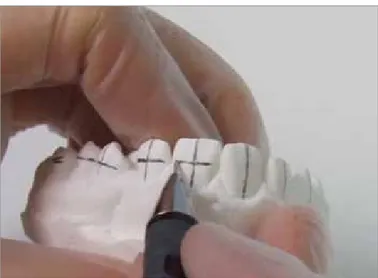
2. The proper location of the brackets is marked with the aid of a fine-tipped pencil and a bracket placement gauge (22). First, the vertical lines, then the horizontal lines, are drawn (Figure 1).
3. A layer of separating medium is applied with a brush and completely dried.
4. According to the clinician’s preference, a chemically-cured, a thermally-cured, or a light-cured resin can be used for the bonding of the brackets to the casts (Figure 2). Excessive res-in must be removed, and the resres-in must be properly polym-erized (22).
If a chemically-cured resin is used, the clinician should wait until the completion of the polymerization according to the
Figure 1. Vertical and horizontal lines for the location of brackets
recommendation of the manufacturer. If a thermally-cured resin is used, the model must be put in an oven of 120-170°C temperature for 15 min for the polymerization (2). For the light-cured resin, a light-emitting diode (LED) curing light must be used from mesial and distal surfaces for an addi-tional 10 s for each tooth as the stone model does not reflect light like enamel.
5. Before the fabrication of the transfer tray, a block-out is re-quired for the undercuts. Block-outs can be made with a wax or viscous silicone (20).
6. The clinician can choose to use a thermoplastic or a sili-cone transfer tray according to the clinical resin. If a light-cured resin is used, a transparent transfer tray is required (Figure 3). After the transfer tray is prepared, the edges should be trimmed. The tray can be divided into two or three parts for easy placement and control. Model and transfer trays should be immersed in warm water for 15 min to dissolve the separating agent, and at the end of this period, the trays could be easily separated from the model.
7. The borders of the tray are cut by 2 mm under the gingival borders. Hard edges are trimmed and corrected with the
help of stone burrs and disks (20). Trays are rinsed with water or cleaned in an ultrasonic cleaner.
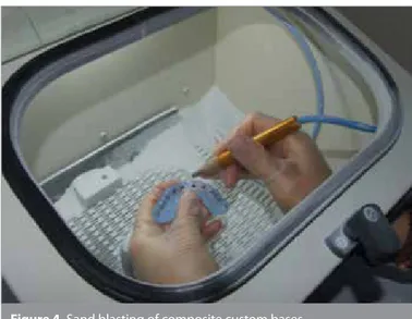
8. Custom composite bases are carefully sanded with 50-µm aluminum oxide particles and cleaned with alcohol (Figure 4) to get rid of oil residues and separating agent residues from the composite base.
9. Trays are dried with air and stored in a dry place until the clinical stage.
Clinical stages
1. Patient’s teeth are cleaned with pumice or a fluoride-free paste. They are washed and dried after 37% phosphoric acid is applied for 15 s. Etching must be applied only to the brackets’ locations to provide easy flash cleaning around the brackets. Some etching templates called Duran maskscan also be used for controlled etching (23). To prepare these masks, the bracket locations are marked with a copying pencil in the stone model and a thermoplastic or an acrylic plate can fabricated. Later, the location of the brackets can be opened with a diamond burr. During bonding, only these areas are etched.
2. After etching, a primer that is compatible with the clinical resin is applied. Trays must be completely fitting and the po-sition of the trays must be confirmed.
3. If the clinical resin is chemically-cured, resin is applied both to the enamel and the custom composite base, and the tray is firmly compressed to the teeth according to the manufac-turer’s recommendations (Figure 5) (22).
4. If the clinical resin is light-cured, adhesive polymerization must be done using a LED light source from the mesial and distal side of each tooth for 10 s. After removing the tray, an additional 5 s of light-curing can be done from the gingival and incisal side of the brackets to ensure complete polymer-ization.
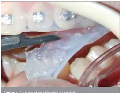
5. The transfer tray can be divided into two parts using a scal-pel for easy removal from the patient’s mouth (Figure 6). 6. The tray is removed using a scaler under the edge of the tray;
excessive resins are cleaned using scaler and tungsten-car-bide burs (22).
7. Initial archwires can be inserted and ligatured. Figure 2. Bonding of brackets to the stone model
Figure 3. Fabrication of transparent transfer tray
Figure 4. Sand blasting of composite custom bases
Is the Indirect Bonding Technique as Solid as the Direct Bonding Technique?
In vitro shear-bond strength studies
The strength of the brackets bonded using the indirect bond-ing technique was investigated in numerous studies. Un-fortunately, it is hard to compare these due to the rapid ad-vancement of resin technology. There are two SBS studies by Hocevar and Vincent (24) and Milne et al. (25) in 1988 and in 1989, respectively, with similar results. In both studies, they used a chemically-cured resin (Concise) for direct and indi-rect bonding groups. They investigated SBS of the groups and found no differences in SBS between the direct and indirect bonding groups.
Yi et al. (26) compared the direct and indirect bonding groups. They used adhesive precoated brackets (APC) and chemical-ly-cured resin (Sondhi Rapid Set) for the indirect bonding tech-nique and APC brackets for the direct bonding techtech-nique. Simi-larly, they found no differences between the SBS values.
Klocke et al. (27, 28) conducted two different studies in 2003. In the first one, they compared SBS values of different custom base resins in the indirect bonding groups with the direct bonding group (27). Chemically-cured (Maximum Cure), thermally-cured (Therma Cure), and light-cured (Transbond XT) resins were used for indirect groups, and there was no difference between the di-rect and indidi-rect groups. In the second study, three different res-in custom bases and clres-inical resres-ins were compared res-in the res-indirect bonding technique, and all the groups had adequate SBS values for clinical use (28).
Polat et al. (29) compared SBS values of three different groups; light-cured resin (Transbond XT) was used for direct bonding in the first group, chemically-cured resin (Custom IQ) was used for indirect bonding in the second group, and chemically-cured res-in (Sondhi Rapid Set) was used for res-indirect bondres-ing res-in the third group. There was no difference between the first and second groups, but the lowest bond strength was obtained for the indi-rect bonding group.
In 2004, Klocke et al. (30) conducted another indirect bonding study, and they investigated the stand-by time of the custom composite bases. In the first group, they used chemically-cured resins (Phase II and Custom IQ), and in the second group, they used light-cured (Transbond XT) and chemically-cured resins (Custom IQ). They pre-aged the custom composite bases for 7, 15, 30, and 100 days and compared the bond strength values. They found that the pre-aging up to 30 days had no effect on the SBS values in the indirect bonding technique.
Polat et al. (31) studied the effects of chlorhexidine varnish in the indirect bonding technique. They found that the chemical-ly-cured resin (Sondhi Rapid Set) group in indirect bonding with chlorhexidine varnish had lower SBS values than that in direct bonding with varnish and in indirect bonding with no varnish. As a summary, they did not suggest the use of a chlorhexidine varnish prior to indirect bonding.
Linn et al. (32) compared the SBS values of the direct and indi-rect bonding groups in 2006. Sixty teeth were divided into three groups; in the first group, light-cured resin (Transbond XT) was used for direct bonding; in the second group, chemically-cured resin (Sondhi Rapid Set) was used for indirect bonding; and in the third group, light-cured resin (Enlight LV) was used for indi-rect bonding. No differences were found among the groups. Daubt et al. (33) investigated the effects of thermocycling in in-direct bonding, and they found that SBS values decreased with thermocycling.
Thompson et al. (34) investigated the effects of the use of flowable resins in indirect bonding, and they found that flowable resins did not improve SBS values for the indirect bonding technique. Viwattanatipa et al. (35) evaluated the bond strength of the ef-fect of different surface preparation techniques on survival prob-abilities of orthodontic brackets bonded to nanofill composite resins in the indirect bonding technique, and they found that the most favorable results were found for aluminum oxide prepara-tion technique.
Figure 5. Application of chemically-cured resin to the composite
custom bases Figure 6. Division of transfer tray by scalpel
Kanashiro et al. (36) investigated the influence of different meth-ods of cleaning custom bases on SBS values of indirect bonding. Methyl methacrylate monomers, acetone, aluminum oxide, and washing agents were used for the cleaning. There were no differ-ences between the groups.
Flores et al. (37) compared the bond strengths of the direct and indirect bonding techniques with a self-etching ion releasing S-PRG filler, also they investigated the effect of thermocycling in their study. They found that SBS values were reducing with ther-mocycling and the SBS values were lower in indirect bonding groups. In addition, the self-etching group had lower SBS values than did the direct groups.
In vitro assessment of adhesive remnant after bracket
debonding in indirect bonding
The determination of the remnant adhesive after debonding is useful to select the proper resin in orthodontics and to predict the removal of the composite resin from the enamel. In 1984, Ar-tun and Bergland (38) introduced the adhesive remnant index (ARI) to assess the amount of resin left on the bracket base area. The score was a 4-point scale, which was determined according to the remaining adhesive at the bracket base; 0=all adhesive left on the bracket base, 1=more than half of the adhesive left on the bracket base, 2=less than half of the adhesive left on the bracket base, and 3=no adhesive left on the bracket base. In 1990, Bisha-ra and Trulove (39) developed a 5-point scale for ARI scores: 1=no adherence of composite on the bracket base, 2=less than 10% of composite remaining on the bracket surface, 3=more than 10% but less than 90% of composite remaining on the bracket sur-face, 4=more than 90% of composite remaining on the bracket surface, and 5=all composite remaining on the bracket base. In the literature on direct and indirect bonding (30,32,33,35,36), the ARI scores were usually 1 and 2 for most of the resins. This shows us that the fracture type is usually cohesive. Therefore, less resin removal is needed after debonding.
In vitro studies of bracket position accuracy in indirect bonding
Koo et al. (40) duplicated the models of the same patient 19 times, and the first model was ideally bonded by one orthodontist. In the second group, there were nine models bonded to a simu-lator and nine different orthodontists directly bonded brackets to these models with a light-cured adhesive (Transbond XT). In the third group, there were nine models bonded to the simula-tor and the same nine orthodontists indirectly bonded brackets with a thermally-cured adhesive (Thermacure). All of the model photos were taken in a standard position, and measurements were taken from the photos. As a result, no differences were found in mesio-distal and angular position of the brackets with both techniques, but the brackets, which were indirectly bond-ed, were in a more proper position in the vertical dimension. Aguirre et al. (41) compared bracket position accuracy in the direct and indirect bonding technique. For both techniques, al-though they achieved similar results, indirect bonding showed favorable results for upper canines and direct bonding for sec-ond premolars.
Hodgeet al. (42) investigated bracket position accuracy in the direct and indirect bonding technique. They found no difference between the two techniques in the vertical, angular, and me-sio-distal position.
Nichols et al. (43) evaluated the repeatability of the bracket posi-tion in the indirect bonding technique in 2013. Five experienced orthodontists bonded 10 models at three different times. Mod-els were scanned using an ICAT scanner and superimpositions of the models were made with a computer. They evaluated the differences of the bracket positions. As a result, every clinician’s bracket position was consistent in them, and the maximum bracket position difference was 1.25 mm between the groups. Castilla et al. (44) compared bracket position accuracy of five different types of indirect bonding transfer trays in 2014. The transfer trays consisted of mono-phase silicone, dual-phase sili-cone, monolayer thermoplastic, double-layer thermoplastic, and a combination of silicone-thermoplastic material. Duplicated 25 models were divided into five groups. Bracket positions were measured using calipers, and it was found that silicone-based trays transferred the brackets more accurately when the error rates were analyzed in all the tray systems.
In Vivo Studies of the Indirect Bonding Technique
In vivo evaluation of bond failures of indirectly bonded
brackets
Polatet al. (29) used two different types of chemically-cured resins (Sondhi Rapid Set vs. Custom IQ) in the indirect bonding technique with a split-mouth design. Fifteen patients were in-cluded in the study and were followed up for 9 months. Molar teeth were not included in the study. A total number of 295 teeth were indirectly bonded, 13 teeth (4%) were debonded, and no significant difference was found between the groups.
Miles et al. (45) used chemically-cured (Maximum Cure) and light-cured flowable (Filtek Flow) resins in the indirect bonding technique. In all, 112 patients were included in the study and were followed up for 6 months. Molar teeth were also included. The failure rate for chemically-cured resin group was 2.9% and that for the light-cured resin group was 2.4%.
Thiyagarajah et al. (46) used light-cured resin (Transbond XT) for both direct and indirect bonding. Thirty-three patients were in-cluded in the study and were followed up for 1 year. Molar teeth were not included in the study. A total number of 273 teeth were indirectly bonded, and the failure rate was 2.2% for indi-rect bonding. There were no statistically significant differences between the direct and indirect bonding groups.
Deahl et al. (47) compared direct and indirect bonding with a prac-tice-based study. A total number of 1368 patients were included in the study, 772 of these patients were directly bonded and 596 of patients were indirectly bonded by five different orthodontists. The failure rate for the direct bonding technique was 1.17% and that for the indirect bonding technique was 1.21%. There were no statistically significant differences between the groups.
Bozelli et al. (48) compared direct and indirect bonding with split-mouth design in 2012. Seventeen patients were included in the study and were followed up for 6 months. In addition, the to-tal clinical times for both techniques were compared. The failure rate for the direct bonding technique was 4.6% and that for the indirect bonding technique was 6.5%, but there were no signif-icant differences between these groups. The clinical time for in-direct bonding was 17 min shorter than that for in-direct bonding. Meniniet al. (49) compared the failure rates of direct and indirect bonding in 2014. A total number of 55 patients were included in the study, 33 patients were directly bonded, 19 were indirectly bonded, and they were followed up for 15 months. The failure rate for the direct bonding technique was 3.54% and that for the indirect bonding technique was 5.79%, but there were no signif-icant differences between these groups.
Vijayakumar et al. (50) used splint-mouth technique for bonding 30 patients with the direct and indirect bonding technique. They followed up the patients for 6 months, and molar teeth were not included in the study. The failure rate for the direct bonding technique was 8.8% and that for the indirect bonding technique was 10.5%, but there were no significant differences between these groups.
In Vivo Studies That Evaluate the Periodontal Tissues after
Indirect Bonding
Zachrisson and Brobakken (21) compared periodontal and plaque indices of patients who were directly and indirectly bonded and found no differences between the groups.
Dalessandri et al. (23) used Duran masks for acid etching to de-crease plaque accumulation in indirect bonding technique and compared this technique with the direct bonding technique. Thirty patients were included in the study, who were bonded with the splint-mouth technique, were followed up for 6 months. They found that teeth that were bonded with Duran masks have lower plaque accumulation in the first 4 months of treatment. Furthermore, these teeth have lower white-spot lesions.
CONCLUSION
The indirect bonding technique is a better method for the accu-rate placement of brackets and for the comfort of both the clini-cian and patient. For this technique to be a success, it is import-ant to work with precision and experience. With the developing technology and progress in dentistry, the technique will become even easier and simplifier.
Peer-review: Externally peer-reviewed.
Author contributions: Concept - Ö.P.Ö.; Design - Ö.P.Ö.; Supervision - Ö.P.Ö.; Data Collection and/or Processing - H.P.; Analysis and/or Interpre-tation - H.P.; Literature Search - H.P.; Writing - H.P.; Critical Reviews - Ö.P.Ö. Conflict of Interest: No conflict of interest was declared by the authors. Financial Disclosure: The authors declared that this study has received no financial support.
REFERENCES
1. Silverman E, Cohen M, Gianelly AA. A universal direct bonding sys-tem for both metal and plastic brackets. Am J Orthod 1972; 62: 236-44. [CrossRef]
2. Thomas RG. Indirect bonding: simplicity in action. J Clin Orthod 1979; 13: 93-106.
3. Miles PG. A comparison of retention rates of brackets with thermal-ly-cured and light-cured custom bases in indirect bonding proce-dures. Aust Orthod J 2000; 16: 115-7.
4. Newman GV. Direct and indirect bonding of brackets. J Clin Orthod 1974; 8: 264-72.
5. Silverman E, Cohen M, Demke RS, Silverman M. A new self-curing hybrid glass ionomer. J Clin Orthod 1997; 31: 315-8.
6. Rajagopal R, Venkatesan A, Gnanashanmugham K, Harish Babu S. A new indirect bonding technique. J Clin Orthod 2004; 38: 600-2. 7. Read MJF, O’Brien KD. A clinical trial of an indirect bonding
tech-nique with a visible lightcured adhesive. Am J Orthod Dentofacial Orthop 1990; 98: 259-62. [CrossRef]
8. Miles PG. Indirect bonding with a flowable light-cured adhesive. J Clin Orthod 2002. 2002; 11: 646-7.
9. Sachdeva R, Frugé JF, Frugé AM, Ingraham R, Petty WD, Bielik KL, et al. SureSmile: a report of clinical findings. J Clin Orthod 2005; 5: 297-314.
10. Gracco A, Tracey S. The insignia system of customized orthodontics. J Clin Orthod 2011; 45: 442-51.
11. Wiechmann D. A new bracket system for lingual orthodontic treat-ment. Part 1: Theoretical background and developtreat-ment. J Orofac Orthop 2002; 63: 234-45. [CrossRef]
12. Dalessandri D, Lazzaroni E, Migliorati M, Piancino MG, Tonni I, Bonet-ti S. Self-ligaBonet-ting fully customized lingual appliance and chair-Bonet-time reduction: a typodont study followed by a randomized clinical trial. Eur J Orthod 2013; 35: 758-65. [CrossRef]
13. Romano R, Geron S, Echarri P. Customized Brackets and Archwires, Lingual and Esthetic Orthodontics. 1st ed. London: Quintessence Publishing; 2011. p. 154-156.
14. Kalange JT. Ideal appliance placement with APC brackets and indi-rect bonding. J Clin Orthod 1999; 33: 516-26.
15. Guenthner TA, Larson BE. Indirect Bonding: A technique for preci-sion and efficiency. Semin Orthod 2007; 13: 58-63. [CrossRef]
16. Kalange JT. İndirect bonding: A comprehensive review of the ad-vantages. World J Orthod 2004; 5: 301-7.
17. Miles PG, Weyant RJ. A clinical comparison of two chemically-cured ad-hesives used for indirect bonding. J Orthod 2003; 30: 331-6. [CrossRef]
18. Boese LR. Fiberotomy and reproximation without lower retention, nine years in retrospec: Part I .Angle Orthod 1980; 50: 88-97. 19. White LW. A new and improved indirect bonding technique. J Clin
Orthod 1999; 33: 17-23.
20. Sondhi A. Efficient and effective indirect bonding. Am J Orthod Dentofacial Orthop 1999; 115: 352-9. [CrossRef]
21. Zachrisson BU, Brobakken BO. Clinical comparison of direct versus indirect bonding with different bracket types and adhesives. Am J Orthod 1978; 74: 62-78. [CrossRef]
22. Kalange JT. Prescription-based precision full arch indirect bonding. Semin Orthod 2007; 13: 19-42. [CrossRef]
23. Dalessandri D, Dalessandri M, Bonetti S, et al. Effectiveness of an in-direct bonding technique in reducing plaque accumulation around braces. Angle Orthod 2012; 82: 313-8. [CrossRef]
24. Hocevar RA, Vincent HF. Indirect versus direct bonding: Bond strength and failure location. Am J orthod Dentofacial Orthop 1988; 94: 367-71. [CrossRef]
25. Milne JW, Andreasen GF, Jakobsen JR. Bond strength comprasion: A simplified indirect technique versus direct placement of brackets. Am J Orthod Dentofacial Orthop 1989; 96: 8-15. [CrossRef]
26. Yi GK, Dunn WJ, Taloumise LJ. Shear bond strength comparison be-tween direct and indirect bonded ortodontic brackets. Am J Orthod Dentofacial Orthop 2003; 124: 577-81. [CrossRef]
27. Klocke A, Shi J, Kahl-Nieke B, Bismayer U. Bond strength with cus-tom base indirect bonding techniques. The Angle Orthod 2003; 73: 176-80.
28. Klocke A, Shi J, Kahl-Nieke B, Bismayer U. In vitro evaluation of a moisture-active adhesive for indirect bonding. The Angle Orthod 2003; 73: 697-701.
29. Polat O, Karaman AI, Buyukyılmaz T. In-vitro evaluation of shear bond strengths and in vitro analysis of bond survival of indi-rect-bonding resins. Angle Orthod 2004; 74: 405-9.
30. Klocke A, Tadic D, Vaziri F, Kahl-Nieke B. Custom base preaging in indirect bonding. Angle Orthod 2004; 74: 106-11.
31. Polat O, Uysal T, Karaman AI. Effects of a Chlorhexidine varnish on shear bond strength in indirect bonding. The Angle Orthod 2005; 75: 1036-40.
32. Linn BJ, Berzins D, Dhuru VB, Bradley TG. A comparison of bond strength between direct- and indirect-bonding methods. The An-gle Orthod 2006; 76: 289-94.
33. Daubt J, Berzins DW, Linn BJ, Bradley TG. Bond strength of direct and indirect bonded brackets after thermocycling. The Angle Orth-od 2006; 76: 295-300.
34. Thompson MA, Drummond JL, BeGole EA. Bond strength analysis of custom base variables in indirect bonding techniques. Am J Or-thod Dentofacial Orthop 2008; 133: 15-20. [CrossRef]
35. Viwattanatipa N, Jermwiwatkul W, Chintavalakorn R, Nanthavanich N, et al. The effect of different surface preparation techniques on the survival probabilities of orthodontic brackets bonded to nano-fill composite resin. J Orthod 2010; 7: 162-73. [CrossRef]
36. Kanashiro LK, Robles-Ruiz JJ, Ciamponi AL, Medeiros IS, Tortama-no A, Paiva JB. Influence of different methods of cleaning custom bases on the shear bond strength of indirectly bonded brackets. J Orthod 2014; 41: 175-80. [CrossRef]
37. Flores T, Mayoral JR, Giner L, Puigdollers A. Comparison of enam-el-bracket bond strength using direct- and indirect-bonding tech-niques with a self-etching ion releasing S-PRG filler. Dent Mater J 2015; 34: 41-7. [CrossRef]
38. Artun J, Bergland S. Clinical trials with crystal growth conditioning as an alternative to acid-etch enamel pretreatment. Am J Orthod 1984; 85: 333-40. [CrossRef]
39. Bishara SE, Trulove TS. Comparisons of different debonding
tech-niques for ceramic brackets: an in vitro study. Part I. Background and methods. Am J of Orthod and Dent Orthoped 1990; 98: 145-53.
[CrossRef]
40. Koo BC, Chung G-H, Vanarsdall RL. Comparison of the accuracy of bracket placement between direct and indirect bonding techniques. Am J Orthod Dentofacial Orthop 1999; 116: 346-51. [CrossRef]
41. Aguirre MJ, King GJ, Waldron JM. Assessment of bracket placement and bond strength when comparing direct bonding to indirect bonding techniques. Am J Orthod Dentofacial Orthop 1982; 82: 269-76. [CrossRef]
42. Hodge TM, Dhopatkar AA, Rock WP, Spary DJ. A randomized clinical trial comparing the accuracy of direct versus indirect bracket place-ment. J Orthod 2000; 31:132-7. [CrossRef]
43. Nichols DA, Gardner G, Carballeyra AD. Reproducibility of bracket positioning in the indirect bonding technique. Am J Orhod Dent Orthod 2013; 144: 770-6. [CrossRef]
44. Castilla AE, Crowe JJ, Moses JR, Wang M, Ferracane JL, Covell DA Jr. Measurement and comparison of bracket transfer accuracy of five indirect bonding techniques. Angle Orthod 2014; 84: 607-14.
[CrossRef]
45. Miles PG, Weyant RJ. A comparison of two indirect bonding adhe-sives. Angle Orthod 2005; 75: 1019-23.
46. Thiyagarajah S, Spary DJ, Rock WP. A clinical comparison of bracket bond failures in association with direct and indirect bonding. J Or-thod 2006; 33: 198-204. [CrossRef]
47. Deahl ST, Salome N, Hatch JP, Rugh JD. Practice-based comparison of direct and indirect bonding. Am J Orthod Dentofacial Orthop 2007; 132: 738-42. [CrossRef]
48. Bozelli JV, Bigliazzi R, Barbosa HA, Ortolani CLF, Bertoz FA, Faltin JK. Comparative study on direct and indirect bracket bonding tech-niques regarding time length and bracket detachment. Dental Press J Orthod 2013; 18: 51-7. [CrossRef]
49. Menini A, Cozzani M, Sfondrini MF, Scribante A, Cozzani P, Gandini P. A 15-month evaluation of bond failures of orthodontic brackets bonded with direct versus indirect bonding technique: a clinical tri-al. Prog Orthod 2014; 15: 70-6. [CrossRef]
50. Vijayakumar RK, Jagadeep R, Ahamed F, Kanna A, Suresh K. How and why of orthodontic bond failures: An in vivo study. J Pharm Bio-allied Sci 2014; 6: 58-9. [CrossRef]