Diagnosis and Treatment Approaches for Intraabdominal
Masses in Adults
Erișkin İntraabdominal Kitlelerinde Tanı ve Tedavi Yaklașımları
Muzaffer Akkoca
1, Serhat Tokgöz
1, Kerim Bora Yılmaz
1, Melih Akıncı
1, Demet Yılmazer
21 Department of General Surgery, University of Health Sciences,
Dışkapı Yıldırım Beyazıt Research and Training Hospital, Ankara/Turkey
2 Department of Pathology, University of Health Sciences, Dışkapı
Yıldırım Beyazıt Research and Training Hospital, Ankara/Turkey
Aim: To evaluate patients who underwent surgery because of an intra-abdominal mass, in respect of symptoms, findings, diagnosis and treatment in accordance with information in literature.
Material and Methods: A retrospective examination was made of patients who underwent surgery because of an intra-abdominal mass between May 2010 and May 2017. For each patient a record was made of age, gender, symptoms, findings, type of mass determined, diagnostic methods used, the organ or tissue of origin, whether or not preoperative biopsy was taken, dimension of the mass, benign or malignant nature of the mass, type of operation performed and pathological diagnosis.
Results: Evaluation was made of 45 patients with an intra-abdominal mass. The patients comprised 18 (40%) males and 27 (60%) females with a mean age of 54.3 years (range, 19-86 years). The most common symptom was abdominal pain in 26 (57.8%) patients, and being able to feel the mass in the abdomen in 15 (33.4%). The intra-abdominal mass could be determined on palpation in 28 (62.2%) as a finding in the physical examination. USG was the most preferred imaging examination method in 36 (80%) patients, followed by CT in 28 (62.2%), MRI in 10 (22.2%) and PET-CT in 2 (4.4%). Preoperative endoscopy was applied to 27 (60%) patients and biopsy to 21 (46.7%). The long axis of the mass was mean 13cm (range, 4-50 cm). Treatment was applied as total excision of the mass in 29 (64.4%) patients, enbloc resection of the mass together with the bowel in 14 (31.1%), together with a distal pancreatectomy in 1 (2.2%) and together with a pancreaticoduodenectomy in 1 (2.2%). The pathology result was reported as benign in 15 (33.3%) patients and as malignant in 30 (66.7%). Of the benign masses, mesenteric cyst was determined most frequently in 5 (11.1%) patients and gastrointestinal stromal tumors (GIST) were determined as the most common malignant masses in 10 (22.2%) patients.
Conclusion: Although intra-abdominal masses originate from different organs and have different clinical and histopathological properties, they are pathologies that require systematic evaluation in respect of diagnosis and treatment approaches.
Key Words: Intra-Abdominal Masses, Physical Examination, Abdominal Ultrasonography, Histopathological Type, Gastrointestinal Stromal Tumor.
Amaç: Bu çalıșma ile; karın içi kitle nedeni ile ameliyat edilen hastaların; semptom, bulgu, tanı ve tedavi sistematiği açısından literatür bilgileri doğrultusunda değerlendirilmesi amaçlandı.
Gereç ve Yöntem: Mayıs 2010- Mayıs 2017 tarihleri arasında karın içi kitle nedeni ile ameliyat edilen hastalar geriye dönük olarak incelendi. Karın içi kitle nedeni ile ameliyat edilen hastaların; yaș, cins, semptom, semptom, bulgu, kitlenin tespit edilme șekli, hangi tanı yöntemi ile tespit edildiği, hangi organ veya dokudan köken aldığı, ameliyat öncesi biyopsi yapılıp yapılmadığı, kitlenin boyutu, kitlenin benign veya malign olușu,hangi ameliyatın yapıldığı ve patolojik tanının ne olduğu belirlendi.
Bulgular: Çalıșmaya dahil edilen karın içi kitleli 45 hastanın ortalama yașının 54,3 (19-86) olduğu 18’inin erkek (%40), 27’sinin ise kadın (%60) olduğu görüldü. En fazla semptom 26 hastada (%57,8) karın ağrısı, 15 hastada ise (%33,4) abdomende ele gelen kitle idi. Fizik muayene bulgusu olarak 28 hastada (%62,2) palpasyonda karın içi kitle tespit edilebildi. Görüntüleme çalıșmalarından en fazla tercih edilen 36 hasta (%80) ile abdomen ultrasonografi (USG) idi. USG’yi 28 (%62,2) hasta ile abdomen bilgisayarlı tomografi (BT), 10 hasta ile (%22,2) abdomen manyetik rezonans görüntüleme (MRG), 2 hasta ile (%4,4) pozitron emisyon tomografi-bilgisayarlı tomografi (PET-BT) takip etmekte idi. Ameliyat öncesi 27 hastaya (%60) endoskopi, 21 hastaya da (%46,7) biyopsi yapıldığı görüldü. Kitlelerin uzun ekseni ortalama 13cm (4cm-50cm) idi. 29 hastanın (%64,4) kitlenin total olarak eksize edilmesi ile, 14 hastanın (%31,1) kitlenin barsak rezeksiyonu ile birlikte enblok rezeke edilmesi ile, 1 hastanın (%2,2) distal pankreatektomi ile, 1 hastanın da (%2,2) pankreatikoduodenektomi ile birlikte tedavi edildiği saptandı. 15 hastanın (%33,3) patoloji sonucu benign, 30 hastanın (%66,7) ise malign olarak raporlandığı görüldü. Benign kitlelerden en sık mezenter kistine (n:5, %11,1), malign kitlelerden ise gastrointestinal stromal tümörlere (GİST) (n:10, %22,2) rastlandı.
Tartıșma ve Sonuç: Karın içi kitleler; farklı organlardan köken alan, farklı klinik ve histopatolojik özelliklere sahip olan kitleler olmakla birlikte tanı ve tedavi yaklașımları açısından sistematik değerlendirilmesi gereken patolojilerdir.
Anahtar Sözcükler: İntraabdominal Kitle, Fizik Muayene, Abdominal Ultrasonografi, Histopatholojik Tip, Gastrointestinal Stromal Tümör.
Received: Nov. 17, 2017 Accepted: Dec. 19, 2017 Corresponding Author:
Muzaffer Akkoca
E-mail: [email protected] Mobil phone: 0 505 649 74 43 Fax/phone: 90 (312) 596 23 12
Department of General Surgery, University of Health Sciences, Dıșkapı Yıldırım Beyazıt Research and Training Hospital, Altındağ/Ankara 06110, Turkey
Intra-abdominal masses are defined as masses located at the edges of the ribs and in the region of the paraspinal muscles limited in the anterior by the iliac wings and the symphysis pubis (1). Generally, intra-abdominal masses are determined as a result of symptoms in the abdomen or in a physical examination or incidentally during radiological imaging performed for another reason. Evaluation of intra-abdominal masses is based on knowledge of the anatomy of the abdomen and which quadrant corresponds to the organs of the abdomen. In a basic anatomic approach, the abdomen is separated into 9 areas of epigastric, umbilical, suprapubic, right hypochondrium, left hypochondrium, right lumbar, left lumbar, right inguinal and left inguinal regions. However, in clinical practice, the terms more often used, especially by surgeons, are the right upper and right lower quadrant, left upper and left lower quadrant, epigastric and hypogastric regions (2).
As these masses are seen in a widely varying range, it is not correct to group them under a single diagnostic category or to apply the same diagnostic and treatment methods. Knowing that there can be several pathologies under the heading of intra-abdominal mass and what type of masses there could be in which regions of the abdomen with which symptoms and findings, and the need for systematic follow-up of which methods are used in the approach to these, demonstrates the need for evaluation of these pathologies under a heading.
There are reports in literature of diagnostic, treatment and follow-up approaches for intra-abdominal masses caused by various diseases. Although these pathologies are more often presented in the form of single case reports, there are also case series and research papers on a specific diagnosis. However, to the best of our
knowledge, there has been no previous study that has examined intra-abdominal masses in general in respect of the symptoms, findings, diagnostic approaches and treatment methods.
The aim of this study was to systematically evaluate cases operated on in our clinic because of intra-abdominal mass in respect of symptoms, findings, diagnosis and treatment in the light of the relevant information in literature.
Material and Method
Approval for the study was granted by the Local Ethics Committee. A retrospective examination was made of patients operated on because of an intra-abdominal mass in the General Surgery Clinic of Ankara Dışkapı Yıldırım Beyazıt Training and Research Hospital between May 2010 and May 2017. The data related to the patients were retrieved from hospital records, operating reports, operation photographs, the epicrisis reports recorded in the hospital automated information system, laboratory, imaging and pathological examination reports. For each patient a record was made of age, gender, symptoms, findings, the type of mass determined, diagnostic methods used, the organ or tissue of origin, whether or not preoperative biopsy was taken, dimension of the mass, benign or malignant nature of the mass, type of operation performed and pathological diagnosis. Data and analyses related to imaging examinations made during diagnosis were taken from the Radiology Clinic and data and analyses related to the histopathological diagnosis from the Pathology Clinic. Cases were excluded from the study if there were no clear findings of a mass in the physical examination or on imaging examination, if there was an endoluminal mass of the gastrointestinal system or if data were incomplete or unavailable.
Results
The study included a total of 45 patients with an intra-abdominal mass, comprising 18 (40%) males and 27 (60%) females with a mean age of 54.3 years (range, 19-86 years). The most common symptom was abdominal pain in 26 (57.8%) patients. A total of 15 (33.4%) patients presented with the complaint of being able to feel the mass in the abdomen (Table 1). In some patients, there was more than one symptom, 1 (2.2%) patient underwent emergency surgery because of intra-abdominal bleeding and in 3 (6.6%) patients, there were no symptoms. The intra-abdominal mass could be determined on palpation in 28 (62.2%) as a finding in the physical examination, and the mass could not be palpated in 17 (37.8%). USG was the most preferred imaging examination method in 36 (80%) patients, followed by abdominal CT in 28 (62.2%), abdominal MRI in 10 (22.2%) and PET-CT in 2 (4.4%) (Figure 1). More than one imaging method was used in some patients. Table 1. Data on demographic and diagnostic
evaluations of the patients.
Number of patients (n) Percentage (%) 45 100 Gender Male Female 27 18 40 60 Symptom Abdominal pain Mass Hemorrhagea None 26 15 1 3 57,8 33,4 2,2 6,6 Physical examination Palpable Not palpable 28 17 62,2 37,8 Imaging methods USG BT MRI PET-BT 36 28 10 2 80 62,2 22,2 4,4 Endoscopy No Yes - Upper GIS - Lower GIS 18 27 9 18 40 60 20 40 Preoperative biopsy No Yes -USG guided -CT guided 24 21 17 4 53,3 46,7 37,8 8,8
According to the data obtained from the operating notes, the long axis of the mass was measured as mean 13cm (range, 4-50 cm). The smallest mass with the long axis of 4 cm was seen to be related to endometriosis and the largest with the long axis of 50 cm was determined as an intra-abdominal mass related to liposarcoma. The origin of the intra-abdominal mass was seen to be the intestinal mesentery in most cases (n:18), followed by the retroperitoneum (n:10), the ovary (n:5), and the abdominal anterior wall (n:4). As treatment, total excision of the mass was applied to 29 (64.4%) patients, enbloc resection of the mass together with the bowel to 14 (31.1%), together with a distal pancreatectomy in 1 (2.2%) and together with a pancreaticoduodenectomy in 1 (2.2%)
(Figure 2). The mean length of hospital stay was 7.2 days (ra nge, 3-16 days) and the mortality rate was determined as 4.4% (n:2) (Table 2). Table 2. Tumor localization and type of
surgery Number of patients (n) Percentage (%) Tumor localization Mesenter Retroperiton Over Anterior wall of abdomen Pancreas Omentum Stomach Duodenum Small intestine Colon 18 10 5 4 2 2 1 1 1 1 40 22,2 11 8,8 4,4 4,4 2,2 2,2 2,2 2,2 Type of surgey Total excision
Enbloc resection of the mass together with the bowel Distal pancreatectomy Pancreaticoduodenectomy 29 14 1 1 64,4 31,1 2,2 2,2
When the pathology results of the patients were examined, the mass was reported as benign in 15 (33.3%) patients and as malignant in 30 (66.7%). The most frequently determined histopathological diagnosis of the benign masses was mesenteric cyst (n:5, 11.1%) and of the malignant masses, gastrointestinal stromal tumors (GIST) (n:10, 22.2%) (Table 3).
Table 3. Histopathologic types and rates of
intraabdominal masses. Benign
Pathologies n (%) Pathologies Malign n (%) Mesenter cyst 5 11,1 GIST 10 22,2 Endometriosys 2 4,4 Liposarcoma 7 15,6 Cyst hydatic 2 4,4 Over tumor 5 11,1 Fibroma 2 4,4 Condrosarcom 2 4,4 Distrofic
calcification 1 2,2 Neuroendocrin tumor 2 4,4 Aberran
pancreas 1 2,2 mesenchimal tumor Malign 2 4,4 Leiomyoma 1 2,2 Lymphoma 1 2,2 Pseudocyst 1 2,2 Schwannoma 1 2,2 Total 15 33,3 Total 30 66,7
Figure 1. a) The CT image of the jejunum-derived GIST, b) MRI image of Schwannom in the retrorectal region c) CT image of over tumor filling
the abdomen
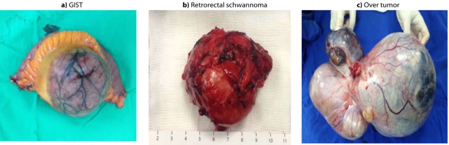
a) GIST b) Retrorectal schwannoma c) Over tumor
Figure 2: a) Post-operative specimen of Gastrointestinal stromal tumor (GIST), b) Post-operative specimen of Schwannom in the retrorectal
region, c) Post-operative specimen of over tumor.
Discussion
Abdominal masses include several different pathological lesions with benign or malignant properties, solid or cystic and which can be different according to age, gender, localization and organ or tissue of origin. For a diagnosis of a pathology with so many different properties, systematic evaluation is necessary for the application of correct treatment. Knowledge of the different pathologies causing abdominal masses and the diagnostic and treatment approaches to these is of particular importance.
The process of diagnosis of an intra-abdominal mass starts with a detailed history and physical examination. These two steps form the basis of the preliminary diagnosis and differential diagnosis by which the causes of the intra-abdominal mass will be determined. The patient must be questioned in detail about symptoms, time since onset, additional gastrointestinal symptoms, known diseases, family history, previous operations and trauma history (3). In the current study, the most frequent symptom on presentation was abdominal pain followed by the complaint of feeling the mass in the abdomen. Although all of the patients in this study had an abdominal mass, only 13 presented with the complaint of the mass, showing that most of the patients were not aware of the abdominal mass. This suggests that masses that have not reached large dimensions can remain asymptomatic or in selected cases, it can manifest with the complaint of abdominal pain. In this result, the size of the mass is undoubtedly important in addition to the localization. It may be difficult for patients to notice masses, especially those with a deep retroperitoneal location or a pelvic location.
According to the study results, although the majority of the patients had complaints of a mass, in the physical
examination an abdominal mass could only be palpated in 28 patients. This result indicates the importance of the physical examination. In the determination of a mass in the abdomen in the physical examination, first it must be attempted to differentiate pathological states from causes such as postural differences which could be normal or swelling caused by obesity, gas distension, pregnancy or fecaloma. Furthermore, it is necessary to determine in the physical examination whether the mass determined requires emergency treatment, the localization, the size, the shape, the texture, the contours, whether or not it is pulsatile and whether or not it is adhering to surrounding tissues. In 1 of the cases in the current study, as a result of the findings of the physical examination of the mass determined in the abdomen, there were seen to be findings of intra-abdominal bleeding and because of hemodynamic instability, the patient was admitted for emergency surgery. This mass was determined to be of small intestine mesenteric origin, and tumor perforation had caused intra-abdominal bleeding. The pathology of the mass was reported to be GIST. In the physical examination, when
diagnosis cannot be made and localization or the origin of the mass cannot be determined or benign- malignant differentiation cannot be made, imaging examinations and biopsy procedures made according to these are extremely important in the diagnosis of intra-abdominal masses. Of the imaging examinations, it is recommended that abdominal ultrasonography (USG) is applied first as it is easily accessible, has no radiation exposure, can be applied rapidly and in a practical manner and can identify the organ of origin with solid cystic differentiation (4). In the diagnostic evaluation in the current study, abdominal USG was applied to 80% of the patients.
However, despite the above-mentioned advantages of USG, because it is dependent on the practitioner’s experience and in some cases where it was insufficient for the images of intra-abdominal masses, abdominal computed tomography (CT) was applied to 28 (62.2%) patients either as the first imaging procedure or after abdominal USG (Figure 1). Abdominal CT is one of the most effective methods in current diagnostic and interventional evaluations of intra-abdominal masses (5). Unlike USG, abdominal CT clearly shows the tissue planes without any effect from gas, fat or bone structures in the abdomen and can provide the surgeon with information on the removability of the mass by showing the borders between the mass and adjacent structures (6). In patients with renal failure or a contra-indication to contrast material containing iodine and in cases of masses with pancreas or retroperitoneal localization, magnetic resonance imaging is preferred (7). To evaluate whether the intra-abdominal
mass originates from hollow organs or obstruction which can occur because of the effect of the mass on the stomach and intestines, gastrointestinal (GIS) system endoscopy may be necessary. To evaluate endoluminal pathologies, lower GIS endoscopy was applied to 40% of the current cases and upper GIS endoscopy to 20%. Following imaging studies revealing the intra-abdominal mass, there may be a need for biopsy to determine the treatment approach. While biopsy under USG guidance is preferred in large, superficial and cystic lesions, biopsy under CT guidance is preferred in masses with a deep location which cannot be visualized on USG as it has high rates of accuracy (8, 9). In our study, preoperative USG and CT guided biopsy rates is similar to the literure.
In the majority of cases, the abdominal mass could be totally removed with intact surgical borders. However, in some patients, resection of the colon or small intestine was necessary to be able to remove the whole mass (Figure 2). The tumour localisation was determined as most frequently in the intestine mesentery followed by retroperitoneal location and ovarian. Of the malignant masses, the most common was determined as GIST followed by liposarcoma and the most common of the benign masses was seen to be mesenteric cysts. GIST, which can originate from any part of the gastrointestinal tract, but primarily the stomach, are rarely seen masses with the potential to be malignant (10,12).
As intra-abdominal masses more often have exophytic growth, they can manifest clinically and the surgical strategy is removal of the mass which will obtain a negative surgical border macroscopically and microscopically (13, 14). Liposarcoma are tumours which demonstrate malignant behaviour with mesenchymal origin from fat tissue. In the treatment of these masses, which can reach a large size without showing symptoms, it is again aimed to completely remove the mass with a negative surgical border (15-18). Mesenteric cysts are rarely seen, can cause symptoms of a mass with abdominal pain and can be removed laparoscopically or with open surgery depending on the symptoms and localisation (19-22). In the current study, apart from these,
several different histopathological types of mass originating from several different organs were seen (Table 3). In conclusion, as intra-abdominal masses
originate from different organs and have different clinical and histopathological properties, they are pathologies that require systematic evaluation in respect of diagnosis and treatment approaches. Although, to the best of our knowledge, this is the first study in literature on this subject, it is a fact that with an increase in the number of cases, many lesions will be able to be seen with different clinical and pathological properties. Therefore, there is a need for further multi-center studies to be able to establish diagnosis and treatment algorithms for intra-abdominal masses.
REFERENCES
1. American College of Surgeons ACS Surgery Principal and Practice. Abdominal masses, p 488-500, 2012.
2. Swartz MH: Textbook of Physical Diagnosis: History and examination, 5th ed. Saunders Elsevier, Philadelphia, 2006, p 479.
3. Brady MS, Gaynor JJ, Brennan MF: Radiation associated sarcoma of bone and soft tissue. Arch Surg 127:1379, 1992. 4. Barker CS, Lindsell DRM: Ultrasound of
the palpable abdominal mass. Clin Radiol 41:98, 1990.
5. Lawler LP, Fishman EK: Three-dimensional CT angiography with multidetector CT data: study optimization, protocol design, and clinical applications in the abdomen. Crit Rev CT 43:77, 2002. 6. Fishman EK, Horton KM: Imaging
pancreatic cancer: the role of multidetector CT with three-dimensional CT angiography. Pancreatology 1:610, 2001.
7. GascinCM, HelmsCA.Lipomas, lipoma
variants,and welldifferentiated liposarcomas (atypical lipomas): results of
MRI evaluations of 126 consecutive fatty masses.AJR Am J Roentgenol. 2004 Mar;182(3):733-9.
8. Gazelle GS, Haaga JR: Guided percutaneous biopsy of intraabdominal lesion. AJR Am J Radiol 153:929, 1989. 9. Caspers JM, Reading CC, McGahan JP, et
al: Ultrasound-guided biopsy and drainage of the abdomen and pelvis. Diagnostic Ultrasound, 2nd ed. Rumack CM, Wilson SR, Charboneau JW, Eds. Mosby, St Louis,1998, p 600.
10. Nishida T, Blay JY, Hirota S, et al. Gastric cancer . Vol. 19. Gastric; 2016. The standard diagnosis, treatment, and follow-up of gastrointestinal stromal tumors based on guidelines; pp. 3–14.
11. Mesenteric stromal tumor: An unusual cause of abdominal mass (Journal in French-English) Tarchouli M, Bounaim A, Boudhas A, et al. Pan Afr Med J. 2015;21:161.
12. A rare case of concomitant huge exophytic gastrointestinal stromal tumor of the stomach and Kasabach-Merritt phenomenon. Watanabe T, Segami K, Sasaki T, et al. World J Surg Oncol. 2007;5:59.)
13. Pinaikul S, et al. Gastrointestinal stromal tumor (GIST): Computed tomographic features and correlation of CT findings with histologic grade. J Med Assoc Thai. 2014;97:1189–1198.
14. McCarter MD, Antonescu CR, Ballman KV et al. American College of Surgeons Oncology Group (ACOSOG) Intergroup Adjuvant Gist Study Team Microscopically positive margins for primary gastrointestinal stromal tumors: analysis of risk factors and tumor recurrence. J Am Coll Surg. 2012;215:53–59.
15. Gronchi A, Miceli R, Shurell E, et al. Outcome prediction in primary resected retroperitoneal soft tissue sarcoma: histology-specific overall survival and disease-free survival nomograms built on major sarcoma center data sets. J Clin Oncol 2013;31:1649–55.
16. Toulmonde M, Bonvalot S, Ray-Coquard I, et al. Retroperitoneal sarcomas: patterns of care in advanced stages, prognostic factors and focus on main histological subtypes: a multicenter analysis of the French Sarcoma Group. Ann Oncol 2014;25:730–4. 17. Wang JH, Lin JT, Hsu CW. Laparoscopic
excision of mesenteric duplication enteric cyst embedded in sigmoid mesocolon mimicking retroperitoneal neurogenic tumor in adults. Surg Laparosc Endosc Percutan Tech 2012;22:e294–6.
18. Challa SR, Senapati D, Nulukurthi TK, Chinamilli J. Mucinous mesenteric cyst of the sigmoid mesocolon: a rare entity. Br Med J Case Rep 2016;pii: bcr2015210411.