Address for Correspondence / Yazışma Adresi: Serkan Akbulut, MD Ankara University Faculty of Medicine, Surgical Oncology Department, Cebeci, Ankara, Turkey. E-mail: [email protected]
©Telif Hakkı 2018 Gazi Üniversitesi Tıp Fakültesi- Makale metnine http://medicaljournal.gazi.edu.tr/ web adresinden ulaşılabilir. ©Copyright 2018 by Gazi University Medical Faculty- Available on-line at web site http://medicaljournal.gazi.edu.tr/
doi:http://dx.doi.org/10.12996/gmj.2018.93
A Rare Cause of Acute Abdomen: Multifocal Calcifying Fibrous Tumor
Nadir Bir Akut Karın Nedeni: Multifokal Kalsifiye Fibröz Tümör
Serkan Akbulut
1, Elife Aşut
2, Kağan Gökçe
3, Hakan Karataş
4 1 Ankara University, Faculty of Medicine, Surgical Oncology Department, Ankara, Turkey2 Health Sciences University, Adana City Education and Research Hospital, Pathology Department, Adana, Turkey 3 Muğla Sıtkı Koçman University, Education and Research Hospital, Surgical Oncology Department, Muğla, Turkey 4 Ermenek State Hospital, Karaman, Turkey
ABSTRACT
Calcifying fibrous tumor (CFT) is a rare benign lesion, mostly located in soft tissues, pleura and peritoneum. Differential diagnosis and total excision of the lesions are important. This is a case of a nineteen years old boy admitted to the emergency service with colic abdominal pain, guarding and rebound tenderness. Imaging modalities revealed nothing except minimal fluid at pelvic area. After 12 hours follow up, due to increament of rebound tenderness and guarding, the patient was taken to laparotomy. At exploration, 7x5x4 cm mass between small intestine mesentery and transverse colon mesentery, three 2x1x1 cm mass at antimesenteric part of terminal ileum, multiple white-gray calcified milimetric lesions at omentum and multiple lymphadenopathies (LAP) at ileum mesentery were determined. Omentum, all described lesions and 2 LAPs were excised. Treatment of CFT is total excision of lesions. No death or no metastasis are declared but few local recurrences are reported. Follow up is important.
Key Words: Calcifying fibrous tumor, acute abdomen, soft tissue tumor
Received: 12.03.2017 Accepted: 09.17.2018
ÖZET
Kalsifiye fibröz tümör (CFT), çoğunlukla yumuşak doku, plevra ve peritonda yerleşen nadir benign bir lezyondur. Ayrıcı tanı ve lezyonların tam eksizyonu önemlidir. Bu yazı kolik karın ağrısı, defans ve rebound hassasiyet bulguları ile acil servise başvuran 19 yaşında bir erkek hasta ilgili vaka takdimidir. Görüntüleme yöntemleri ile pelvik bölgede az miktarda sıvı dışında bulgu yoktu. Oniki saat takip sonrasında, rebound hassasiyet ve defansın artması üzerine hasta acil laparotomiye alındı. Eksplorasyonda ince barsak ve transvers kolon mezoları arasında 7x5x4 cm’lik kitle, terminal ileumun antimezenterik kısmında 3 adet 2x1x1 cm’lik kitle, omentum üzerinde çok sayıda milimetrik beyaz-gri kalsifiye lezyonlar ve ileum mezosunda çok sayıda lenfadenopati (LAP) saptandı. Omentum, tüm tanımlanan lezyonlar ve 2 adet LAP eksize edildi. CFT tedavisi lezyonların tamamen eksizyonudur. Ölüm ve metastaz bildirilmemiştir, birkaç lokal rekürrens raporlanmıştır. Takip önemlidir. Anahtar Sözcükler: Kalsifiye fibröz tümör, akut karın, yumuşak doku tümörü Geliş Tarihi: 03.12.2017 Kabul Tarihi:17.09.2018
INTRODUCTION
Calcifying fibrous tumor (CFT) mostly affecting children and young adults, slightly more common in females is a rare benign soft tissue tumor. In 1988 Rosenthal et al. (1) described this tumor as childhood fibrous tumor with psammoma bodies and in 1993 Fetsch et al. (2)termed it as calcifying fibrous pseudotumor. Since 2002 World Health Organisation (WHO) classified it as calcifying fibrous tumor. CFTs are well-demarcated, unencapsulated, spherical to lobulated masses and microscopically characterized by paucicellular, hyalinized collagen, psammomatous or dystrophic calcification and lymphoplasmacytic infiltration. These tumors are mostly located in soft tissue, pleura and peritoneum (3). Rarely multifocal lesions and cases admitting with acute abdomen are also reported in the literature (4,5). Here we report a case of multifocal calcifying fibrous tumor presented with acute abdomen.
CASE REPORT
A 19-year-old male patient admitted to emergency unit with a complaint of colic pain on umblicus and at bilateral lower quadrants lasting for 3 days. He had a history of appendectomy 2 years ago in which pathological examination was reactive follicular lymphoid hyperplasia. On physical examination there was bilateral lower quadrant guarding and rebound tenderness. He had loss of appetite, but no urgency, vomitting or change of bowel habits. USG and contrast CT reports described only a small amount of free fluid in the pelvis. There was mild leukocytosis (11,5x10³ /µl) and mild CRP increase (1,5 mg/dl), urinalysis was normal. After 12 hours follow up because of increased guarding and rebound tenderness, patient was taken for laparotomy.
On exploration there was a 7x5x4 cm mass extending between small intestine mesentery and transverse mesocolon, three 1x2x1 cm nodular lesions on antimesenteric serosa of terminal ileum (Figure 1), multiple calcified milimetric lesions on omentum, multiple lymphadenopathies in small intestine mesentery and minimal free serous fluid in rectovesical area.
Figure 1. Nodular lesions on antimesenteric serosa of terminal ileum and mesentery.
All the lesions, omentum and 2 lymph nodes in small intestine mesentery were totally excised, and the patient was dismissed on the postoperative 5th day. Pathological examination of the lesions were calcifying fibrous tumor and 2 reactive lymph nodes (Figure 2-4).
Figure 2. Psammomatous calcifications of calcifying fibrous tumor were noted (Hematoxylin Eosin stain x40).
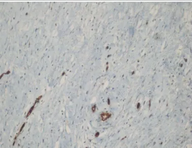
Figure 3. No immunohistochemical staining with CD 117 was detected (CD 117 x200).
Figure 4. CD 34 immunonegativity was observed (CD 34 x200).
DISCUSSION
Although described as a soft tissue tumor of childhood cases presenting at different age groups and with various localisations have been reported recently. Chorti et al. mentioned trimodal age distribution; with first peak in the first 4 years of life, a second peak in the mid-20s and a third peak in the mid-30s; by analysing 104 articles about CFT since the description of disease (6).
Intraabdominal CFTs are usually asymptomatic and detected incidentally by imaging modalities. Non-calcified and well demarcated lesions of CFT can be diagnosed by USG, CT and MRI. In addition to non-calcified well circumscribed lesions, punctate, clustered, amorphous or band like calcifications can also be seen on CT, various echogenicities due to scattered calcifications can be observed by USG (4,5).
In pathological examination well demarcated, unencapsulated lesions containing abundant hyalinized collagen, lymphocytic infiltrations and psammomatous calcifications are characteristic features of CFTs. As in our case CD 34 and CD 117 immunonegativity is significant in the differential diagnosis with GIST and other neoplastic lesions (5).
Treatment of CFT is total excision of the lesions. Only 6 local recurrences are reported and no deaths or metastasis are declared in the literature. In our case, after 18 months follow up, there is no recurrence. Follow up is important. Conflict of interest
No conflict of interest was declared by the authors.
REFERENCES
1. Childhood fibrous tumor with psammoma bodies. Clinicopathologic features in two cases. Rosenthal NS, Abdul-Karim FW. Arch Pathol Lab Med. 1988;112:798-800.
2. Calcifying fibrous pseudotumor. Fetsch JF, Montgomery EA, Meis JM. Am J Surg Pathol. 1993;17:502-8.
3. Montgomery E: Calcifying fibrous tumor. Fletcher CDM, Unni KK, Mertens F, editors. World Health Organization Classification of Tumors, pathology and genetics of tumours of soft tissue and bone. Lyon:IARC Press; 2002;77-8. 4. Calcifying fibrous pseudotumor of lungs. Özkan S, Demirağ F, Yekeler E, Karaoğlanoğlu N. Turk J Med Sci. 2014;44:901-3.
5. Calcifying fibrous tumor: a case report. Gamsızkan M, Yıldırım C, Daş K, Günhan Ö. Turk Patoloji Derg. 2015;31:141-4.
6.Calcifying Fibrous Tumor: Review of 157 Patients Reported in International Literature. Chorti A, Papavramidis TS, Michalopoulos A. Medicine (Baltimore). 2016;95: e3690.