8 D’Ambrosio L, Dara M, Tadolini M, et al. Tuberculosis elimination: theory and practice in Europe. Eur Respir J 2014; 43: 1410–1420.
9 Esposito S, D’Ambrosio L, Tadolini M, et al. ERS/WHO Tuberculosis Consilium assistance with extensively drug-resistant tuberculosis management in a child: case study of compassionate delamanid use. Eur Respir J 2014; 44: 811–815.
10 Codecasa LR, Ciconali G, Mazzola E. Managing an XDR-TB outbreak: the public health face of the medal. Eur Respir J 2015; 45: 292–294.
11 Smit PW, Haanperä M, Rantala P, et al. Molecular epidemiology of tuberculosis in Finland, 2008–2011. PLoS One 2013; 8: e85027
12 Rangaka MX, Wilkinson KA, Glynn JR, et al. Predictive value of interferon-γ release assays for incident active tuberculosis: a systematic review and meta-analysis. Lancet Infect Dis 2012; 12: 45–55.
13 Hinks TS, Varsani N, Godsiff DT, et al. High background rates of positive tuberculosis-specific interferon-γ release assays in a low prevalence region of UK: a surveillance study. BMC Infect Dis 2012; 12: 339.
14 Bradshaw L, Davies E, Devine M, et al. The role of the interferon gamma release assay in assessing recent tuberculosis transmission in a hospital incident. PLoS One 2011; 6: e20770.
15 Fox GJ, Barry SE, Britton WJ, et al. Contact investigation for tuberculosis: a systematic review and meta-analysis. Eur Respir J 2013; 41: 140–156.
Eur Respir J 2015; 45: 276–279 | DOI: 10.1183/09031936.00125914 | Copyright ©ERS 2015
False-negative interferon-
γ release
assay results in active tuberculosis:
a TBNET study
To the Editor:
Tuberculosis is one of the leading causes of morbidity and mortality worldwide [1]. Rapid identification of contagious tuberculosis patients and effective treatment are necessary to prevent the spread of Mycobacterium tuberculosis, the causative bacterium of the disease. Although interferon-γ release assays (IGRAs) have been developed for the diagnosis of latent infection with M. tuberculosis, these assays are sometimes used as adjunctive tests in the diagnostic workup for active tuberculosis, despite poor specificity [2].
A systematic review and meta-analysis [2] found a pooled sensitivity for the diagnosis of culture-proven active tuberculosis of 81% and 92% of the QuantiFERON Gold in-tube test (QFT-GIT) (Qiagen, Dusseldorf, Germany) and the T-SPOT.TB test (Oxford Immunotec, Oxford, UK), respectively. Thus, approximately 8–19% of patients have a negative IGRA result when presenting with active tuberculosis. Several risk factors were associated with negative IGRA results including immunodeficiency, young or advanced age, a negative tuberculin skin test (TST) result, extrapulmonary tuberculosis, disseminated tuberculosis, concomitant tuberculosis treatment and smoking. However, these studies were limited by an observational design implemented in single centres and most of them did not include large numbers of patients with culture-confirmed tuberculosis.
An international, multicentre, retrospective, cross-sectional study was performed by the Tuberculosis Network European Trials Group (TBNET) (www.tb-net.org) to identify risk factors associated with false-negative IGRA results in patients with active tuberculosis.
Clinical data and laboratory results from patients enrolled at 25 participating centres with a confirmed diagnosis of active tuberculosis (i.e. positive M. tuberculosis culture and/or positive M. tuberculosis-specific nucleic acid amplification assay) who had a routine IGRA investigation by the T-SPOT.TB test or the QFT-GIT, as part of the diagnostic evaluation between April 2006 and May 2011, were retrospectively recorded on a standardised anonymous questionnaire. For each patient with a negative IGRA test result, two tuberculosis patients with a positive IGRA result admitted directly before and after the patient with a negative IGRA result were included as controls. Immunocompromised patients were defined as patients with at least one of the following medical conditions: HIV infection, treatment with immunosuppressive
drugs, diabetes mellitus, rheumatoid arthritis, malignancy and/or history of solid-organ or stem cell transplantation.
Logistic regression analysis was carried out to assess potential independent variables associated with a negative IGRA test result in patients with active tuberculosis. All variables with a p-value <0.1 were included in the multivariate analysis. A p-value <0.05 was considered to be statistically significant. Statistical analyses were conducted using Stata 9.0 (StataCorp, College Station, TX, USA).
Data were collected from 771 patients. Of these, 107 patients were excluded because of missing clinical data (n=55), missing nucleic acid amplification testing and/or culture confirmation of tuberculosis (n=21), indeterminate test results (n=17), missing control patients (n=9), identified false data entry (n=3) or patient data duplication (n=1).
For the final analysis, 221 tuberculosis patients with a negative IGRA test result and 442 control tuberculosis patients with a positive IGRA test result (total of 664 patients) were included. 32 tuberculosis patients had a negative T-SPOT.TB, 182 tuberculosis patients had a negative QFT-GIT, and seven tuberculosis patients had both a negative T-SPOT.TB and a negative QFT-GIT test result. The median age (interquartile range) of the patients was 41 (28–56) years in the QFT-GIT and 41 (30–53) years in the T-SPOT.TB group. 26.3% and 27.3% TB patients of the QFT-GIT and T-SPOT.TB test groups, respectively, were immunocompromised.
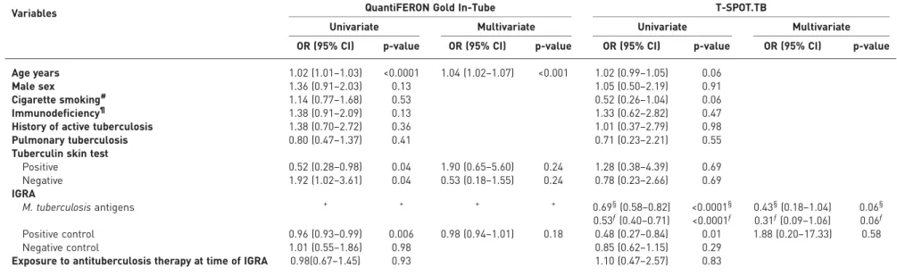
Age resulted the only significant variable associated with a negative QFT-GIT test in patients with tuberculosis in multivariate analysis (OR 1.04, 95% CI 1.02–1.07; p<0.0001) (table 1). Mean±SDage of the cases was 46.94±17.76 years and mean age of the control patients 41.16±16.24 ( p<0.001). Immunocompromised patients did not have a significantly increased risk of a false-negative QFT-GIT test (OR 1.38, 95% CI 0.91–2.09; p=0.13). None of the different diseases with immunodeficiency when analysed independently were associated with an increased risk of a false-negative IGRA test result. In contrast to the QFT-GIT, age was not recognised as a predictor for a negative T-SPOT.TB test results in the univariate analysis (OR 1.02, 95% CI 0.99–1.05; p=0.06). Similar to the QFT-GIT, immunocompromised patients did not have a significant higher probability of a false negative T-SPOT.TB test (OR 1.33, 95% CI 0.62–2.82; p=0.47).
Older age was previously recognised as a risk factor for false-negative IGRA test results and the interferon (IFN)-γ concentration obtained in reaction to the 6-kDa early secretory antigenic target (ESAT-6) or the 10-kDa culture filtrate protein seems (CFP-10) to decrease gradually with age [3]. This can explain the difference between the tests in terms of statistical significance of the variable age as a risk factor for a negative result. The T-SPOT.TB test requires a specific number of peripheral blood mononuclear cells in the assay so that smaller amounts of IFN-γ can be detected, whereas the QFT-GIT uses whole blood without any standardisations of the number of mononuclear cells.
There is also evidence that false-negative IGRA results can be observed more often in younger children. However, because there were no children aged <5 years enrolled in this study, we were unable to address this possible relationship.
In contrast to previous investigations, immunodeficiency, concomitant tuberculosis treatment, disseminated tuberculosis, extrapulmonary tuberculosis and smoking could not be identified as risk factors for false-negative IGRA test results.
Apart from the association with older age, it remains unclear why some individuals with active tuberculosis have unidentifiable M. tuberculosis-specific adaptive immune responses at the time of tuberculosis diagnosis. Results from previous studies have suggested different aetiologies for false-negative IGRA test results that were not evaluated in this study.
During tuberculosis, progression the natural cytokine balance is altered while the bacterial load increases, potentially influencing the performance of IGRA tests [4, 5]. Decreased M. tuberculosis-specific immune responses, especially IFN-γ production [4–7], have been attributed to the immunomodulatory action of CD4+CD25+FoxP3+ regulatory T (Treg)-cells, which expand in the course of active tuberculosis. To support speculatively this experimental hypothesis, TST reactions are reversely related to the frequency of Treg-cells in the peripheral blood [8].
TABLE 1Logistic regression analysis of potential independent variables associated with negative interferon-γ release assay (IGRA) results in patients with active
tuberculosis
Variables QuantiFERON Gold In-Tube T-SPOT.TB
Univariate Multivariate Univariate Multivariate
OR (95% CI) p-value OR (95% CI) p-value OR (95% CI) p-value OR (95% CI) p-value Age years 1.02 (1.01–1.03) <0.0001 1.04 (1.02–1.07) <0.001 1.02 (0.99–1.05) 0.06
Male sex 1.36 (0.91–2.03) 0.13 1.05 (0.50–2.19) 0.91
Cigarette smoking# 1.14 (0.77–1.68) 0.53 0.52 (0.26–1.04) 0.06
Immunodeficiency¶ 1.38 (0.91–2.09) 0.13 1.33 (0.62–2.82) 0.47
History of active tuberculosis 1.38 (0.70–2.72) 0.36 1.01 (0.37–2.79) 0.98 Pulmonary tuberculosis 0.80 (0.47–1.37) 0.41 0.71 (0.23–2.21) 0.55 Tuberculin skin test
Positive 0.52 (0.28–0.98) 0.04 1.90 (0.65–5.60) 0.24 1.28 (0.38–4.39) 0.69 Negative 1.92 (1.02–3.61) 0.04 0.53 (0.18–1.55) 0.24 0.78 (0.23–2.66) 0.69 IGRA M. tuberculosis antigens + + + + 0.69§(0.58–0.82) <0.0001§ 0.43§(0.18–1.04) 0.06§ 0.53ƒ(0.40–0.71) <0.0001ƒ 0.31ƒ(0.09–1.06) 0.06ƒ Positive control 0.96 (0.93–0.99) 0.006 0.98 (0.94–1.01) 0.18 0.48 (0.27–0.84) 0.01 1.88 (0.20–17.33) 0.58 Negative control 1.01 (0.55–1.86) 0.98 0.85 (0.62–1.15) 0.29
Exposure to antituberculosis therapy at time of IGRA 0.98(0.67–1.45) 0.93 1.10 (0.47–2.57) 0.83
#: past or present.¶: e.g. HIV infection, malignancy, transplantation, diabetes mellitus or rheumatoid arthritis.+: 6-kDa early secretory antigen target (ESAT-6), 10-kDa culture filtrate protein (CFP-10) and TB7.7; values≤0.34 predict data perfectly (the output of the logistic regression analysis did not define any specific odds ratios because there is a complete overlap between a specific outcome and the values of the interferon-γ responses lower than 0.35).§: ESAT-6.ƒ: CFP-10.
T-cells, whereas the predominant production of IFN-γ in active tuberculosis occurs at the site of infection [12–14]. It has been observed that a substantial number of tuberculosis patients with negative TST test results at the time of presentation develop positive TST results on antituberculosis therapy.
Our study has limitations. IGRA test results were analysed in retrospect and quantitative test results were not collected. Apart from demographic parameters, immunological mechanisms or genetic causes of false-negative IGRA results could not be studied. Nevertheless, this is the largest study to evaluate false-negative IGRA responses in active tuberculosis to date.
In conclusion, apart from advanced age, we could not identify risk factors for false-negative IGRA results in patients with active tuberculosis. As IGRAs cannot distinguish latent M. tuberculosis infection from tuberculosis, there is need to improve immunodiagnostic methods to distinguish different stages of M. tuberculosis infection [15].
@ERSpublications
TB outbreak investigation can be enhanced by using whole genome sequencing, IGRA and social network analysishttp://ow.ly/AzxfH
Veerle de Visser1, Giovanni Sotgiu2, Christoph Lange3,4,5, Martine G. Aabye6,7, Marleen Bakker8, Filippo Bartalesi9,
Kristian Brat10, Cynthia B.E. Chee11, Keertan Dheda12, Jose Dominguez13, Fusun Eyuboglu14, Maha Ghanem15, Delia Goletti16, Asli Gorek Dilektasli17, Lorenzo Guglielmetti18, Won-Jung Koh19, Irene Latorre13, Monica Losi20,
Monica Polanova21, Pernille Ravn22, Felix C. Ringshausen23, Rudolf Rumetshofer24, Maria Luiza de Souza-Galvão25, Steven Thijsen1, Graham Bothamley26and Aik Bossink1, for the TBNET1
1Dept of Pulmonary Disease, Diakonessenhuis, Utrecht, The Netherlands.2Epidemiology and Medical Statistics Unit,
Dept of Biomedical Sciences, University of Sassari-Research, Medical Education and Professional Development Unit, AOU Sassari, Sassari, Italy. 3Division of Clinical Infectious Diseases, German Center for Infection Research (DZIF),
Research Center Borstel, Borstel, Germany.4Dept of Medicine, University of Namibia School of Medicine, Windhoek, Namibia.5International Health/Infectious Diseases, University of Lubeck, Lubeck, Germany.6Clinical Research Unit,
University of Copenhagen, Hvidovre Hospital, Copenhagen Denmark.7National Institute of Medical Research, Mwanza, Tanzania. 8Erasmus Medical Centre, Rotterdam, The Netherlands. 9SOD Malattie Infettive e Tropicali, Azienda
Ospedaliero-Universitaria Careggi, Florence, Italy.10University Hospital Brno, Brno, Czech Republic.11Tan Tock Seng Hospital, Singapore.12University of Cape Town, Cape Town, South Africa.13Microbiology Dept, Institut d’Investigació
Germans Trias i Pujol, Universitat Autònoma de Barcelona, Ciber Enfermedades Respiratorias, Barcelona, Spain.
14Baskent University Faculty of Medicine, Dept of Pulmonary Diseases, Ankara, Turkey.15Dept of Chest Diseases and
Tuberculosis, Assiut University Hospital, Assiut, Egypt.16IRCCS Instituto Nazionale Malattie Infettive“L. Spallanzani”, Rome, Italy.17Uludag University Faculty of Medicine, Dept of Pulmonary Diseases, Bursa, Turkey.18Unità Operativa
Complessa di Malattie Infettive, University of Verona, Verona, Italy. 19Samsung Medical Center, Sungkyunkwan University School of Medicine, Seoul, South Korea.20University of Modena and Reggio Emillia, Modena, Italy.21The
National Institute of TB, Respiratory Diseases and Thoracic Surgery, Vyšné Hágy, Slovakia.22
Department of Pulmonary and Infectious Diseases, University of Copenhagen, Hillerød Hospital, Denmark.23Hannover Medical School, Dept of
Respiratory Medicine, Hannover, Germany.24Treatment Center for Tuberculosis, Dept of Respiratory and Critical Care Medicine, Otto Wagner Hospital, Vienna, Austria. 25Drassanes Tuberculosis Unit, Vall d’Hebron Teaching Hospital,
Barcelona, Spain.26Dept of Respiratory Medicine, Homerton University Hospital, London, UK.
Correspondence: Veerle de Visser, Dept of Pulmonary Disease, Diakonessenhuis, Bosboomstraat 1, Utrecht, The Netherlands. Email: [email protected]
Received: June 30 2014 | Accepted after revision: Sept 29 2014 | First published online: Oct 30 2014 Conflict of interest: Disclosures can be found alongside the online version of this article at erj.ersjournals.com
References
1 World Health Organization. Global tuberculosis report 2013. Geneva, World Health Organization, 2013.
2 Sester M, Sotgiu G, Lange C, et al. Interferon-γ release assays for the diagnosis of active tuberculosis: a systematic review and meta-analysis. Eur Respir J 2011; 37: 100–111.
3 Hang NT, Lien LT, Kobayashi N, et al. Analysis of factors lowering sensitivity of interferon-γ release assay for tuberculosis. PLoS One 2011; 6: e23806
4 Kobashi Y, Mouri K, Yagi S, et al. Clinical utility of the QuantiFERON TB-2G test for elderly patients with active tuberculosis. Chest 2008; 133: 1196–1202.
5 Vanham G, Toossi Z, Hirsch CS, et al. Examining a paradox in the pathogenesis of human pulmonary tuberculosis: immune activation and suppression/anergy. Tuber Lung Dis 1997; 78: 145–168.
10 Arend SM, Geluk A, van Meijgaarden KE, et al. Antigenic equivalence of human T-cell responses to Mycobacterium tuberculosis-specific RD1-encoded protein antigens ESAT-6 and culture filtrate protein 10 and to mixtures of synthetic peptides. Infect Immun 2000; 68: 3314–3321.
11 Lange C, Pai M, Drobniewski F, et al. Interferon-γ release assays for the diagnosis of active tuberculosis: sensible or silly? Eur Respir J 2009; 33: 1250–1253.
12 Losi M, Bossink A, Codecasa L, et al. Use of a T-cell interferon-γ release assay for the diagnosis of tuberculous pleurisy. Eur Respir J 2007; 30: 1173–1179.
13 Jafari C, Ernst M, Strassburg A, et al. Local immunodiagnosis of pulmonary tuberculosis by enzyme-linked immunospot. Eur Respir J 2008; 31: 261–265.
14 Jafari C, Ernst M, Diel R, et al. Rapid diagnosis of smear negative tuberculosis by bronchoalveolar-lavage enzyme-linked immunospot. Am J Respir Crit Care Med 2006; 174: 1048–1054.
15 Chegou NN, Heyckendorf J, Walzl G, et al. Beyond the IFN-γ horizon: biomarkers for immunodiagnosis of infection with Mycobacterium tuberculosis. Eur Respir J 2014; 43: 1472–1486.