Review Article
Therapeutic role of rifampicin in Alzheimer
’s disease
Burak Yulug,
MD,
1,2* Lütfü Hanoglu,
MD,
1,2Mehmet Ozansoy,
PhD,
2,3Dogan Is
ık,
MD,
4Ulkan Kilic,
PhD,
5Ertugrul Kilic,
PhD2,3and Wolf Rüdiger Schabitz,
MD6Departments of1Neurology,2Restorative and Regenerative Medicine,3Physiology, International School of Medicine, 4
Psychiatry, Istanbul Medipol University,5Department of Medical Biology, Faculty of Medicine, University of Health Sciences, Istanbul, Turkey, and6Department of Neurology, Bethel, EVKB, Bielefeld, University of Münster, Münster, Germany
Rifampicin exerts significant brain protective func-tions in multiple experimental models. Here we summarize the underlying mechanisms of the neuroprotective and pro-cognitive effects of rifam-picin that are mediated by its anti-inflammatory, anti-tau, anti-amyloid, and cholinergic effects. Beyond suggesting that rifampicin shows strong brain protective effects in preclinical models of Alzheimer’s disease, we also provide substantial
clinical evidence for the neuroprotective and pro-cognitive effects of rifampicin. Future neuroimag-ing studies combined with clinical assessment scores are the following steps to be taken in this field of research.
Key words: Alzheimer’s disease, amyloid beta clear-ance, neuroprotection, oligomeric amyloid hypothe-sis, rifampicin.
A
LZHEIMER’S DISEASE (AD) IS the most impor-tant cause of a progressive decline of cognitive and memory functions among older people. It is a progressive neurodegenerative disease and ulti-mately leads to disturbance of memory and cogni-tive function, finally resulting in dementia.1 Clinically, there are different stages of dementia that typically progress slowly from mild (early) to severe (late stage).1 It is important to note that neurode-generative changes in the affected regions of the Alz-heimer’s brain begin years before any signs of the disease are prominent. This latent period, which can last for years, is called preclinical AD, representing the target for any neuroprotective/neuropreventive approach. A therapeutic approach effectively block-ing this neurodegenerative cascade would show huge benefits in the progress of this disease. The research of recent years has led to an improved pre-cision in detection of early forms of AD by variousmetabolic measurements, such as glucose positron emission tomography (PET) and amyloid PET.
In this respect, improvement of early stage diagno-sis has shifted focus more and more towards protec-tion of neuronal cell populaprotec-tions in this early period of neurodegeneration. Although the classical‛time is brainʼ concept of stroke cannot be generalized to the relatively slow neurodegenerative nature of AD, it is important to note that early intervention in the criti-cal period involving a preventive approach would provide important benefits. In this context, it has already been shown that early molecular interactions guided by early onset of neuroinflammation play an important role in the progress of AD.2–5Considering all these factors, it is not unreasonable to assume that there are some basic pathophysiological similarities between acute stroke and the early period of AD. In connection with this, an acute therapeutic approach similar to the fibrin dissolving therapy in stroke can also be considered for the early period of AD, which is characterized with accumulated amyloid fibrils. Accordingly, recent studies have revealed that the microglia-driven oxidative pathway modulates the amyloid beta (Aß) metabolism through the key mediators of neuroinflammation during AD.3–5 *Correspondence: Burak Yulug, MD, Department of Neurology,
Istanbul-Medipol University, Goztepe cıkısı No.1, Istanbul 34214, Turkey. Email: [email protected]
Received 26 September 2017; revised 6 December 2017; accepted 17 December 2017.
In contrast to the conventional Aß concept, the alternative oligomeric amyloid hypothesis, which is closely linked to early neuroinflammation, enables us to investigate the acute/subacute neuroprotective effect of some anti-inflammatory and anti-oxidant drugs that have already been shown to be neuropro-tective in cerebrovascular diseases.6,7 Regarding this, antibiotics were demonstrated to counteract these processes, reduce reperfusion failure, free radical for-mation, and ultimately reduce ischemic damage.6,7
Rifampicin is one of several neuroprotective anti-biotics that may simultaneously modulate the neu-roinflammatory response and Aß metabolism in AD.6 Our argument to involve rifampicin is that it combines an anti-inflammatory and anti-oxidative property that is capable of crossing the blood–brain barrier (BBB) with neuroprotective properties that help by limiting inflammation and oxidative stress. However, despite considerable literature on the neu-roprotective effect of rifampicin, there are limited and controversial data on the therapeutic role of rifampicin in AD. This might be due to the lack of understanding of pathophysiological underpinnings of AD. Notably, there is a growing body of patho-physiological evidence suggesting that there is cross talk between amyloid metabolism, free radical injury, and neuroinflammation. In light of these findings, we aimed to provide an update on the neu-roprotective role of rifampicin in AD. Beyond sum-marizing the conventional and alternative amyloid hypothesis, we have also evaluated the most signi fi-cant Aß clearance mechanisms that may play an important role in mediating the neuroprotective role of rifampicin in AD. To that end, we have been focused on the AD-relevant anti-inflammatory, anti-oxidant, anti-amyloidogenic effects of rifampicin.
CONVENTIONAL AMYLOID
HYPOTHESIS
The neuropathological feature of AD is defined as the accumulation of extracellular senile plaques and intracellular neurofibrillary tangles associated with significant neuronal cell death and related brain atrophy.8–11 It has been shown that the complex enzymatic cleavage of transmembrane amyloid-precursor protein (APP) leads to the generation of Aß from APP.12,13Moreover, in a healthy brain, the clearance of the amyloid load is critical for the maintenance of normal brain function while the dis-turbance in the balance between protein fragment
production and its clearance12,13may result in amy-loid accumulation closely linked to free radical pro-duction.14,15 This suggests that in addition to amyloid accumulation, inflammation and oxidative injury play major roles in AD pathogenesis. In this respect, the latest research suggests that micromolar concentrations of Aß in contrast to picomolar con-centrations within the brain cause neurotoxicity and subsequent neurodegeneration.2,14 During this phase, it has already been demonstrated that microglia-driven inflammatory response that is due to Aß aggregation increases over time as the disease progresses.2,15 These findings suggest that there is a cross talk between the amyloid and tau-related neu-rotoxicity and neuroinflammation.
EARLY INFLAMMATION AND
OLIGOMERIC AMYLOID HYPOTHESIS:
A NEW CROSS TALK?
In contrast to the conventional Aß cascade hypothe-sis, which plays a critical role in sustaining neuroin-flammation in the later and chronic stages, the oligomeric amyloid hypothesis proposes that toxic amyloid oligomers may trigger glial activation in the early stages of inflammation.16,17 Based on these findings, it is not unreasonable to assume that cog-nitive dysfunction, beginning early in the disease, can be attributed to oligomer-induced disruption of synaptic plasticity at its earliest, which is followed by oligomer-induced neuronal degeneration with later stages. These Aß oligomers ranging from dimers to higher polymers have been identified in AD brain extracts while experimental studies indi-cate that these oligomers may impair excitatory
syn-apses through a receptor complex involving
mGluR5 and NMDA receptors leading to specific synaptic dysfunction, which also includes the inhibi-tion of LTP.16–19More generally, it has already been revealed that Aβ oligomers initiate several neuro-pathological processes involving the axonal dysfunc-tion, tau hyperphosphoryladysfunc-tion, dysregulation of Ca2+ homeostasis, oxidative stress, mitochondrial damage, energy depletion, endoplasmic reticular stress, proteasome inhibition, cell cycle re-entry, acti-vation astrocytes, and microglia, which are character-ized by prominent neuroinflammation and directly correlate to brain dysfunction in AD.16 Taken together, based on its critical role for early neuroin-flammatory response and related neuronal dysfunc-tion through microglial activadysfunc-tion and increased
levels of inflammatory markers (i.e., inducible nitric oxide, nitric oxide, and tumor necrosis factor-α),17it can be hypothesized that early memory loss in AD can be reversed with feasible anti-inflammatory, anti-oligomeric, and neuroprotective agents, such as rifampicin. Thus, studies have already revealed that rifampicin inhibits Aß oligomerization to produce monomeric components that form less toxic insolu-blefibrils.
ROLE OF PERMEABILITY
GLYCOPROTEIN AND LIPOPROTEIN
RECEPTOR-RELATED PROTEIN-1
TRANSPORTERS AND BBB
CLEARANCE MECHANISMS IN AD
Aβ-peptide is at the center of the disease pathogene-sis and is responsible for the generation of amyloid plaques through proteolytic cleavage by β- and γ-secretases20–23 which are important neuropatho-logical hallmarks of AD20–23 Depending on the cleavage site byγ-secretase, Aβ40and Aβ42differ not only in their length but also in their production and cleavage rate.24 During this process, Aβ40 is pro-duced at a significantly greater rate than Aβ42, while the critical point for the neurodegenerative process is determined by thefinal levels of free Aβ, which is related to the balance between generation, aggrega-tion, degradaaggrega-tion, and clearance from the brain.20–24 Thefinal concentrations of Aβ also showed a signifi-cant correlation with synaptic pathology and the presence of dementia.22 Recent evidence indicates that disturbed clearance of Aβ from the brain also plays an important role in sporadic and familial forms of AD.25,26The homeostasis of Aβ is critically dependent on its clearance, indicating that sufficient amounts of Aβ should be eliminated from the brain to maintain the critical balance between the produc-tion and clearance of Aβ proteins. Accordingly, recent studies also suggest that the endocytic elimi-nation via transmembrane protein lipoprotein receptor-related protein-1 plays an important role in the clearance of Aβ from the brain. It has already been demonstrated that low- density lipoprotein receptor-related protein 1 (LRP1) reveals a diverse modifying effect on amyloid trafficking in the brain.27,28 Additionally, the efflux transporter per-meability glycoprotein (P-gp), which is located on brain capillary endothelial cells in the BBB and belongs to the adenosine-triphosphate-binding cas-sette B1 transporter family contributes to the
elimination of Aβ1–40 and Aβ1–42 from the brain across the BBB.29–32 In agreement with this, recent evidence suggests that decreased transport activity of P-gp is associated with increased levels of Aβ in aged humans and in the specific brain regions affected in AD patients.29 These findings together suggest the important role of Aß transport across the BBB in the pathogenesis of AD.
THE NEUROPROTECTIVE EFFECT OF
RIFAMPICIN IN AD
Preclinical studies
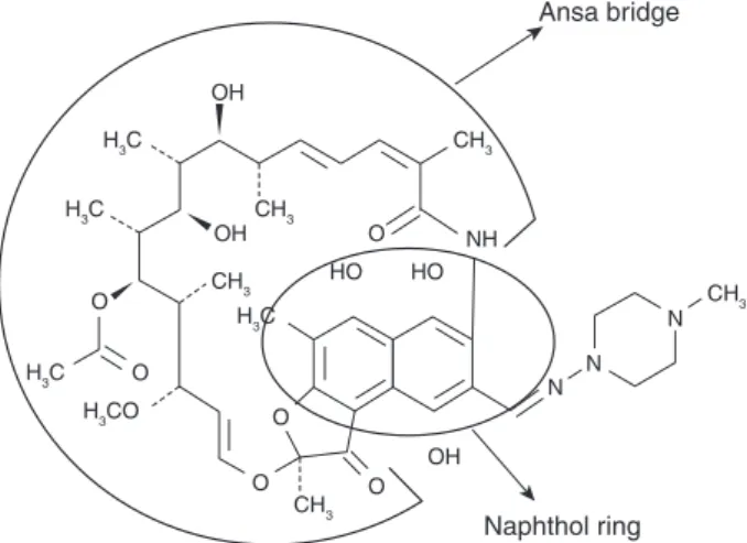
Rifampicin is a broad-spectrum antibiotic and belongs to the fermentation products of Nocardia mediterranei.4–6,33–35 Rifampicin consists of a naphtho-hydroquinone chromophore spanned by a lipophilic aliphatic ansa chain33 (Fig. 136) that is mainly responsible for the transport of the drug across the BBB into the brain parenchyma.34,35 Rifampicin reaches maximal serum concentration 1–4 h after application and its plasma half-time is 2–5 h.34,35 Beyond its conventional anti-infectious effect, rifampicin also exerts significant neuroprotec-tive effect in various experimental studies. The neu-roprotective effect of rifampicin has already been shown in various neurodegenerative diseases, including Parkinson’s disease and multisystem atro-phy. The underlying mechanism of action includes increased dopaminergic cell survival, decreased
alpha synuclein toxicity, and MPP+-induced
H3C CH3 CH3 CH3 CH3 CH3 H3C H3C H3CO H3C OH OH HO HO NH O O O O OH O O N N N Ansa bridge Naphthol ring
Figure 1. Structural formula of rifampicin, including the ansa bridge and naphthol ring, with oxygen atoms and hydroxyls (modified from Campbell et al.36with permission).
apoptosis, which are associated with the stabiliza-tion of the mitochondrial and endoplasmic reticular stress via modulation of critical cytoprotective chap-eron and anti-apoptotic proteins (i.e., glucose-regulated protein 78 and Bcl-2).37–42 Moreover, there is rapidly replicating evidence showing that rifampicin may attenuate the free radical injury and decrease the neuroinflammation that finally result with significant neuroprotective effect.43 For instance, a recent study indicated that rifampicin may decrease toll-like receptor 2 (TLR2) and mitogen-activated protein kinases (MAPK) via pressing the nuclear factor-kappa B, which may sup-port the potential anti-inflammatory role of rifampicin in Parkinson’s disease.42,44,45
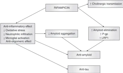
Considering all this valuable evidence, we pre-ferred to summarize the neuroprotective mecha-nisms of rifampicin (Fig. 2) that are most relevant to the pathogenesis of AD. These include its inhibi-tory activity on free oxygen radicals, tau and Aβ pro-tein accumulation, microglial activation, apoptotic cascades, and its most recently defined stimulating effect on brain Aß clearance through LRP1 and P-gp. It has already been shown that free radical produc-tion plays an important role in the generaproduc-tion of Aß.14,15Accordingly, studies have revealed the thera-peutic role of antioxidative agents in Aß-plaque-related neurotoxicity in AD. The first in vitro study was conducted by Tomiyama et al., who demon-strated that rifampicin inhibited aggregation and fibril formation of synthetic Aβ1–40peptide and pre-vented neurotoxicity in a dose-dependent manner in rat pheochromocytoma PC12 cells.46 The authors
used the electron spin resonance technique com-bined with a cytotoxicity assay and demonstrated that rifampicin revealed significant anti-oxidative effects via its hydroquinone (or naphtho-quinone) ring while the main responsible anti-scavenger (hydroxyl radical anti-scavenger) activity was attributed to the hydroxyl group at position C-1 of the naphtho-hydroquinone.33 Interestingly, those authors also demonstrated that rifampicin was 10–100-fold more effective than vitamins in inhibit-ing Aß aggregation. Another study also showed that the ansa chain of rifampicin was not essential for the Aß aggregation inhibitory activities while its lipophi-licity contributed significantly to transport of the drug molecule into the brain in vivo.35 The majority of in vitro studies confirmed the anti-amyloid effect of rifampin, including inhibition of amyloid fibril formation.47,48 These findings together suggest that both the rifampicin-mediated inhibition of Aß aggre-gation and its free radical scavenging effects could be essential for the therapeutic role of rifampicin in AD. These study results are in agreement with previ-ous findings, suggesting that the anti-amyloid effect of rifampicin includes radical-scavenging activity associated with decreased peptide aggregation, amyloid-binding, and amyloid–cell interaction. Interestingly, further studies evaluating the anti-amyloid effect of rifampicin on amylin fibrin aggre-gation and related toxicity have revealed that the inhibitory effect was induced by their binding to peptidefibrils rather than by their intracellular anti-oxidant action.49–51These study results suggested the presence of an additional mechanism, other than
Anti-inflammatory effect ↓ Oxidative stress ↓ Neutrophilic infiltration ↓ Microglial activation Anti-oligomeric effect RIFAMPICIN ↑ Cholinergic transmission
↓ Amyloid aggregation ↑ Amyloid elimination↑ P-gp ↑ LRP1
Anti-amyloid
Anti-tau
Figure 2. The underlying mecha-nisms of the neuroprotective role of rifampicin in Alzheimer’s disease.
scavenging of free radicals, by which rifampicin and its analogs may decrease the Aß toxicity. These pre-clinical data were confirmed by Lieu et al.,49 who demonstrated that rifampicin inhibited amyloid fibrillogenesis of lysozyme in a concentration-dependent manner utilizing a hen egg-white lyso-zyme model, which is a protein-unfolding model that is widely used in crystallography. The subse-quent cell culture studies usingfluorescence spectro-metric analysis showed that rifampicin displayed significant anti-oxidant and anti-amyloid activity, including the inhibition of preformedfibril accumu-lation.49,51In their molecular-based neuropreventive study for AD, Umeda et al. have very recently revealed that rifampicin conferred significant activity against the accumulation of Aß and tau oligomers in various transgenic mice models.52 The administra-tion of rifampicin for 1 month resulted in significant decrease in amyloid and tau toxicity associated with improved synapse loss and microglial activation. Moreover, rifampicin also improved memory loss and inhibited apoptotic cascades, including the cas-pase 3 activation and cytochrome c release in the hippocampus.52 Furthermore, those authors also revealed that rifampicin stimulated the restoration of autophagy-lysosomal function. Although there were some minor differences in amyloid deposition in the different transgenic mice models (i.e., tauopathy, amyloid oligomer, and AD model) used in this study, the results indicated that rifampicin exerted significant inhibitor activity on tau and amyloid olig-omer accumulation, tau hyperphosphorylation, microglial activation, and apoptotic cascades, which were positively correlated to the neurocognitive out-comes.52These findings together suggest a potential role of rifampicin as a neuroprotective agent but also as an effective neuropreventive agent in Alzheimer’s and other neurodegenerative diseases.
From another point of view, recent evidence strongly suggests that impaired clearance of Aß across the BBB might lead to the formation of Aß brain deposits and AD progression.53–55 The efflux transporter P-gp may play an important role in the elimination of Aβ1–40 and Aβ1–42 from the brain across the BBB.53–55 In agreement with this, further studies have demonstrated that decreased intracellu-lar accumulation of A1–40 is associated with P-gp upregulation caused by rifampicin56,57 These find-ings are consistent with the correlation between in vitro concentration-dependent increase in P-gp expression and activity by rifampicin.58 Moreover, a
very recent mice study has shown that the upregula-tion of low-density LRP1 and P-gp at the BBB by rifampicin and caffeine enhanced brain amyloid clearance.59This suggested the presence of a possible transporter/receptor that plays an important role in amyloid clearance, which is upregulated by rifampi-cin.60 Accordingly, a very recent study by Kaur et al. has revealed that rifampicin significantly improved memory dysfunction and locomotor impairment in a rat dementia model.61By evaluating the underlying mechanisms of neuroprotection, they have interest-ingly shown that rifampicin significantly reduced the oxidative stress, neutrophilic infiltration, amyloid deposition, and acetylcholinesterase activity that was reversed with the inhibition of Pregnane X receptors (PXR), which mediate the upregulation of P-gp expression. Beyond suggesting the anti-oxidative, anti-inflammatory, and amyloid-lowering effects of rifampicin, this study also indicated that rifampicin could enhance cholinergic neurotransmission through its anti-cholinesterase activity. Additionally, rifampicin’s recently defined agonist activity on PXR may provide further rationale for the potential role of PXR in mediating the procognitive and neuropro-tective effect of rifampicin in the pathophysiol-ogy of AD.
It should also be noted that there are very rare in vitro studies indicating that rifampicin does not inhibit Aß protein aggregation and neurotoxicity in pancreatic islet cells and cerebral cortical neu-rons.62,63Despite these conflicting in vitro data, there are rapidly replicating in vivo and in vitro studies that are supporting a possible relation between P-gp dys-regulation and cognitive improvement in patients with AD. These findings together indicate that in addition to oxidative injury and fibril aggregation, targeting the expression of P-gp by rifampicin could be an effective strategy in decreasing the progres-sion of AD.
Clinical studies
In contrast to the preclinical data, only a few clinical studies have evaluated the effects of rifampicin in patients with AD regarding efficacy and outcome. Namba et al. examined 16 brains from leprosy patients without dementia and compared the neuro-fibrillary tangles and senile plaques in 140 Japanese non-demented elderly subjects through immunohis-tochemical staining.64 Interestingly they have reported that non-demented elderly leprosy patients
who were on rifampicin treatment for years showed an unusual absence of senile plaques in their brains compared with age-matched controls.64Surprisingly, these results could not be replicated with the follow-ing work showfollow-ing that rifampicin does not affect the prevalence of AD in leprosy patients.62 Subsequent studies that were designed to understand the causal antidementia effect of rifampicin also failed to pro-vide clear clinical data. Loeb et al. have confirmed the anti-dementia effects of oral rifampicin (300 mg/daily for 3 months) resulting in a signi fi-cant increase in cognitive function in 101 mild– moderate AD patients measured by a Standardized Alzheimer’s Disease Assessment Scale – Cognitive Subscale score.65 Unfortunately, these promising data could not be confirmed in a study that included a 12-month rifampicin treatment.66 In this study, short-term treatment effect might have been overruled by a lack of long-term treatment effect of rifampicin.
In line with this, Iizuka et al. described that pre-ventive effect of rifampicin needs at least 450 mg daily for 1 year even during the pre-dementia stage.67 In their interesting retrospective FDG-PET study, rifampicin therapy in the predementia stage significantly improved the metabolic (posterior cin-gulate gyrus) and cognitive decline in the long-term follow up. Rifampicin dose and treatment (450 mg/ day for ≥12 months) significantly improved the FDG uptake in the posterior cingulate gyrus, which was reflected in Mini-Mental State Examination scores. These findings together suggested that the failure in clinical trials with rifampicin is probably due to the late timing and insufficient dosage of the treatment and indicate the importance of higher doses and longer treatment duration of preventive treatment. In this respect, adverse events (i.e., drug resistance and gastrointestinal side-effects) associ-ated with long-term use can easily be overcome with combined antibiotic treatment strategy and quick dose adjustment due to the short half-life of rifam-picin. Moreover, it should also be remembered that different clinical study designs, as well as some well-known systematic translation failures of preclinical findings into the human situation, could be respon-sible for inconsistent clinical results.
CONCLUSION
Besides its anti-infectious properties, rifampicin also exerts strong brain protective effects in various
models of neurodegeneration and brain trauma. Fur-thermore, pilot clinical studies indicate that patients with AD may benefit from rifampicin treatment. Despite these positive study results, clinical con fir-matory evidence remains scarce and results are inconsistent.61–67To overcome the above-mentioned barriers to consistent clinical data, well-designed ran-domized clinical research with larger experimental pharmacological combination studies is needed. This approach should also be combined with prospective functional neuroimaging data (etc., amyloid-PET) and clinical assessment scores. This multimodal strategy would help us to enlighten the clinical rele-vance of the neuroprotective effect of rifampicin, which may also offer a significantly low-cost alterna-tive to the long-duration of expensive antidementia treatment, especially in third-world countries.
DISCLOSURE STATEMENT
There is no conflict of interest in this study.
AUTHOR CONTRIBUTIONS
B.Y.: Conception and design of the study; acquisi-tion and analysis of data. L.H.: Drafting the manu-script and figures. M.O.: Drafting the manuscript. U.K.: Acquisition and analysis of data; drafting the figures. E.K.: Acquisition and analysis of data; draft-ing the manuscript. W.R.S.: Conception and design of the study; acquisition and analysis of data; draft-ing the manuscript.
REFERENCES
1. Geldmacher DS, Whitehouse PJ. Evaluation of dementia. N. Engl. J. Med. 1996; 335: 330–336.
2. Bolós M, Perea JR, Avila J. Alzheimer’s disease as an inflammatory disease. Biomol. Concepts 2017; 8: 37–43. 3. Berkinbosch F, Biewenga J, Brouns M, Rozemuller JM,
Strijbos P, van Dam A-M. Cytokines and inflammatory proteins in Alzheimer’s disease. Res. Immunol. 1995; 143: 657–663.
4. Chao CC, Hu S, Sheng WS, Bu D, Bukrinsky MI, Peterson PK. Cytokine-stimulated astrocytes damage human neurons via a nitric oxide mechanism. Glia 1996; 16: 276–284.
5. McGeer PL, McGeer EG. The amyloid cascade-inflammatory hypothesis of Alzheimer disease: Implica-tions for therapy. Acta Neuropathol. 2013; 126: 479–497. 6. Yulug B, Hanoglu L, Kilic E, Schabitz WR. Rifampicin: An
antibiotic with brain protective function. Brain Res. Bull. 2014; 107: 37–42.
7. Stock ML, Fiedler KJ, Acharya S et al. Antibiotics acting as neuroprotectants via mechanisms independent of their anti-infective activities. Neuropharmacology 2013; 73: 174–182. 8. Schaeffer EL, Figueiro M, Gattaz WF. Insights into
Alzhei-mer disease pathogenesis from studies in transgenic animal models. Clinics (Sao Paulo) 2011; 66 (Suppl. 1): 45–54. 9. Estus S, Tucker HM, van Rooyen C et al. Aggregated
amyloid-beta protein induces cortical neuronal apoptosis and concomitant “apoptotic” pattern of gene induction. J. Neurosci. 1997; 17: 7736–7745.
10. Hardy JA, Higgins GA. Alzheimer’s disease: The amyloid cascade hypothesis. Science 1992; 256: 184–185.
11. Philipson O, Lord A, Gumucio A, O’Callaghan P, Lannfelt L, Nilsson LN. Animal models of amyloid-beta-related pathologies in Alzheimer’s disease. FEBS J. 2010; 277: 1389–1409.
12. Cummings JL. Alzheimer’s disease. N. Engl. J. Med. 2004; 351: 56–67.
13. Morrison AS, Lyketsos C. The pathophysiology in Alzhei-mer’s disease and directions in treatment. Adv. Stud. Nurs. 2005; 3: 256–270.
14. Dyrks T, Dyrks E, Hartmann T, Masters C, Beyreuther K. Amyloidogenicity of beta A4 and beta A4-bearing amy-loid protein precursor fragments by metal-catalyzed oxi-dation. J. Biol. Chem. 1992; 267: 18210–18217.
15. Hensley K, Carney JM, Mattson MP et al. A model for beta-amyloid aggregation and neurotoxicity based on free radical generation by the peptide: Relevance to Alzheimer disease. Proc. Natl. Acad. Sci. U. S. A. 1994; 91: 3270–3274.
16. Ferreira ST, Klein WL. The Aβ oligomer hypothesis for synapse failure and memory loss in Alzheimer’s disease. Neurobiol. Learn. Mem. 2011; 96: 529–543.
17. White JA, Manelli AM, Holmberg KH, Van Eldik LJ, Ladu MJ. Differential effects of oligomeric and fibrillar amyloid-beta 1-42 on astrocyte-mediated inflammation. Neurobiol. Dis. 2005; 18: 459–465.
18. Zhao WQ, Santini F, Breese R et al. Inhibition of calcineurin-mediated endocytosis and alpha-amino-3-hydroxy-5-methyl-4-isoxazolepropionic acid (AMPA) receptors prevents amyloid beta oligomer-induced synap-tic disruption. J. Biol. Chem. 2010; 285: 7619–7632. 19. Renner M, Lacor PN, Velasco PT et al. Deleterious effects
of amyloid beta oligomers acting as an extracellular scaf-fold for mGluR5. Neuron 2010; 66: 739–754.
20. De Strooper B, Saftig P, Craessaerts K et al. Deficiency of presenilin-1 inhibits the normal cleavage of amyloid pre-cursor protein. Nature 1998; 391: 387–390.
21. Martin JB. Molecular basis of the neurodegenerative dis-orders. N. Engl. J. Med. 1999; 340: 1970–1980.
22. Lue LF, Kuo YM, Roher AE et al. Soluble amyloid beta pep-tide concentration as a predictor of synaptic change in Alz-heimer’s disease. Am. J. Pathol. 1999; 155: 853–862. 23. Campion D, Dumanchin C, Hannequin D et al.
Early-onset autosomal dominant Alzheimer disease: Prevalence,
genetic heterogeneity, and mutation spectrum. Am. J. Hum. Genet. 1999; 65: 664–670.
24. Deane R, Zlokovic BV. Role of the blood-brain barrier in the pathogenesis of Alzheimer’s disease. Curr. Alzheimer Res. 2007; 4: 191–197.
25. Zlokovic BV. The blood-brain barrier in health and chronic neurodegenerative disorders. Neuron 2008; 57: 178–201. 26. Zlokovic BV. Clearing amyloid through the blood-brain
barrier. J. Neurochem. 2004; 89: 807–811.
27. Sagare A, Deane R, Bell RD et al. Clearance of amyloid-beta by circulating lipoprotein receptors. Nat. Med. 2007; 13: 1029–1031.
28. Shibata M, Yamada S, Kumar SR et al. Clearance of Alz-heimer’s amyloid-ss(1–40) peptide from brain by LDL receptor-related protein-1 at the blood-brain barrier. J. Clin. Invest. 2000; 106: 1489–1499.
29. Vogelgesang S, Cascorbi I, Schroeder E et al. Deposition of Alzheimer’s beta-amyloid is inversely correlated with P-glycoprotein expression in the brains of elderly non-demented humans. Pharmacogenetics 2002; 12: 535–541. 30. Deo AK, Borson S, Link JM et al. Activity of
P-glycopro-tein, a beta-amyloid transporter at the blood-brain bar-rier, is compromised in patients with mild Alzheimer disease. J. Nucl. Med. 2014; 55: 1106–1111.
31. Hartz AM, Miller DS, Bauer B. Restoring blood-brain bar-rier P-glycoprotein reduces brain amyloid-beta in a mouse model of Alzheimer’s disease. Mol. Pharmacol. 2010; 77: 715–723.
32. Wang W, Bodles-Brakhop AM, Barger SW. A role for P-glycoprotein in clearance of Alzheimer amyloidβ-peptide from the brain. Curr. Alzheimer Res. 2016; 13: 615–620. 33. Tomiyama T, Shoji A, Kataoka K et al. Inhibition of
amy-loid beta protein aggregation and neurotoxicity by rifam-picin: Its possible function as a hydroxyl radical scavenger. J. Biol. Chem. 1996; 271: 6839–6845.
34. Acocella G. Clinical pharmacokinetics of rifampicin. Clin. Pharmacokinet. 1978; 3: 108–127.
35. Mindermann T, Landolt H, Zimmerli W, Rajacic Z, Gratzl O. Penetration of rifampicin into the brain tissue and cerebral extracellular space of rats. J. Antimicrob. Che-mother. 1993; 31: 731–737.
36. Campbell EA, Korzheva N, Mustaev A et al. Structural mechanism for rifampicin inhibition of bacterial RNA polymerase. Cell 2001; 104: 901–912.
37. Lin D, Jing X, Chen Y et al. Rifampicin pre-treatment inhibits the toxicity of rotenone-induced PC12 cells by enhancing sumoylation modification of α-synuclein. Bio-chem. Biophys. Res. Commun. 2017; 485: 23–29.
38. Low PA, Robertson D, Gilman S et al. Efficacy and safety of rifampicin for multiple system atrophy: A randomised, double-blind, placebo-controlled trial. Lancet Neurol. 2014; 13: 268–275.
39. Ubhi K, Rockenstein E, Mante M et al. Rifampicin reduces alpha-synuclein in a transgenic mouse model of multiple system atrophy. Neuroreport 2008; 19: 1271–1276.
40. Xu J, Wei C, Xu C et al. Rifampicin protects PC12 cells against MPP+-induced apoptosis and inhibits the expres-sion of an alpha-Synuclein multimer. Brain Res. 2007; 1139: 220–225.
41. Li J, Zhu M, Rajamani S, Uversky VN, Fink AL. Rifampicin inhibits alpha-synuclein fibrillation and disaggregates fibrils. Chem. Biol. 2004; 11: 1513–1521.
42. Bi W, Zhu L, Wang C et al. Rifampicin inhibits microglial inflammation and improves neuron survival against inflammation. Brain Res. 2011; 13: 12–20.
43. Kilic U, Kilic E, Lingor P, Yulug B, Bähr M. Rifampicin inhibits neurodegeneration in the optic nerve tran-section model in vivo and after 1-methyl-4-phenylpyridinium intoxication in vitro. Acta Neuropathol. 2004; 108: 65–68.
44. Kim SK, Kim YM, Yeum CE, Jin SH, Chae GT, Lee SB. Rifampicin inhibits the LPS-induced expression of Toll-like Receptor 2 via the suppression of NF-kappaB DNA-binding activity in RAW 264.7 cells. Korean J. Physiol. Pharmacol. 2009; 13: 475–482.
45. Bi W, Zhu L, Jing X, Liang Y, Tao E. Rifampicin and Par-kinson’s disease. Neurol. Sci. 2013; 34: 137–141.
46. Tomiyama T, Asano S, Suwa Y et al. Rifampicin prevents the aggregation and neurotoxicity of amyloid beta protein in vitro. Biochem. Biophys. Res. Commun. 1994; 204: 76–83. 47. Tomiyama T, Kaneko H, Kataoka K, Asano S, Endo N. Rifampicin inhibits the toxicity of pre-aggregated amyloid peptides by binding to peptide fibrils and preventing amyloid–cell interaction. Biochem. J. 1997; 322: 859–865. 48. Findeis MA. Approaches to discovery and characterization of inhibitors of amyloid beta-peptide polymerization. Biochim. Biophys. Acta 2000; 1502: 76–84.
49. Lieu VH, Wu JW, Wang SS, Wu CH. Inhibition of amyloid fibrillization of hen egg-white lysozymes by rifampicin and p-benzoquinone. Biotechnol. Prog. 2007; 23: 698–706. 50. Ono K, Hasegawa K, Yoshiike Y, Takashima A,
Yamada M, Naiki H. Nordihydroguaiaretic acid potently breaks down pre-formed Alzheimer’s beta-amyloid fibrils in vitro. J. Neurochem. 2002; 81: 434–440.
51. Ono K, Hamaguchi T, Naiki H, Yamada M. Anti-amyloidogenic effects of antioxidants: Implications for the prevention and therapeutics of Alzheimer’s disease. Biochim. Biophys. Acta 2006; 1762: 575–586.
52. Umeda T, Ono K, Sakai A et al. Rifampicin is a candidate preventive medicine against amyloid-β and tau oligo-mers. Brain 2016; 139: 1568–1586.
53. Lam FC, Liu R, Lu P et al. Beta-amyloid efflux medi-ated by p-glycoprotein. J. Neurochem. 2001; 76: 1121–1128.
54. Cirrito JR, Deane R, Fagan AM et al. P-glycoprotein de fi-ciency at the blood–brain barrier increases amyloid-beta deposition in an Alzheimer disease mouse model. J. Clin. Invest. 2005; 115: 3285–3290.
55. Kuhnke D, Jedlitschky G, Grube M et al. MDR1-P-glycoprotein (ABCB1) mediates transport of Alzheimer’s
amyloid-β peptides: Implications for the mechanisms of Aβ clearance at the blood–brain barrier. Brain Pathol. 2007; 17: 347–353.
56. Brückmann S, Brenn A, Grube M et al. Lack of P-glycoprotein results in impairment of removal of beta-amyloid and increased intraparenchymal cerebral beta-amyloid angiopathy after active immunization in a transgenic mouse model of Alzheimer’s disease. Curr. Alzheimer Res. 2017; 14: 656–667.
57. Abuznait AH, Cain C, Ingram D, Burk D, Kaddoumi A. Up-regulation of P-glycoprotein reduces intracellular accumulation of beta amyloid: Investigation of P-glycoprotein as a novel therapeutic target for Alzheimer’s disease. J. Pharm. Pharmacol. 2011; 63: 1111–1118. 58. Abuznait AH, Patrick SG, Kaddoumi A. Exposure of
LS-180 cells to drugs of diverse physicochemical and therapeutic properties up-regulates P-glycoprotein expression and activity. J. Pharm. Pharm. Sci. 2011; 14: 236–248.
59. Kanekiyo T, Bu G. The low-density lipoprotein receptor-related protein 1 and amyloid-β clearance in Alzheimer’s disease. Front. Aging Neurosci. 2014; 6: 93.
60. Qosa H, Abuznait AH, Hill RA, Kaddoumi A. Enhanced brain amyloid-β clearance by rifampicin and caffeine as a possible protective mechanism against Alzheimer’s dis-ease. J. Alzheimers Dis. 2012; 31: 151–165.
61. Kaur P, Sodhi RK. Memory recuperative potential of rifampicin in aluminum chloride-induced dementia: Role of pregnane X receptors. Neuroscience 2015; 288: 24–36.
62. Endoh M, Kunishita T, Tabira T. No effect of anti-leprosy drugs in the prevention of Alzheimer’s disease and beta-amyloid neurotoxicity. J. Neurol. Sci. 1999; 165: 28–30. 63. Meng F, Marek P, Potter KJ, Verchere CB, Raleigh DP.
Rifampicin does not prevent amyloidfibril formation by human islet amyloid polypeptide but does inhibit fibril thioflavin-T interactions: Implications for mechanistic stud-ies of beta-cell death. Biochemistry 2008; 47: 6016–6024. 64. Namba Y, Kawatsu K, Izumi S, Ueki A, Ikeda K. Neuro
fi-brillary tangles and senile plaques in brain of elderly lep-rosy patients. Lancet 1992; 340: 978.
65. Loeb MB, Molloy DW, Smieja M et al. Randomized, con-trolled trial of doxycycline and rifampin for patients with Alzheimer’s disease. J. Am. Geriatr. Soc. 2004; 52: 381–387.
66. Molloy DW, Standish TI, Zhou Q, Guyatt G. DARAD Study Group. A multicenter, blinded, randomized, facto-rial controlled tfacto-rial of doxycycline and rifampin for treat-ment of Alzheimer’s disease: The DARAD trial. Int. J. Geriatr. Psychiatry 2013; 28: 463–470.
67. Iizuka T, Morimoto K, Sasaki Y et al. Preventive effect of rifampicin on Alzheimer disease needs at least 450 mg daily for 1 year: An FDG-PET follow-up study. Dement. Geriatr. Cogn. Dis. Extra 2017; 7: 204–214.