DOI: 10.5455/annalsmedres.2019.05.271 2019;26(8):1600-4
Clinical and gait analysis of isolated soft tissue release
surgery in crouch gait patients
Baris Gulenc1, Shavkat Kuchimov2, Yener Temelli3 1Istanbul Medipol University, Department of Orthopaedic, Istanbul, Turkey 2Bogazici University, Department of Biomedical Engineering, Istanbul, Turkey
3Istanbul University, Istanbul Faculty of Medicine, Department of Ortopaedics and Traumatology, Istanbul, Turkey Copyright © 2019 by authors and Annals of Medical Research Publishing Inc.
Abstract
Aim: The aim of this study was to evaluate whether isolated soft tissue release operation provides clinical and kinematic improvement
in crouch gait, which is one of the major walking problems in cerebral palsy (CP) patients.
Material Methods: This retrospective study included 32 limbs of 16 patients aged between 4 and 12 years with crouch gait walking
pattern due to cerebral palsy between January 2004 and December 2013. All patients underwent multilevel isolated soft tissue release surgery. The preoperative and postoperative GMFSC level, clinical findings and gait analysis data of the patients were recorded. Pre-operative and post-Pre-operative data, which are obtained at post-op 6th month clinical examination and gait analysis, was compared.
Results: A total of 114 lower extremity soft tissue segments (mean 7.1 segments) were operated. The most common surgery was
medial hamstring lengthening (22). GMFCS score was 2.38±0.5 before operation, 1.69±0.4 at postoperative controls,and popliteal angle decreased from 65.2±11.9 to 60.15 (p <0.05) . There was no significant difference in the range of motion and joint forces of the hips, knees and ankle joints. According to the kinematic data, the minimum hip flexion (15.22±16.45 vs 7.33±14.34 p<0.05), initial contact knee flexion (33.25±14.26 vs 24.46±8.84 p<0.05) and maximum knee flexion in the swing phase (49.5±12 vs 43±11 p<0.05) were declined and midstance foot ankle dorsiflexion (-3.02±1 vs. 3.75±7 p<0.05) was increased. When the temporospatial data were compared, it was observed that cadence (104.71±38.5 vs 89.59±35.31 p <0.05), double support time (161.3±90.2 vs 150.09±81.91 p <0.05) decreased after surgery. While decrease in cadence was statistically significant, there was no significant difference between the double support time periods. However, step length (mm) (655.14±247.7 vs 767.5±307 p <0.05), the walking speed (m/s) (0.656±0.268 vs 0.667±0.267 p>0.05) and step width (mm)(144.41±50.61 147.94±87.8 p> 0.05) increased in postoperative evaluation.
Conclusions: Single-session isolated soft tissue release surgeries have borth positive effect on clinical and functional outcomes in
selected CP patients with crouch gait walking disorder. Priority should be to correct bone deformities in patients with bone deformity.
Keywords: Multilevel isolated soft tissue surgery; crouch gate; cerebral palsy.
Received: 18.05.2019 Accepted: 02.07.2019 Available online: 29.08.2019
Corresponding Author: Baris Gulenc, Istanbul Medipol University, Department of Orthopaedic, Istanbul, Turkey
E-mail: [email protected]
INTRODUCTION
Cerebral palsy (CP) is a neurological disorder which develops as a result of non-progressive encephalopathy in first years of life (1,2). Due to first motor neuron damage, increase in muscle tone (spasticity) causes permanent muscle stiffness and bone deformities in time (3,4). As a result of these extremity deformities, functional regression and walking problems occur.
Crouch gait is one of the most common gait disorders in spastic cerebral palsy disease (5). It is characterized by excessive flexion of knee hip and dorsiflexion of ankle in the stance phase. The gravity reaction force passes
through the knee posteriorly as a result of walking in this posture. This situation causes hip, knee and ankle extensor muscles fail to balance knee against gravity (6,7). In patients with knee flexion above 30°, it is known that the forces to stabilize the knee during the stance phase increase by more than 50% (8). Tertiary bone deformities that occur after contracture often require surgical treatment. Fascial release of the affected muscle groups may be sufficient during the contracture stage. In the last years, all bone and soft tissue correcting surgeries (SEMLS) are routinely preferred for correction of gait. In randomized clinical controlled trials, SEMLS
surgeries have been reported to be effective in correcting deformities secondary to CP in terms of both cost and recovery rate (9-10). Combination of bone surgery and soft tissue surgery causes problems in evaluating isolated soft-tissue surgery results in these patients. For this reason, the number of publications evaluating frequently preferred soft tissue surgery, especially in patients with cerebral palsy, is limited.
In this study, we aimed to evaluate isolated soft tissue release surgery with clinical and gait analysis results in children with crouch gait walking.
MATERIAL and METHODS
16 (11 M/ 5 F) patients with cerebral palsy and crouch gait who underwent soft-tissue release surgery in Istanbul University Istanbul Faculty of Medicine between 2002 and 2012 were included in our retrospective study. All patients had spastic diparetic palsy-diagnosed with crouch gait walking pattern and preoperative gross motor function classification level (GMFCS) was 1, 2 and 3.
Patients with other neurom disease patients with spastic diparesis, advanced deformities requiring bone surgery, patients who had hip derotation surgery, those with GMFCS 4 and above, and patients with less than one year follow-up were not included in the study.
Among the soft tissue surgeries, hip adductors and flexor release, medial and lateral hamstring release, Vulpius release and rectus transfer are the most preferred soft tissue surgeries. The hip flexor release was performed at the pelvic rim level in 8 patients, and at the lesser trochanter level in 20 patients.
While preoperative clinical and gait analysis data were recorded retrospectively, all patients were called for the last clinical and gait analysis evaluation after surgery. Soft tissue surgery information of patients was recorded. Physical evaluation included hip, knee and ankle range of motion, strength of lower extremity major muscle groups from 0 to 5, Thomas test for hip, popliteal angle and knee and Silverskiöld test for ankle contractures were noted. All physical evaluation was performed with an expert orthopedist and physiotherapist.
Three dimensional gait analysis
Three dimensional gait analysis was assessed using the BTS motion analysis system (Elite Eliclinic, BTS, Milan, Italy) consisting of six cameras and two force plates. A total of 20 passive markers were placed on the pelvis, thigh, shank and foot segments of the patients and the walking videos were transferred to 3D simulation and converted to numerical data. A number of the patients enrolled in the study were found to have an appropriate gait, based on analysis before surgery and at a minimum of 2 years after surgery. Data on temporospatial parameters (stance phase %, swing phase %, double support time %, cadence, gait velocity, step length, stride length, step width) and kinematic data (mean pelvic tilt, mean pelvic rotation, peak hip flexion in swing, mean foot progression angle
(FPA) in mid-stance, maximum knee flexion in swing) were collected, and preoperative values were compared with those at the last follow-up.
Statistical analysis
Statistical analysis was carried out using the Student t test for parametric data, the Mann–Whitney U (Wilcoxon test) test for non-parametric data and the chi-square test for categorical data, as appropriate (SPSS v18.0;NY). The Kolmogov–Smirnov and Shapiro–Wilk tests were used for normalization. A p value of B0.05 was considered to be significant.
RESULTS
Results of 114 soft tissue surgery results in 16 patients (32 extremities) evaluated. The mean age at the time of surgery was 10.37 (4-19). The most preferred surgeries were hip flexor release and medial hamstring release surgery (28).
Clinical examination
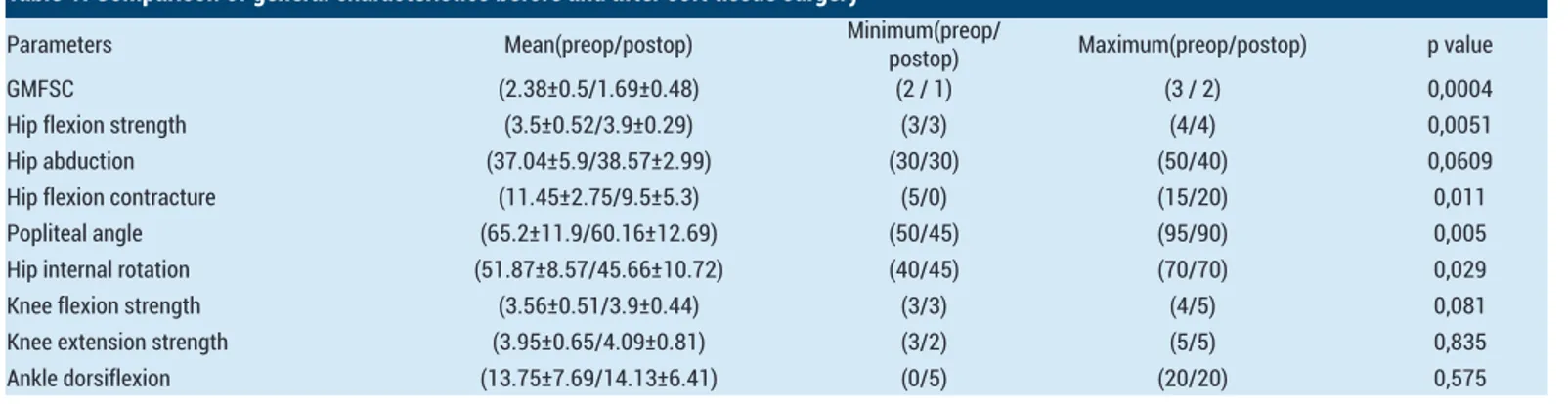
According to pre-operative and post-operative results comparison hip flexion contracture (11.45±2.75 vs 9.5±5.3, p=0.011), popliteal angle (65.2±11.9 vs 60.16±12.69 p<0.005), hip internal rotation (51.87±8.5 vs 45.66±10.72 p<0.029) were decreased significantly. All data were statistically significant (Table-1).
The preoperative and postoperative GMFCS (2.38±0.51 vs 1.69±0.48 p<0.05) showed a significant improvement in the classification of the patients after surgery (Table-1). When the muscle strengths evaluated manually hip flexion (3.5±0.52 vs 3.9±0.29 p>0.05), knee flexion (3.56 ±0.51 vs 3.9±0.44 p>0.05) / extension (3.95±0.65 vs 4.09±0.81 p>0.05) and foot dorsiflexion strength inceased. (3.95±0.65 vs 4.09±0.81 p>0.05). These datas weren’t statistically significant(Table-1).
Kinematics
Kinematic data were compared for hip sagittal ROM (41.08±10.99 vs 39.39±7.93 p <0.05), knee sagittal ROM (45.95±13.12 vs 41.75±12.07 p<0.05) hip coronal ROM (14.2±7.63 vs 12.97±5.31 p<0.05) and there was a decrease in the scores but this difference was not statistically significant. There was no statistically significant difference in the ankle ROM (17.77±4.63 vs 17.78±5.83 p <0.05) although there was minimal postoperative increase (Table-2).
Joint Strength (N*m/kg)
When the joint strengths of the patients were compared before and after surgery (-0.5 /-0.05 vs 0.57/ 0.76 p<0.05), the strength of the knee (-0.05/0.5 vs 0.024/0.001 p>0.05) and ankle strength increased. (-0.01/-0.6 vs 0.194/0.184 p>0.05), but this increase is not statistically significant (Table-3).
Effect on crouch gait
Comparison in crouch gait before and after surgery; minimum hip flexion (15.22±16.45 vs 7.33 ±14.34 p <0.05), initial contact knee flexion (33.25±14.26 vs 24.46±8.84 p<0.05), swing phase maximum knee flexion (49.5±12.29
vs 43.02±11.6 p<0.05) were decreased and the difference was statistically significant. Mid-stance ankle dorsiflexion (-3.02±11.6 vs 3.75±7.76) was observed to increase statistically significantly (Table-2).
Temporal parameters
When the temporospatial data were compared, the cadence (104.71±38.5 vs 89.59±35.31 p<0.05) indicating the number of steps per minute, and double support time (161.3±90.2 vs 150.09±81.91 p<0.05) decreased after the
surgery. Change in cadence was statistically significant but the change in double support time was not significant. However, the step length (mm) (655.14±247.7 vs 767.5±307 p<0.05), the walking speed (m/s) (0.656±0.268 vs 0.667±0.267 p>0.05) and the step width (mm (144.41±50.61 147.94±87.8 p>0.05) increased after surgery. While the increase in the step length was statistically significant, the increase in walking speed and step width values were not statistically significant (Table-4).
Table 1. Comparison of general characteristics before and after soft tissue surgery
Parameters Mean(preop/postop) Minimum(preop/postop) Maximum(preop/postop) p value
GMFSC (2.38±0.5/1.69±0.48) (2 / 1) (3 / 2) 0,0004
Hip flexion strength (3.5±0.52/3.9±0.29) (3/3) (4/4) 0,0051
Hip abduction (37.04±5.9/38.57±2.99) (30/30) (50/40) 0,0609
Hip flexion contracture (11.45±2.75/9.5±5.3) (5/0) (15/20) 0,011
Popliteal angle (65.2±11.9/60.16±12.69) (50/45) (95/90) 0,005
Hip internal rotation (51.87±8.57/45.66±10.72) (40/45) (70/70) 0,029
Knee flexion strength (3.56±0.51/3.9±0.44) (3/3) (4/5) 0,081
Knee extension strength (3.95±0.65/4.09±0.81) (3/2) (5/5) 0,835
Ankle dorsiflexion (13.75±7.69/14.13±6.41) (0/5) (20/20) 0,575
Table 2. Comparison of kinematik parameters before and after soft tissue surgery
Parameters (kinematic) Mean(preop/postop) Minimum(preop/postop) Maximum(preop/postop) p value
Hip sagital ROM 41.08±10.99 vs 39.39±7.93 (26/28) (58.6/59.2) 0,86
Knee sagital ROM 45.95±13.12 vs 41.75±12.07 (26.3/20.86) (73.98/66.39) 0,945
Ankle ROM 17.77±4.63 vs 17.78±5.83 (11.4/10.2) (28.77 /37.29) 0,103
Pelvik Tilt 10.41±6.11 vs 9.5±5.8 (3.02 / 2.7) (22.2/31.9) 0,793
Hip coronal plane ROM 14.2±7.63 vs 12.97±5.31 (4.44/4.68) (29.35/22.87) 0,892 Pelvic rotation (-0.158±7.96) vs 0.5±6.61 (-10.7 /-9.92) (12.82/13.81) 0,857 Initial contact knee flexion (33.25±14.26 / 24.46±8.84) (9/-1) (67/41) 0,0029 Maximum knee flexion at swing phase (49.5±12.29/43.02±11.6) (30.5/22) (84/66) 0,024 Mid-stance ankle dorsiflexion (-3.02±11.61/3.75±7.76) (-19/-10) (23/19) 0,047
Minimum hip flexion (15.22±16.45/7.33±14.34) (-18/-20) (46/32) 0,0079
Table 3. Comparison of kinematic parameters ( Joint power) before and after surgery
Parameters (kinematic) Mean(preop/postop) Minimum(preop/postop) Maximum(preop/postop) p value Hip Power (N*m/kg) (0.2±0.27/0.23±0.22) (-0.5/-0.05) (0.57/0.76) 0,506 Knee power (N*m/kg) (-0.16±0.18/-0.13±0.11) (-0.05/0.5) (0.024/0.001) 0,945 Ankle power (N*m/kg) (0.067±0.065/0.009±0.148) (-0.01/-0.6) (0.194/0.184) 0,425
Table 4. Comparison of temporospatial parameters before and after surgery
Parameters (temporospatial) Mean(preop/postop) Minimum(preop/postop) Maximum(preop/postop) p value Cadence (step/min) (121.5±27.61/106.7±13) (80/90) (161/133) 0,237 Step lenght (cm) (770.5±159.3/798.8±130.5) (465/512) (1001/1134) 0,111 Step width (cm) (159.2±34.9/156±73.4) (102/19) (222/279) 0,461 Walking speed (m/sn) (0.78±0.22/0.71±0.21 (0.4/0.42) (1.02/1.01) 0,725 Double support time (sn) (131.3±86.7/135.7±65.2) (11/10) (265/255) 0,488 Foot progression angle (-8.84±11.7/-17.3±10.8) (-27/-42.9) (19.11/0) 0,029
DISCUSSION
In this study, we aimed to evaluate isolated soft-tissue release surgeries with clinical and 3-dimensional walking analysis of patients with crouch gait related gait problems. We found a significant improvement in physical examination data after soft tissue surgery. Kinematic data showed that there was no significant change in the range of motion, while knee flexion and ankle dorsiflexion in gait phases improved and the crouch gait walking pattern decreased clinically.
In temporospatial data, we found a significant decrease in cadence and increase in the walking speed and step length of the patients.
Joint problems that occur after spastic cerebral palsy cause deterioration in the walking pattern of the patients. While the spasticity problem in early period can be treated by physical therapy and botulinum toxin injection, surgical treatment comes first in the patients who develop contracture and bone deformities (11)
SEMLS are soft tissue and bone interventions that correct all deformities in patients in a single surgical session. The results of single-session multiple surgeries are similar, providing a significant improvement in clinical and gait parameters (12-14)
Crouch gait is a common gait disorder in cerebral palsy patients with knee, hip flexion and increased ankle dorsiflexion (5). Increased knee flexion in the stance phase causes an increase in knee loads. In patients, the step length decreases, and the amount of energy required for walking increases. In knee flexion, a 10-degree increase in the stance phase leads to compensatory flexion of the hip and ankle (15,16). In cases where it is not treated, it may lead to an increase in walking problem due to overloading of the knee and hip (2).
Hamstring release surgery is one of the most preferred surgeries in patients with crouch gait. Thus, anterior pelvic tilt increases in patients and maximum knee flexion in the middle of the stance. (17) In patients with normal hamstring length, as a result of release surgery may cause hip and knee flexion and increase crouch gait, it should be evaluated by physical examination before surgery (18). At this stage, the development of bone deformities and radiological anomalies is inevitable (19).
We know that isolated hamstring release surgeries do not provide adequate improvement in patients with fixed flexion deformity. In these patients, distal femur extension osteotomy (DFEO) may help to provide adequate improvement. Novacheck et al. reported that patients with cerebral palsy who had knee flexion contracture between 10 and 30 degrees were definite indications for DFEO (20). In the early period after soft tissue surgery, the functional status of patients may worsen. Saraph et al. reported in
their study evaluating 32 patients who underwent SEMLS, that the results of the early three-year evaluation were not suitable for determining long-term results. They also revealed that the long term results should be evaluated at least 3 years after the surgery for the most predictive evaluation. (21)
The number of studies evaluating isolated soft tissue releases in patients with cerebral palsy who have problems walking, is limited. Adolfsen et al. evaluated 31 patients as clinically and radiologically who underwent isolated multilevel soft tissue surgery due to walking problems in the sagittal plane. Surgical results showed that the first contact knee flexion decreased and the walking pattern improved significantly in crouch gait patients. (22) We have achieved similar results in our study.
First contact knee flexion is increased in crouch and jump gait and it has been reported that achieving normal values after surgery improves walking function.
After soft tissue release surgeries, in the stance phase extensor knee moment decreases and it reduces the load in knee thus, walking in long term facilitates. Improvement of the load transfer around the knee is important to solve long-term problems in crouch gait (23,24). However, bone problems such as genu valgum and increased femoral anteversion should not be ignored in knee pain in crouch gait (25).
One of the positive aspects of this study is that is one of a limited number of articles evaluating isolated soft tissue release surgeries in patients with crouch gait. It is also important to evaluate the patients not only clinical but also in detail with 3-dimensional gait analysis and to show long-term follow-up results.
One of the important negative aspects of the study is that it is a retrospective study. Low number of patients is another deficiency. The problem was the low number of patients who underwent isolated soft-tissue procedure and the difficulty to reach these patients retrospectively. CONCLUSION
Isolated soft tissue surgery is one of the important treatment options in patients with cerebral palsy crouch gait. After the evaluation of a good physical examination and gait analysis, surgery can be done in selected patients. When applied to appropriate patients, both physical examination parameters and walking pattern can provide significant improvement in long term. It should be kept in mind that soft tissue surgeries alone will not be sufficient in patients with bone deformity.
Competing interests: The authors declare that they have no competing interest.
Financial Disclosure: There are no financial supports
Ethical approval: Ethical approval was obtained by the local ethics committee and informed consent forms were obtained from all participants.
Baris Gulenc ORCID: 0000-0002-4565-2315 Shavkat Kuchimov ORCID: 0000-0003-1481-8447 Yener Temelli ORCID: 0000-0001-6622-0645
REFERENCES
1. Oskoui M, Coutinho F, Dykeman J, et al. An update on the prevalence of cerebral palsy: a systematic review and meta-analysis. Dev Med Child Neurol 2013;55:509-19.
2. O'Sullivan R, Horgan F, O'Brien T, French H. The natural history of crouch gait in bilateral cerebral palsy: A systematic review. Res Dev Disabil 2018 80:84-92.
3. Andersson C, Mattsson E. Adults with cerebral palsy: a survey describing problems, needs, and resources, with special emphasis on locomotion. Dev Med Child Neurol 2001:43:76-82.
4. Falisse A, Bar-On L, Desloovere K, et al. A spasticity model based on feedback from muscle force explains muscle activity during passive stretches and gait in children with cerebral palsy. PLoS One 2018;13:
5. Rethlefsen SA, Blumstein G, Kay RM, et al. Prevalence of specific gait abnormalities in children with cerebral palsy revisited: influence of age, prior surgery, and gross motor function classification system level. Dev Med Child Neurol 2017;59:79-88.
6. Rodda JM, Graham HK, Nattrass GR, et al. Correction of severe crouch gait in patients with spastic diplegia with use of multilevel orthopaedic surgery. J Bone Joint Surg Am 2006,88:2653-64.
7. Joseph B, Reddy K, Varghese RA, et al. Management of severe crouch gait in children and adolescents with cerebral palsy. J Pediatr Orthop 2010;30:832-9.
8. Perry J, Antonelli D, Ford W. Analysis of knee-joint forces during flexed-knee stance. J Bone Joint Surg Am 1975;57:961-7.
9. Putz C, Döderlein L, Mertens EM, et al. Multilevel surgery in adults with cerebral palsy. Bone Joint J 2016;98:282-8. 10. Lamberts RP, Burger M, du Toit J, et al. A systematic review
of the effects of single-event multilevel surgery on gait parameters in children with spastic cerebral palsy. PLoS One 2016;18:11.
11. Freeman M. Cerebral Palsy.2005. Springer Science NY 12. Thomason P, Selber P, Graham HK. Single event multilevel
surgery in children with bilateral spastic cerebral palsy: a 5 year prospective cohort study. Gait Posture 2013;37:23-8.
13. Nene AV, Evans GA, Patrick JH. Simultaneous multiple operations for spastic diplegia. Outcome and functional assessment of walking in 18 patients. The Journal of Bone and Joint Surgery 1993;75:488–94.
14. Saraph V, Zwick EB, Zwick G, et al. Multilevel surgery in spastic diplegia: evaluation by physical examination and gait analysis in 25 children. J Pediatr Orthop 2002;22:150-7. 15. Rose J, Gamble JG, Medeiros J, et al. Energy cost of
walking in normal children and in those with cerebral palsy: comparison of heart rate and oxygen uptake. J Pediatr Orthop 1989;9:276-9.
16. Temelli Y, Akalan NE. Treatment approaches to flexion contractures of the knee. Acta Orthop Traumatol Turc 2009;43:113-20
17. DeLuca PA, Ounpuu S, Davis RB, Walsh JH. Effect of hamstring and psoas lengthening on pelvic tilt in patients with spastic diplegic cerebral palsy. J Pediatr Orthop 1998;18:712-8.
18. Kedem P, Scher DM. Evaluation and management of crouch gait. Curr Opin Pediatr 2016;28:55-9.
19. Kerr Graham H, Selber P. Musculoskeletal aspects of cerebral palsy. J Bone Joint Surg Br 2003;85:157-66. 20. Novacheck TF, Stout JL, Gage JR, et al. Distal femoral
extension osteotomy and patellar tendon advancement to treat persistent crouch gait in cerebral palsy. Surgical technique. J Bone Joint Surg Am 2009;91 Suppl 2:271-86. 21. Saraph V, Zwick EB, Auner C, et al. Gait improvement surgery
in diplegic children: how long do the improvements last?. J Pediatr Orthop 2005;25:263-7.
22. Adolfsen SE, Ounpuu S, Bell KJ, et al. Kinematic and kinetic outcomes after identical multilevel soft tissue surgery in children with cerebral palsy. J Pediatr Orthop 2007;27:658-67.
23. Steele KM, Demers MS, Schwartz MH, et al. Compressive tibiofemoral force during crouch gait. Gait Posture 2012;35:556-60.
24. Rodda JM, Graham HK, Nattrass GR, et al. Correction of severe crouch gait in patients with spastic diplegia with use of multilevel orthopaedic surgery. J Bone Joint Surg Am 2006;88:2653-64.
25. Rethlefsen SA, Nguyen DT, Wren TA, et al. Knee pain and patellofemoral symptoms in patients with cerebral palsy. J Pediatr Orthop 2015,35:519-22.