BIAS CORRECTION IN FINDING COPY
NUMBER VARIATION WITH USING READ
DEPTH-BASED METHODS IN EXOME
SEQUENCING DATA
a thesis
submitted to the department of computer engineering
and the graduate school of engineering and science
of bilkent university
in partial fulfillment of the requirements
for the degree of
master of science
By
Fatma Balcı
August, 2014
I certify that I have read this thesis and that in my opinion it is fully adequate, in scope and in quality, as a thesis for the degree of Master of Science.
Assist. Prof. Can Alkan(Advisor)
I certify that I have read this thesis and that in my opinion it is fully adequate, in scope and in quality, as a thesis for the degree of Master of Science.
Assist. Prof. Bu˘gra Gedik
I certify that I have read this thesis and that in my opinion it is fully adequate, in scope and in quality, as a thesis for the degree of Master of Science.
Assist. Prof. ¨Ozlem C¸ avu¸s
Approved for the Graduate School of Engineering and Science:
Prof. Dr. Levent Onural Director of the Graduate School
ABSTRACT
BIAS CORRECTION IN FINDING COPY NUMBER
VARIATION WITH USING READ DEPTH-BASED
METHODS IN EXOME SEQUENCING DATA
Fatma Balcı
M.S. in Computer Engineering Supervisor: Assist. Prof. Can Alkan
August, 2014
Medical research has striven for identifying the causes of disorders with the ultimate goal of establishing therapeutic treatments and finding cures since its early years. This aim is now becoming a reality thanks to recent developments in whole genome (WGS) and whole exome sequencing (WES). Despite the decrease in the cost of sequencing, WGS is still a very costly approach because of the need to evaluate large number of populations for more concise results. Therefore, sequencing only the protein coding regions (WES) is a more cost effective alterna-tive. With the help of WES approach, most of the functionally important variants can be detected. Additionally, single nucleotide polymorphisms (SNPs) that are located within coding regions are the most common causes for Mendelian diseases (i.e. diseases caused by a single mutation). Moreover, WES approaches require less analysis effort compared to whole genome sequencing approaches since only 1% of whole genome is sequenced. Besides the advantages, there are also some shortcomings that need to be addressed such as biases in GC−content and probe efficiency. Although there are some previous studies on correcting GC−content related issues, there are no studies on correcting probe efficiency effect. In this thesis, we provide a formal study on the effects of both GC−content and probe efficiency on the distribution of read depth in exome sequencing data. The cor-rection of probe efficiency will make it possible to develop new CNV discovery methods using exome sequencing data.
Keywords: Copy number variations, read depth, bias correction, GC content, exome sequencing, next-generation sequencing, probe efficiency, DNA sequencing.
¨
OZET
D˙IZ˙I DER˙INL˙I ˘
G˙I Y ¨
ONTEM˙I KULLANILARAK KOPYA
SAYISI FARKLILIKLARINI TESP˙IT ETMEDE EKZOM
D˙IZ˙ILEME DATALARINDA VAROLAN ETK˙ILER˙IN
D ¨
UZELT˙ILMES˙I
Fatma Balcı
Bilgisayar M¨uhendisli˘gi, Y¨uksek Lisans Tez Y¨oneticisi: Yrd. Do¸c. Can Alkan
A˘gustos, 2014
˙Insanlı˘gın varolu¸sundan bu yana tıptaki ara¸stırmalar, hastaları tedavi ede-bilmek ve hastalıkların ¸caresini bulaede-bilmek adına bunların altında yatan sebep-leri bulmak i¸cin yapılmı¸stır. Bu ama¸c, son zamanlarda t¨um genom ve t¨um ek-zom dizilemede ya¸sanan geli¸smeler sayesinde ger¸cekle¸stirilebilmektedir. Dizileme maliyetlerinde ya¸sanan azalmalara ra˘gmen, daha do˘gru sonu¸clar elde edebilmek adına ¸cok sayıda insan genomunun dizilenme ihtiyacı oldu˘gundan t¨um genom dizileme halen y¨uksek maliyetli bir y¨ontemdir. Bu sebeple, sadece protein kod-layan b¨olgeleri dizileyen t¨um ekzom dizileme y¨ontemi nispeten daha az maliyetli bir alternatiftir. T¨um ekzom dizileme yakla¸sımlarının yardımıyla, fonksiyonel ¨
onem ta¸sıyan varyantların ¸co˘gu bulunabilmektedir. Buna ek olarak, Mendeliyen (tek mutasyon kaynaklı) hastalıkların en b¨uy¨uk sebebi olan tek n¨ukleotid polimor-fizmlerinden, ekzon b¨olgelerinde yer alanlar da bulunabilmektedir. Ayrıca t¨um ekzom dizilemeye dayalı yakla¸sımlar, insan genomun sadece %1´lik kısmını kap-sadı˘gından di˘ger yakla¸sıma g¨ore analiz yaparken daha az ¸caba gerektirmektedir. Ancak do˘gru sonu¸clar elde edebilmek i¸cin ekzom dizileme datasında varolan prob etkinli˘gi ve GC i¸ceri˘gi gibi sapma etkilerinin duzeltilmesi gerekmektedir. Bun-lardan GC i¸ceri˘gi sapmasını d¨uzeltmek icin yapılmı¸s bazı ¸calı¸smalar bulunmak-tadır. Ancak literat¨urde, prob etkinli˘gi sapmasını d¨uzeltmek amacıyla yapılan bir ¸calı¸sma bulunmamaktadır. Bu tezde ekzom dizileme datasına ait dizi derinlemesi da˘gılımında varolan prob etkinlili˘gi ve GC i¸ceri˘gi sapmaları ¨uzerinde ¸calı¸sılmı¸stır. Prob etkinli˘gi sapmasının d¨uzeltilmesiyle birlikte, ekzom dizileme datası kullanan yeni kopya sayısı varyantı bulma metotları geli¸stirilmek m¨umk¨un olacaktır. Anahtar s¨ozc¨ukler : Kopya sayısı farklılıkları, dizi derinli˘gi, etki d¨uzeltme, GC i¸ceri˘gi, ekzom dizileme, yeni nesil dizileme, prob verimlili˘gi, DNA dizileme.
Acknowledgement
Foremost, I would like to express my sincere gratitude to my advisor Assist. Prof. Can Alkan for the continuous support of my research, for his patience, motivation, enthusiasm, and immense knowledge. His guidance helped me in all the time of research and writing of this thesis. I could not have imagined having a better advisor and mentor.
Besides my advisor, I would like to thank the rest of my thesis committee: Assist. Prof. Bu˘gra Gedik and Assist. Prof. ¨Ozlem C¸ avu¸s for their support.
My special thanks goes to Basri Kahveci for his endless patience and faith. I couldn’t be finished with this work without his support.
I would like to thank TUBITAK for offering me the scholarship opportunity, through grant 112E135.
I thank my hardworking friend Elif Dal in Alkan Lab. I also thank my friend Havva G¨ulay G¨urb¨uz for all the fun we have had in the last two years.
Last but not the least, I would like to thank my brother Ahmet Balcı for his endless support. I wouldn’t be who I am without him. I would like to thank my parents Makbule Balcı and Zeki Balcı for supporting me spiritually throughout my life.
Contents
1 Introduction 1 1.1 Motivation . . . 2 1.2 Problem Statement . . . 3 1.3 Contributions . . . 4 2 Background 6 2.1 DNA Sequencing . . . 6 2.1.1 DNA . . . 6 2.1.2 Gene . . . 7 2.1.3 Chromosome . . . 82.2 DNA Sequencing Technologies . . . 9
2.2.1 Sanger Sequencing (First-Generation Sequencing Technology) 9 2.2.2 Next-Generation Sequencing (Second-Generation Sequenc-ing Technology) . . . 10
2.2.3 Next Next Generation Sequencing (Third - Generation Se-quencing Technology) (Single Molecule SeSe-quencing) . . . . 15
CONTENTS vii
2.2.4 Fourth-Generation Sequencing Technology (Nanopore
Se-quencing) . . . 16
2.3 Genome Sequencing . . . 17
2.3.1 Whole Genome Sequencing . . . 18
2.3.2 Whole-Exome Sequencing . . . 18 2.4 Genomic Variations . . . 20 2.4.1 Single Nucleotide . . . 20 2.4.2 2 bp to 1,000 bp . . . 20 2.4.3 1 kb to Submicroscopic . . . 21 2.4.4 Microscopic to Subchromosomal . . . 22
2.4.5 Whole Chromosomal to Whole Genome . . . 23
2.5 The Effects of Copy Number Variations on Human Health and Phenotype . . . 23
3 Finding Copy Number Variations in Exome Sequencing Data 26 3.1 Four-Step Procedure . . . 26
3.1.1 Mapping . . . 27
3.1.2 Correcting Biases and Normalization . . . 27
3.1.3 Estimation of Copy Number . . . 30
3.1.4 Segmentation . . . 30
3.2 Methods . . . 30
CONTENTS viii
3.2.2 Split read-based methods . . . 33
3.2.3 Read depth-based methods . . . 34
3.2.4 Assembly-based methods . . . 36
3.2.5 Hybrid approaches . . . 37
4 Related Works 38 4.1 Whole Genome Sequencing . . . 38
4.1.1 Summarizing and Correcting the GC Content Bias in High-Throughput Sequencing . . . 38
4.2 Whole Exome Sequencing . . . 41
4.2.1 Copy Number Variation Detection and Genotyping from Exome Sequencing Data . . . 41
4.2.2 Discovery and Statistical Genotyping of Copy Number Variation from Whole Exome Sequencing Depth . . . 41
5 Description of the Experiments 44 5.1 Data . . . 44
5.2 Mapping . . . 45
5.2.1 Mapping of Reads to the Reference: MrsFAST-Ultra . . . 45
5.2.2 Calculation of read depth: Bedtools . . . 46
5.3 Correcting Biases and Normalization . . . 47 5.3.1 Calculation of Correlation Coefficients for Each Exon Region 51 5.3.2 Calculation of Correlation Coefficients for Each Gene Region 54
CONTENTS ix
5.3.3 Finding optimum span parameter of LOESS method . . . 61
6 Conclusion 68
6.1 Future Work . . . 69
A Glossary 76
B Length measurements 78
List of Figures
2.1 DNA structure . . . 7
2.2 Human chromosomes . . . 8
2.3 Workflow of the Sanger Sequencing Method . . . 10
2.4 Workflow of the next-generation sequencing . . . 11
2.5 454/Roche Machines . . . 12
2.6 ABI Solid Machine and its procedure . . . 13
2.7 Illumina Machines . . . 14
2.8 IonTorrent Machines . . . 15
2.9 Pacific Biosciences Machine (SMRT) . . . 16
2.10 MinION and GridION System . . . 17
2.11 Comparison of the size of whole genome and whole exome that are found on human genome . . . 17
2.12 Sequencing . . . 18
2.13 Workflow of whole exome sequencing . . . 19
LIST OF FIGURES xi
2.15 Tiger with down syndrome . . . 24
3.1 Four-step procedure to find CNVs in WES data . . . 26
3.2 High and low coverage . . . 29
3.3 Multiple vs. unique mapping . . . 29
3.4 Classification of CNV detection methods . . . 31
3.5 Paired-end mapping-based methods . . . 33
3.6 Split read-based methods . . . 33
3.7 Calculation depth of coverage . . . 35
3.8 Read depth-based method . . . 35
3.9 Assembly-based methods . . . 36
5.1 Read depth and probe efficiency for each exon . . . 52
5.2 Read depth and probe efficiency for each exon (0.24<GC Content<0.47) . . . 53
5.3 Read depth and GC content for each exon . . . 54
5.4 Read depth and probe efficiency for each gene . . . 56
5.5 Read depth and probe efficiency for each gene (0.24<GC Content<0.47) . . . 57
5.6 HG00629 . . . 58
5.7 HG01191 . . . 58
5.8 HG01437 . . . 59
LIST OF FIGURES xii
5.10 NA19707 . . . 60 5.11 NA19723 . . . 60 5.12 NA20766 . . . 61 5.13 Smoothed read depth and probe efficiency by LOESS method for
each gene (HG00629 (Span=0.001) ) . . . 62 5.14 Smoothed read depth and probe efficiency by LOESS method for
each gene (HG00629 (Span=0.05) ) . . . 62 5.15 Smoothed read depth and probe efficiency by LOESS method for
each gene (HG00629 (Span=0.9) ) . . . 63 5.16 Smoothed read depth and probe efficiency by LOESS method for
each gene (HG00629 (Span=0.005) ) . . . 63 5.17 Smoothed read depth and probe efficiency by Robust LOESS
method for each gene (HG00629 (Span=0.005) ) . . . 64 5.18 Smoothed read depth and probe efficiency by LOESS method for
each gene . . . 66 5.19 Smoothed read depth and probe efficiency by LOESS method for
List of Tables
2.1 Comparison of the DNA sequencers . . . 10 2.2 Comparison of WES and WGS . . . 20
3.1 Applicability of the tools to the methods . . . 32
4.1 Summary of bioinformatics tools for CNV detection using WGS data. This table is adapted from [1]. . . 40 4.2 Summary of bioinformatics tools for CNV detection using WES
data. This table is adapted from [1]. . . 43
5.1 Correlation between read depth, probe efficiency and GC content for each exon . . . 51 5.2 Correlation between read depth, probe efficiency and GC content
for each exon (0.24<GC Content<0.47) . . . 52 5.3 Correlation between read depth, probe efficiency and GC content
for each gene . . . 55 5.4 Correlation between read depth, probe efficiency and GC content
LIST OF TABLES xiv
5.5 Correlation between read depth, probe efficiency and GC content for each smoothed gene data . . . 64 5.6 Correlation between read depth, probe efficiency and GC content
Chapter 1
Introduction
Although giant strides have been made in recent years in the field of bioinfor-matics, there remains an open question as to find copy number variation (CNV) more accurately to better understand the underlying genetic causes of several diseases, such as autism and schizophrenia. There are four basic sequence signa-tures that can be used to identify CNV (see Section 3.2); but the read depth-based method is the most reliable with whole exome sequencing (WES) data. However, there are several errors in coverage, named biases, introduced in exome capture, which prevent this method to work accurately, as they dramatically alter the read depth distribution properties, and they fail to provide accurate results data because of these biases.
GC-content and probe capture efficiency are two causes of the biases in exome sequencing data. Some studies exist in the literature to correct the GC-content bias, although most have limitations. Moreover, there isn’t any work about probe capture efficiency. This thesis characterizes the effects of GC-content affecting exome sequencing read depth distribution; and demonstrates that the correc-tion of GC-content and probe capture efficiency simultaneously works better in smoothing the depth distribution. Our study described in the following chapters attempts to decrease the biases in WES data through identifying the effects of both GC-content and probe capture efficiency in the read depth distribution for accurately characterizing CNV. We also provide an insight into how to correct
for these biases using a well-known statistical smoothing technique, called locally weighted scatterplot smoothing (LOESS). After this error correction step, it will be easier to apply more standard CNV identification algorithms to better discover CNV using WES data.
In chapter 2, we present background information to help better understand the biological and technical concepts. We define the biases, and provide compar-isons of CNV discovery algorithms, and provide the basic steps of these methods in Chapter 3. In Chapter 4, we discuss the related previous studies. The descrip-tion of the experiment is given in detail in Chapter 5. Finally, we evaluate our formulations and source of bias characterizations in Chapter 6.
1.1
Motivation
Obtaining accurate knowledge of nucleic acid composition is crucial to all life sciences. Deciphering DNA sequences started to shed light on novel biological functions and phenotypic differences, which increased the demand for highly ef-ficient sequencing technologies. As a consequence, an era of synthetic genomics and personalized medicine is expected to start within the next few years.
The aim of this thesis is to help to find copy number variation (CNV) accu-rately in whole exome sequencing (WES) data by calculating read depth (RD) to be able to get rid of the analysis burden of whole genome sequencing (WGS) data.
The new cost-efficient and high throughput strategies for DNA sequencing are now the leading power house of discoveries in life sciences. The new sequenc-ing technologies, commonly referred to as high throughput sequencsequenc-ing (HTS), or next-generation sequencing (NGS) started to appear in 2007. The advantages of HTS platforms are many: Cost of sequencing is reduced 10,000-fold, while data throughput is increased 30-fold per base per day. Despite the improvements, data generated with HTS platforms are more difficult to analyze without a priori in-formation, however the availability of whole genome assemblies for humans and
all major model organisms has strengthened the potential utility of HTS.
In addition, general progress in technology across other related fields, includ-ing chemistry, nucleotide biochemistry, computation, data storage, and others, helped make better use of the HTS data. However, we still need to improve algorithms to better characterize genomic variation. Although the methods to discover and genotype single nucleotide polymorphisms (SNPs) are maturing, ac-curate detection of copy number variation (CNV) is still lacking, but there exist some algorithms with different strengths and weaknesses in the literature. [2]
1.2
Problem Statement
Depth of coverage is the average number of reads representing a given nu-cleotide in the reconstructed sequence. Most of the CNV discovery methods using WGS data perform statistical tests based on a Poisson model, in which reads are assumed to be distributed uniformly across the genome, since the sequence reads are assumed to be chosen randomly from the genome. It means RD in a region should follow a Poisson distribution with mean directly proportional to the size of the region and to the copy number. However, this assumption hardly holds even for normal genomes due to the biases mentioned in Chapter 3.
content and probe capture efficiency are two sources of bias in data. GC-content is the percentage of guanine (G) and cytosine (C) bases in a genomic region. GC-abundance is heterogeneous across the genome and often correlated with functionality so it affects each region differently and needs to be corrected. Exome sequencing involves exon-capture step by which the coding regions are selected from the total genome DNA by means of hybridization. The characteris-tics of the probe, such as length, conformation and abundance on the solid phase, are of relevance in determining the capture efficiency. This capture efficiency is different for each probe due to the characteristics. Probe capture efficiency de-termines whether if an exon will be captured or not and the length of the region captured. Therefore, it also needs to be corrected.
CNV discovery methods with using WES data is mostly affected by GC-content and probe capture efficiency biases that can only be corrected locally with using smoothing methods to be able to reveal data points including CNV in the graph.
Although there are lots of computational methods and tools in the field to find CNV by using read depth-based (RD-based) methods, all of them suffer from various types of biases. After the work completed in this thesis, almost all of the tools will work more reliably by enhancing data.
1.3
Contributions
Learning more about the relationship between copy number variation (CNV) and exome sequencing, could help understanding the effects of CNV on humans and lead to huge improvements on comprehending of the underlying causes of some important diseases and phenotypic changes of humans. Although CNV can affect the other species, we are only interested in human genomes, since most available whole exome sequencing (WES) data are generated from human samples. There is a problem in finding the underlying causes of some important diseases such as schizophrenia and cancer.
Despite the great number of researches on finding CNV, most of them is based on finding CNV in whole genome sequencing (WGS) data. This problem has negatively impacted by the magnitude of the WGS data because a human genome is approximately comprised of 3 billion nucleotides. There are some powerful tools available to find CNV in WGS data. It seems like that they can be used for whole exome sequencing (WES) data, but the usage of these tools is not possible due to some biases. A possible cause of this problem is the difference between probe efficiencies. A study which investigates to understand the relationship between probes and read depth by improving an algorithm could remedy to use WES data with some healing.
in the samples’ genomes. We benefit from the 1000 Genomes data and Agilent Sure Select Capture Kit data with this aim. After reducing some noises in data, such as sex chromosomes, we calculate the correlation coefficient to understand the relationship between read depth and probe efficiency. We demonstrated that there is an important relationship between read depth and probe efficiency.
At the end of this process, we should normalize the data with the help of locally weighted scatterplot smoothing (LOESS) method because each bases in WES data are generally affected by the regions that are close to them. Therefore, we need to evaluate each DNA regions locally. We need to correct data as much as possible to be able to develop statistical approaches with using WES data. The hardest part of this process is to separate bias-based and CNV-based deviations in the data.
People who are working in the field of bioinformatics, patients with genetic diseases, doctors who want to understand the underlying causes of genetic dis-eases, and scientists who work in the related fields of science may benefit from this thesis. If successful tools we plan to develop using the methods presented in this thesis may also be used in clinical sequencing tests that we expected to be used in all hospitals within the next few years.
Chapter 2
Background
2.1
DNA Sequencing
2.1.1
DNA
The hereditary material in humans and almost all other organisms is called as deoxyribonucleic acid (DNA). Human DNA is mostly found in the cell nucleus, but it can also be found in the mitochondria in a small amount. Adenine (A), cytosine (C), guanine (G), and thymine (T) are the coding elements of DNA, nucleotides.
Adenine and thymine are called as purines, whereas cytosine and guanine are called as pyrimidines. Each nucleotide consists of a phosphate group, a 5-carbon sugar (deoxyribose), and a nitrogen containing base attached to the sugar. These four types of nucleotides differ only in the nitrogenous base. The order of these nucleotides determines the information to build and maintain an organism. Nucleotides are located in two long strands that form a spiral, double helix. Almost each cells in our body has the same DNA. Approximately, 3 billion bases exist in human DNA.
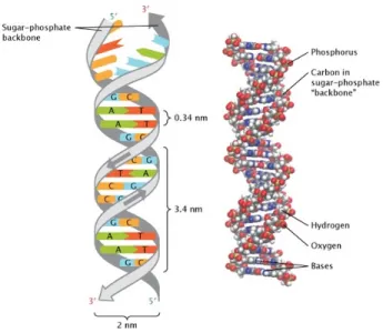
Figure 2.1: The grey ribbons that represent the sugar-phosphate backbone have arrows that run in opposite directions to indicate that the two strands of the helix are arranged in an anti-parallel manner. The upper end of one strand is labeled five prime (5’), and the lower end of the same strand is labeled three prime (3’). The nucleotide bases are shown as differently colored rectangles. The nucleotide guanine (G), shown in blue, binds with the nucleotide cytosine (C), shown in orange and the nucleotide adenine (A), shown in green, binds with the nucleotide thymine (T), shown in red. Gold spheres represent phosphorus atoms, grey spheres represent carbon atoms, white spheres represent hydrogen atoms, red spheres represent oxygen atoms, and blue spheres represent nitrogen atoms. This figure is adapted from [3].
2.1.2
Gene
The principle physical and functional unit of heredity is called as a gene. A gene is used as a template to make protein molecules and they are made up from DNA. The number of genes found in a human body has been estimated between 20.000 and 25,000 by the Human Genome Project. There are two copies of genes in human genomes. One of them is inherited from mother, whereas another is from father. Almost all genes are common between humans except 1% of the
genes.
2.1.3
Chromosome
The thread-like structures in which DNA located in human body are called as chromosomes. 46 chromosomes are found in each human cell. 23 of them are inherited from mother, whereas the remaining 23 of them are inherited from father. Humans have 22 pairs of autosomes and one pair of sex chromosomes, the X and Y.
Autosomes are roughly ordered due to their size. The largest chromosome is Chromosome 1 which has approximately 2,800 genes. Moreover, the smallest chromosome is Chromosome 22 which has approximately 750 genes. These genes are providing instructions for making proteins. Changes in the structure or num-ber of copies of a chromosome can cause problems with health and development, but it doesn’t have to cause any problems.
Figure 2.2: Human chromosomes. This figure is adapted from [4].
A pair of sex chromosomes are found in each human normally. Females have two X chromosomes, whereas male have one X and one Y chromosome.
2.2
DNA Sequencing Technologies
Nucleic acid sequencing is a way to determine the exact order of the DNA. The usage of nucleic acid sequencing has become accessible for researchers in the past decade. These sequencing techniques are key tools in many fields ranging from archeology, genetics, anthropology, biotechnology, forensic sciences to molecular biology. As the first major foray into DNA sequencing, The Human Genome Project is completed in 2004 at the cost of approximately $3 billion. It was a 14-year-long endeavor. The project is completed with using Sanger sequencing which is developed in 1975 by Frederick Sanger.
There are different kinds of platforms for DNA sequencing in the market. Four generations of DNA sequencing technologies can be distinguished by their nature and the kind of output they provide. The field of DNA sequencing technology development has a rich and diverse history.
2.2.1
Sanger Sequencing (First-Generation Sequencing
Technology)
Sanger sequencing method had become the gold standard for 30 years after its discovery in 1977. This method uses DNA polymerase which makes use of inhibitors that terminate the newly synthesized chains at specific residues.
DNA to be sequenced can be prepared in two different ways, shotgun de novo sequencing or targeted resequencing. The output of both methods is an amplified template. Then, template denaturation, primer annealing, and primer extension are performed in a cycle sequencing. Primer is an oligonucleotide complementary to target DNA and leads the DNA extension. With the help of fluorescently labeled ddNTPS, each round of primer extension is halted. Labeled ddNTPs in its current form are mixed with regular, non-labeled, and non-terminating nucleotides in a cycle sequencing reaction. The label on the terminating ddNTP of any fragment corresponds to the nucleotide identifying its terminal position. To separate sequences by length and to provide subsequent interrogation of the
terminating base capillary electrophoresis is applied. Software provides DNA sequences and also their error probabilities for each base-call. [5] [6]
Figure 2.3: Workflow of the Sanger Sequencing Method. This figure is adapted from [7].
2.2.2
Next-Generation Sequencing (Second-Generation
Sequencing Technology)
Machine Capacity Speed Read Length Cost Per Base
454 Roche 35-700 Mb 10-23 hours 400-700 bp 714/14285 × 10−8 e SOLiD 90-180 Gb 7-12 days 75 bp 3/5 × 10−8 e Illumina 6-600 Gb 2-14 days 100-250 bp 2/333 × 10−8 e Ion Torrent 20 Mb-1 Gb 4-5 hours 200 bp 100/10000 × 10−8 e PacBio 1 Gb 30 minutes 3,000 bp 60/80 × 10−8 e Table 2.1: Comparison of the DNA sequencers. This table is adapted from [8].
After the completion of the Human Genome Project, cheaper and faster sequenc-ing methods are demanded in the market. This demand has revealed the devel-opment of next-generation sequencing (NGS) methods. NGS is used for a fast, affordable, and through way to determine the underlying genetic causes of dis-eases. Millions of fragments of DNA from a single sample can be sequenced in unison with NGS. Massively parallel sequencing technology performed in NGS facilitates high-throughput sequencing. With this technology an entire genome are sequenced in less than ten days. In addition to these advantages of NGS, the cost required for a whole human genome has decreased with this technology. It is also minimizing the need for the fragment-cloning methods which are frequently used in Sanger sequencing.
Figure 2.4: Workflow of the next-generation sequencing. This figure is adapted from [8].
After the appearance of the first 2nd generation sequencer in 2005, several second generation sequencers followed this emergence in the market. They are working conceptually similar although they have differences in sequencing bio-chemistry as well as in how the array is sequenced.
2.2.2.1 454/Roche
The first next-generation sequencing technology was 454 pyrosequencing. 454/Roche sequencing method consists of library preparation, emulsion PCR,
and pyrosequencing.
454 is based on the ”sequencing by synthesis principle” which means taking the single stranded DNA to be sequenced and sequencing its complementary in an enzymatic way. In this method the activity of DNA polymerase is monitored by another enzyme, chemiluminescene. When the complementary is bound by the single-stranded sequenced DNA, light is produced. Sequencing is completed by the produced chemiluminescent signals [6].
Figure 2.5: 454/Roche Machines. This figure is adapted from [8].
2.2.2.2 ABI Solid
Emulsion PCR is used to generate the clonal sequencing features in the se-quencing process of ABI Solid sese-quencing technology. Di-base sese-quencing tech-nique in which two nucleotides are read via sequencing by ligation is used at each step of the sequencing process.
Although there are 16 base combinations of di-bases are possible, 4 dyes are used by the system. Therefore, 4 di-bases are represented by a single color. All bases are interrogated twice by the sequencing machine. Each following base can be derived in this way if the previous base is known. Moreover, a misidentified color can change all of the following bases in the translation. ABI Solid is rarely preferred nowadays. [9]
Figure 2.6: ABI Solid Machine and its procedure. This figure is adapted from [8].
2.2.2.3 Illumina
Although better platforms exist in the market, Illumina is the market leader due to the lower prices. Data produced by Illumina’s machine is also used in this project. Illumina sequencing is comprised of library preparation, clustering, and sequencing processes.
In the first part, DNA simultaneously fragment and tag the extracted and purified DNA with adapters. Reduced cycle amplification adds the sequencing primary binding sites, indices, and regions that are complementary to the flow cell oligos after the ligation of adapters.
Each fragment molecule is amplified isothermally in the clustering part. The flow cell is a glass line with lanes. Each lanes is a channel coded with the com-posed of two types of oligos. Complementary oligo to the adapter region on one of the fragment strands, enables hybridization. A polymerase creates a comple-ment of the hybridized fragcomple-ment. The double stranded molecule is denatured and the original template is washed away. The strands are amplified the bridge amplification clonally. In this process the strands pulls over and the adapter
region hybridizes to the second type of the oligo on the flow cell. The comple-mentary strand is generated by polymerases forming a double stranded bridge which is then denatured resulting in two single stranded copies of the molecule. The process is then simultaneously repeated for millions of clusters resulting for all fragments. Then, the reverse strands are washed away.
Figure 2.7: Illumina Machines. This figure is adapted from [8].
In the last part, sequencing, first sequencing primer is extended to produce the first read. One of the 4 fluorescently tagged nucleotide is incorporated based on the sequence of the template. After each incorporation, a light source is used and a characteristic fluorescent signal is emitted. This process is known as ”sequencing by synthesis”. The length of a read is determined by the cycle number and the base call is determined by the emission wave length. Hundreds of millions of clusters are sequenced in a massively parallel process. This entire process generates billions of reads representing all the fragments. [10] [11]
2.2.2.4 IonTorrent
Workflow of Ion Torrent is comprised of library preparation, emulsion PCR, and semiconductor sequencing processes. A hydrogen ion is naturally released as a by-product, when a nucleotide is incorporated into a strand of DNA by a polymerase.
Ion Torrent works on the principle of detection of these hydrogen ion releases in a massively parallel manner. These ions are detected on ion-semiconductor sequencing chips. Ion Torrent technology creates a direct connection between the chemical and digital events. [12]
Figure 2.8: IonTorrent Machines. This figure is adapted from [8]
2.2.3
Next Next Generation Sequencing (Third -
Gener-ation Sequencing Technology) (Single Molecule
Se-quencing)
Single molecule sequencing has ability to resequence the same molecule multi-ple times for improved accuracy and the ability to sequence molecules that cannot be readily amplified because of extremes of GC content, secondary structures, and other reasons. The main focus of the molecule sequencing technology is gener-ally on read length, error rate, and throughput. Potential for lower cost, higher throughput, improved quantitive accuracy, and increased read lengths are offered by single molecule sequencing. [13]
2.2.3.1 Pacific Biosciences (SMRT)
Pacific Biosciences (SMRT) that comprised of library preparation and sequenc-ing processes is an example of ssequenc-ingle molecule sequencsequenc-ing. Pacific Biosciences also developed a ”sequencing by synthesis” approach using fluorescently labeled
nucleotides.
Figure 2.9: Pacific Biosciences Machine (SMRT). This figure is adapted from [8].
DNA is constrained to a small volume in a zero-mode wave guide and flu-orescently labeled nucleotide near the DNA polymerase is measured because of low capability of light penetration. Each nucleotide has a characteristic incor-poration time which helps improving base calls. However, the raw error rate is significantly higher than any other current sequencing technology. Therefore, it creates challenges for variation detection. [13]
2.2.4
Fourth-Generation Sequencing Technology (Nanopore
Sequencing)
The results of fourth-generation sequencing technologies have not been suf-ficiently evaluted yet because this is the newest sequencing technology and it requires time. There is limited information about this technology for now.
2.2.4.1 Oxford Nanopore
Oxford Nanopore follows two parallel strategies. ”Exonuclease sequencing” is the first one which based on exonuclease digestion of a single-stranded template into nucleotides that are fed into a nearby protein nanopore in a lipid membrane. ”Strand sequencing” is the second strategy which is like feeding thread through the eye of a needle.
Figure 2.10: MinION and GridION System.This figure is adapted from [8].
Detailed information about nanopore sequencing is not given here because it requires time to be evaluated sufficiently. [8] [14]
2.3
Genome Sequencing
Genome sequencing can be divided into two categories that is defined below as whole genome sequencing (WGS) and whole exome sequencing (WES).
Figure 2.11: Comparison of the size of whole genome and whole exome that are found on human genome. This figure is adapted from [15].
2.3.1
Whole Genome Sequencing
Whole genome sequencing (WGS) is a laboratory method that reads the exact sequence of all DNA bases in an entire genome. The genome contains an indi-vidual’s entire genetic code which means all of their genetic information. This entire genetic code includes protein coding regions (exons as well as the areas of the genetic code that do not give instructions for making proteins which are non-coding DNA sequences (introns).
Figure 2.12: Sequencing
2.3.2
Whole-Exome Sequencing
All genomic regions coding for proteins and untranslated regions flanking them form the exome of an organism. The exome comprises just over 1% of the genome and 230,000 exons. It provides sequence information for protein-coding regions.
Most of the inherited disorders are believed to reside on the coding regions so laboratories can focus exclusively on exon regions. It gives an opportunity to eliminate the tremendous mass of non-coding DNA in the genome. The major advantage of using WES instead of WGS data is using only 1-2% of human
genome. The cost for sequencing is lowered significantly in this way. [1] [16] The possibilities for analyzing exome has changed with NGS. In the past years exome sequencing is widely used in gene discovery and the identification of disease-causing mutations in pathogenic presentations in that the exact genetic cause is not known. Rare mutations that change the function of a protein which is the cause of most Mendelian and non-Mendelian diseases.
Almost half of the reported CNVs overlap with exon regions in DNA. There-fore, usage of exome sequencing data is more sensible in terms of time and cost efficiency. Most of the essential information about life mechanism of a human body can be obtained with exome sequencing data so redundant data are not necessarily processed.
Even though the reads have a non-uniform distribution, data can be improved by the work discussed in this thesis and some related works [10] [17].
Figure 2.13: Workflow of whole exome sequencing. This figure is adapted from [18].
WES WGS
Target 50 Mb 3 Gb
Depth of Coverage Biased Poisson
Mapping Fast Slow
Analysis time Short Long RD-based CNV discovery tools [1] 12 tools 15 tools
Cost $900 $5,000
Usage in CNV Discovery Less common due to biases Common
Table 2.2: Comparison of WES and WGS
2.4
Genomic Variations
Single nucleotide variants (SNPs), small insertions or deletions (indels), copy number variations (CNVs), and large structural variants are called together as genomic variation. [1]
2.4.1
Single Nucleotide
• Base change - substitution - point mutation: Substitution is a type of mutation where one base pair is replaced by a different base pair. • Insertion - deletions (indels): Indel describes relative gain or loss of a
segment in a genomic sequence.
2.4.2
2 bp to 1,000 bp
• Microsatellites: These sequences are composed of non-coding DNA and they are not parts of genes. These are used as genetic markers to follow the inheritance of genes in families.
• Minisatellites: These are generally situated near genes and they are re-peated segments of the same sequence of multiple triplet codons. Moreover, minisatellites are useful as linkage markers due to their highly polymorphic nature.
• Indels: Indel describes gain or loss of a segment in a genomic sequence. • SNP/SNV: SNPs are a type of polymorphism which involves variation of
a single base pair.
• Variable Number Tandem Repeats - VNTRs: Linear arrangement of multiple copies of short repeated DNA sequences that vary in length and are highly polymorphic, making them useful as markers in linkage analysis.
2.4.3
1 kb to Submicroscopic
Figure 2.14: Classes of structural variation. This figure is adapted from [8].
• Copy number variants (CNVs): Copy number variants comprise of deletions, insertions, and duplications. There is another term that is called as CNP creating confusion. CNP is a CNV that occurs in more than 1%
of the population. CNVs have been observed in the comparison of two or more genomes.
– Copy number gain (insertions or duplications): A sequence alteration whereby the copy number of a given region is greater than the reference sequence.
– Copy number loss (deletions): A sequence alteration whereby the copy number of a given region is less than the reference sequence. • Segmental duplications: A segment of DNA >1 kb in size that occurs
in two or more copies per haploid genome, with the different copies sharing >90% sequence identity.
• Translocation: A region of nucleotide sequence that has translocated to a new position.
• Inversion: A continuous nucleotide sequence is inverted in the same posi-tion.
• CNV regions (CNVRs): Merging of independently ascertained, but overlapping, genomic segments creates the representation of a CNV locus.
2.4.4
Microscopic to Subchromosomal
• Segmental aneusomy: Disorder that results from the inappropriate dosage of crucial genes in a genomic segment.
• Chromosomal deletions - losses: In this disorder, entire chromosomes, or large segments of them, are missing.
• Chromosomal insertions - gains: In this disorder, entire chromosomes, or large segments of them, are duplicated.
• Chromosomal inversions: In this disorder, entire chromosomes, or large segments of them, are altered.
• Intrachromosomal translocations: A segment breaks off the chromo-some and rejoins it at a different location.
• Heteromorphisms: A chromosome pair with some homology but differing in size, shape, or staining properties. Homologous chromosome pair which are not morphologically identical (eg the sex chromosomes).
• Fragile sites: A chromosomal region that has a tendency to break.
2.4.5
Whole Chromosomal to Whole Genome
• Interchromosomal translocation: Recombination resulting from inde-pendent assortment.
• Isochromosome: A chromosome with two genetically and morphologically identical arms.
• Marker chromosomes: A structurally abnormal chromosome in which no part can be identified.
• Aneuploidy : This is the state of having an abnormal number of chromo-somes. Down syndrome is an example of aneuploidy.
• Aneusomy: is the condition in which an organism is made up of cells that contain different numbers of chromosomes. [19] [2] [20]
2.5
The Effects of Copy Number Variations on
Human Health and Phenotype
A human carries two copies of most genes. One copy comes from mother genome and one copy comes from father genome. Occasionally alterations in a chromosome can lead to the gain or a loss of one copy. A deletion can occur when a fragment of DNA is lost. It can occur either during copying or when the genes are shuffles during meiosis. A duplication can occur whereby which we gain an
additional copy of a gene by the same mechanisms. Deletions and duplications of greater than 1,000 nucleotides are called copy number variants.
Figure 2.15: Tiger with down syndrome
A difference in the copy number of a gene can increase or decrease the level of that genes activity so it may cause diseases, phenotypic changes or nothing. For instance, when a copy of a gene is deleted, the cell may produce half as much protein as compared to a normal cell. There are many diseases that are cause by changes in gene copy number.
Next-generation sequencing technology is used to detect copy number varia-tions in both healthy and diseased people. Although copy number variavaria-tions do not necessarily have a negative effect on human health, large number of CNVs have an association with a disease or directly involve in. The most well-known health problem because of CNV is Down Syndrome, which is caused by having an extra copy of chromosome 21.
Some of the most known health problems due to CNVs are autism, schizophre-nia, Turner syndrome, cancer, neuropsychiatric disorders, and obesity. Rare CNVs may account for 15% of cases of pediatric neurodevelopmental diseases Backenroth et al [21]. Although both rare and common CNVs are thought to carry substantial risk for disease, much recent activity has focused on the role played in disease by rare CNVs, given the smaller cohort sizes required to attain statistical significance for identifying highly penetrant risk-associated rare CNVs.
As another example, a recent study found that severe obesity is often associated with a significant burden of large rare CNVs.
To comprehend the underlying causes of these in a less costly way, an efficient sequencing approach is needed. Whole exome sequencing, the most cost-effective way which is known, has the potential to rapidly detect copy number variations that cause these things mentioned above in human coding regions.
WES has been commonly used in not only the detection of pathogenic variants for Mendelian diseases, but also discovery of susceptible loci for complex diseases. By using these kinds of approaches mentioned in this thesis, underlying causes of some diseases and phenotypes and also the treatments of CNV-based diseases can be found. [22] [19]
Chapter 3
Finding Copy Number Variations
in Exome Sequencing Data
The steps required to find copy number variations in exome sequencing data consist of mapping, correcting biases and normalization, estimation of copy num-ber and segmentation parts. The main aim of the thesis is correcting some of the biases in exome sequencing data, especially GC-content and probe efficiency.
3.1
Four-Step Procedure
3.1.1
Mapping
Although human genome has a continuous structure to read, sequencing ma-chines can only read a few hundred DNA letters at a time. These short DNA letter sets are aligned to the reference genome. This causes some mapping problems when these small reads are used for constructing whole part wanted. Mapping is the first step to find CNVs so the problems in mapping affect the reliability of the results. The choice of single or multiple mapping affects is one of these effects. This choice can change copy number in some regions. It can either cause unexpected increases or decreases in copy numbers.
Mapping are also affected by repeats in mapping part. Approximately 45% of a human genome is repeats such as LINEs, SINEs, retrovirus-like elements, and DNA transposon fosils. RepeatMasker program is available to identify repeats for known ones, but this cannot identify all of these repeats.
3.1.2
Correcting Biases and Normalization
Systematic false positive and false negative results of exome sequencing are identified. Mapping and systematic sequencing errors cause false positives. These false positives are removed by comparing each sample against previously se-quenced exomes so using more samples help to improve data. On the other hand, low overall coverage, poor capture efficiency, and difficulty in unambigu-ously aligning repetitive regions cause false negative results.
Other challenges also exist in this field. Reads have non-uniform distribution and exome is not a continuous search space so this does not allow researchers to use statistical approaches easily due to its sparse structure. This is generally caused by different efficiency of exon capture probes. Another challenge is resolu-tion limited by distance between exons. Some other challenges will be explained below.
Probe capture efficiency
Exome sequencing involves exon-capture step by which the coding regions are selected from the total genome DNA by means of hybridization, either to a microarray or in solution. The characteristics of the probe, such as length, conformation and abundance on the solid phase, are of relevance in determining the capture efficiency. Exon capture techniques are not efficient exactly because 3%-5% of the exons can’t be captured and sequenced. [16]
GC-content
GC-content is the percentage of guanine and cytosine bases in a genomic region. It is higher in protein coding regions than intron regions of a genome. GC abundance is heterogeneous across the genome and often correlated with functionality.
GC content bias correction is a necessity for read-depth based copy number detection tools. It was previously shown that there is a positive correlation be-tween read depth and GC-content of a region. Average read depth of a region has a unimodal relationship with its GC-content. Regions that have extremely high or low GC-content might be excluded from the analysis. Bins with high or low GC-content have lower mean read depth than bins with medium GC-content due to the difference in probe efficiency and sequencing.
Most of the sequencing tools are affected by this bias so there are few tools to correct GC-content bias.
Coverage
Multiple copies of a genome are randomly broken into small fragments. Chunks of these fragments are sequenced, generating reads. Reads are merged in regions that they overlap. Coverage is defined as the average number of reads overlapping each base.
The majority of the reads form the final consensus sequence. The higher the coverage of a consensus sequence segment, the more confident you can be in the
accuracy of that segment.
Figure 3.2: High and low coverage
Coverage is calculated by the formula given below:
average coverage = number of reads × read length exon
Mapping
Next-generation sequencing generates short reads that are mapped to a ref-erence genome and some of these reads, multi-reads, are not uniquely mapped to the reference. The number of multi-reads depends on read lengths, allowed number of mismatches, and choosing paired-end or single-end sequencing. On the other hand, the presence of repetitive regions has also an effect on multi-mapping.
Polymerase Chain Reaction (PCR) Process
The PCR is an in vitro method for the enzymatic synthesis of specific DNA sequences, using two oligonucleotide primers that hybridize to opposite strands and flank the region of interest in the target DNA. In short, it represents a form of ”in vitro cloning” that can generate, as well as modify, DNA fragments of defined length and sequence in a simple automated and cyclic reaction.
One of the major cause of distortion in WES data is the PCR process. Less reads are created when genomic fragments have lower PCR rates, which is also affected by GC%.
3.1.3
Estimation of Copy Number
In the third step of read depth-based methods, the aim is to estimate accurate copy numbers along the chromosome to determine gain or loss with the normalized read depths. This step changes due to the sample size and selected path.
3.1.4
Segmentation
Segmentation is the process that combines all the reads from same continuous region into a segment with determined boundaries. An ideal segmentation ap-proach will merge adjacent data points with same copy number into one segment and divide regions with different copy numbers into different segments. The copy number state should also be evaluated for each region. The challenging part of the segmentation is to separate random effects from copy number variations. [1]
3.2
Methods
Due to the increasing demand for copy number variation (CNV) detection, a lot of algorithms have been appeared in the field. Choice of using whole genome
sequencing-based (WGS-based) tools or whole exome sequencing-based (WES-based) tools are mostly based on need in a research. However, WES-based tools are getting more popular due to its efficiency in both cost and time.
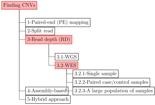
Here is the table depicting types of variations that can be found by specific tools. Read depth-based (RD-based) method is the main concern of this thesis and duplications and deletions are the variation types that we want to find.
Finding CNVs
1-Paired-end (PE) mapping 2-Split read
3-Read depth (RD)
3.1-WGS 3.2-WES
3.2.1-Single sample
3.2.2-Paired case/control samples 3.2.3-A large population of samples 4-Assembly-based
5-Hybrid approach
Figure 3.4: Classification of CNV detection methods
Some of the popular tools for CNV detection with using WES data are CON-DEX [23], CONIFER [24], CONTRA, Control-FREEC, ExoCNVTest [25], ExomeCNV, ExomeDepth [26], PropSeq [27], SeqGene, VarScan2, and XHMM [28].
SV classes Read pair Read depth Split read
deletion yes yes yes novel sequence insertion yes no yes mobile element insertion yes no yes inversion yes no yes interspersed duplication yes yes yes tandem duplication yes yes yes
Table 3.1: Applicability of the tools to the methods
The quantitive relationship between true copy number and depth is distorted by target-specific and sample-specific biases in capturing, PCR amplification, se-quencing efficiency, and in silico read mapping, GC-content of the targets, target size, sequence complexity, proximity to segmental duplications, single nucleotide polymorphisms(SNPs), DNA concentration, hybridization temperature, experi-mental sample batching, and various indeterminate factors. Hence, RD-based methods using WGS data are not applicable to WES data if the extra biases are not accounted for. Moreover, the assumption of normal distribution may no longer be valid due to the biases regarding read depth distribution and most of the CNV breakpoints could not be detected due to the discontinuation of genomic regions. Lastly, the widely applied segmentation algorithms to merge windows in WGS may not be applicable due to the non-continuous distributions of the reads in WES data.
3.2.1
Paired-end mapping (PEM)-based methods
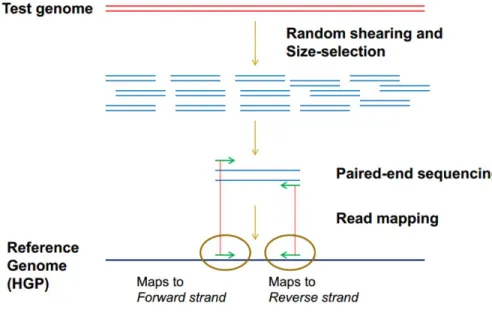
Finding CNV using NGS data was first made by PEM methods that can only applied to paired-end reads. PEM-based method is based on identification of CNVs from discordantly mapped paired-reads whose distances are significantly different from the predetermined average insert size.
Figure 3.5: Paired-end mapping-based methods
In addition to insertions and deletions, this is also used for identification of mobile element insertions, inversions, and tandem duplications. However, this is not applicable for insertion events which are larger than the average insert size.
There are two different approaches in PEM methods which are the clustering approach in which predefined distance is provided and the model-based approach in which statistics is used to define a distance.
Unlike WGS data, the non-random nature of reads from WES limit the appli-cability of PEM-based methods for CNV discovery. For instance, the insert size for paired-end reads in WES data may not be long enough to detect CNV. The PE reads should span the CNV breakpoints, but they may not be within exons, thus not captured.
3.2.2
Split read-based methods
Split read-based (SR-based) methods are conceptually used to find insertions and deletions (indels). These methods use read pairs. First read is aligned to the reference genome uniquely while the other read fails to map or maps partially to the reference genome.
The breakpoints of structural variations (SVs) are provided due to these un-mapped or partially un-mapped reads. The incompletely un-mapped reads are splitted into fragments in SR-based methods. First and last parts of these fragments are aligned to the reference genome and the exact start and end positions are found. SR-based methods can only be applied to the unique regions based on read length.
In contrast to whole genome sequencing (WGS) data, whole exome sequencing (WES) results in nonuniform read depth (RD) between the captured regions and systematic biases that affect data strongly between batch of samples. The split reads should also span the CNV breakpoints, but they may not be within exons, thus not captured. These biases and the sparse nature of the capture make WES unsuitable for well-known CNV detection algorithms [29] [30] [24].
3.2.3
Read depth-based methods
This is the most common approach to estimate copy number due to the ac-cumulation of next generation sequencing (NGS) data. The most appropriate method between these 5 categories to find copy number variations (CNVs) with the help of whole exome sequencing (WES) data is the based on read depth (RD). Depth of coverage, also known as coverage, is calculated by counting the number of reads which cover each base and then calculating their average for each target. RD-based methods are rely on the correlation between the depth of coverage in a genomic region and the copy number of that region. If there is higher count than expected, then there is a duplication in a region. On the contrary, there is a deletion if there is lower count than expected in a region. Exact copy numbers, CNV in complex genomic region classes, and large insertions can be found with the usage of RD-based methods.
Figure 3.7: Calculation depth of coverage
Two major next-generation sequencing approaches, WES and WGS, are used to detect CNVs. If WGS-based tools are used theoretically, the full spectrum of variant can be detected. However, WES-based tools are more effective in terms of time and cost efficiency.
Figure 3.8: Read depth-based method
RD-based methods are classified into 3 categories in terms of the sample size: single sample, paired case/control samples, and a large population of samples. In single sampled-studies the aim is to estimate the read depth distribution us-ing mathematical models and detect the regions with abnormal depth different from the overall distribution. In the second type of method, paired case/control samples, control sample are thought as a reference genome and copy numbers in case sample are reported as relative copies compared to the control sample. These copy numbers are not exact copy numbers. For large population of samples cases, the overall mean of the read depth from multiple samples are used to detect the discordant copy numbers in each sample. This method generally reflects the
exact copy numbers.
CNV discovery from WES data is challenging because of the non-contiguous nature of captured exons (All exons cannot be detected by current technologies). This challenge is compounded by the complex relationship between read depth and copy number which is affected by biases in targeted genomic hybridization, sequence factors such as GC-content, and batching of samples during collection and sequencing. Approaches based on Gaussian and other popular distributions used in CNV detection with using WGS data are not working on CNV detection tools with using WES data due to biases and indeterminate factors.
3.2.4
Assembly-based methods
Firstly, contigs are constructed by using DNA fragments and compared them to the reference genome as a guide. The genomic regions with discordant copy numbers are determined in this way.
Figure 3.9: Assembly-based methods
These kinds of methods propose an unbiased approach to detect novel variants from single base to large structural variations, but these are generally used for the other small-sized genomes due to their huge demand on computational resources. In AS-based methods, reads should also span the CNV breakpoints, but they may not be within exons, thus not captured. These biases mentioned above and the sparse nature of the capture make WES unsuitable for AS-based methods.
3.2.5
Hybrid approaches
Two or more approaches mentioned above are used together in this approach. Though there has been a great progress in each of these approaches mentioned above, none of them is able to detect all variants in a genome precisely.
Each of the methods above has its own advantages and disadvantages so taking advantages creates need to combine some of these methods to increase the performance in detecting variants and reduce false positive rates. [1]
Chapter 4
Related Works
4.1
Whole Genome Sequencing
4.1.1
Summarizing and Correcting the GC Content Bias
in High-Throughput Sequencing
GC content bias is explained as the dependence between read coverage and GC content found on Illumina sequencing data. This bias is not consistent between samples and there is no consensus to find the best method to remove this bias in a single sample. In this paper, a model that produces predictions at base pair level, allow strand-specific GC-effect correction regardless of the downstream smoothing or binning is presented.
Most current correction methods follow a common path. Fragment counts and GC counts are binned to a chosen bin-size. A curve that describes the condi-tional mean fragment count per GC value is estimated. This curve determines a predicted count for each bin based on the bin’s GC. These obtained predictions can be used to normalize the original signal directly. These methods do not use any prior knowledge about the effect whereas they remove most of the GC effect. A descriptive approach to investigate the common structures that is found in
GC curves of DNA sequencing data is described in this work. The effect of GC on fragment count for many high coverage human genomes which are from different labs is studied. Copy numbers for normal genomes change rarely so observed variability in fragment count may always be attributed to technical effects rather than biological effects. A single position model is used to estimate the effect of GC on the fragment counts.
LOESS method is used by them. First, read depth and GC content are calculated for each bin. The GC bias curve is determined by loess regression of count by GC on a random sample of 10000 high mappability (> 0.9) bins. The smoothness parameter, also known as span, for the LOESS should be tuned to produce curves that are smoothing but still capture the main trend in data. This parameter is estimated as 0.3 in this work.
Unlike the other bias correction methods, such as BEADS, predicted fragment rates for the genomic location rather than for the observed reads are generated. [31]
T o o ls Description SeqSeq Detecting CNV brea kp oin ts using massiv ely parallel sequence data CNV-seq Iden tifying CNVs using the difference of observ ed cop y n u m b er ratios RD Xplorer Detecting CNVs throu gh ev en t-wise testing algorithm on normalized read depth of co v erage BIC-seq Using the Ba y esian in formation criterion to detect C NV s based on uni quely mapp ed reads CNAseg Using flo w cell-to-flo w cell v ariabilit y in cancer and con trol samples to reduce false p o si tiv es cn.MOPS Mo deling of read depths across samples at eac h genomic p osition using mixture P oisson mo del Join tSLM P opulation-based approac h to detect common CNVs using read depth data ReadDepth Using breakp oin ts to increase the resolution of CNV detection from lo w-co v erage reads rSW-seq Iden tifying CNVs b y co mparing matc hed tumor and con trol sample CNVnator Using mean-shift ap proac h an d p erforming m ultiple-bandwidth partitioning and GC correction CNVnorm Iden tifying con tamination lev el with normal ce lls CMDS Disco v ering CNVs from m ultiple sam ples mrCaNaV ar A to ol to detect large segmen tal duplications and insertions CNV eM Predicting CNV bre akp oin ts in base-pair resolution cn vHMM Using HMM to detect CNV T able 4.1: Summary of bioinformatics to ols for CNV detection using W GS data. This table is a dapted from [1].
4.2
Whole Exome Sequencing
4.2.1
Copy Number Variation Detection and Genotyping
from Exome Sequencing Data
Copy number inference from exome reads (CoNIFER) is a novel method that uses singular value decomposition (SVD) normalization to discover rare genic copy number variants (CNVs) as well as genotype copy number polymorphic (CNP) loci with high sensitivity and specificity from whole exome sequencing (WES) data. It can be used to discover disruptive genic CNVs that are missed by standard approaches reliably. In this study both read depth (RD) data from WES data with SVD methods to discover rare CNVs and genotype known CNP regions from HapMap samples are combined.
The workflow of CoNIFER starts with dividing fastq-formatted WES reads into non-overlapping 36 base paired-sets and aligning them to the targeted re-gions. These reads are aligned by allowing up to two mismatches per each read set. Reads per thousand bases per million reads sequenced (RPKM) values are calculated and then these are transformed into ZRPKM values, RPKM values transformed into standardized z-scores, based on the median and standard devi-ation of each exon across all samples. ZRPKM values are used as input for the SVD transformation. The strongest k singular values that depends on data are removed. SVD normalization is used to overcome coverage biases introduced by the capture and sequencing of exomes. [24]
4.2.2
Discovery and Statistical Genotyping of Copy
Num-ber Variation from Whole Exome Sequencing Depth
Exome hidden Markov model (XHMM) is a statistical tool that uses princi-pal component analysis (PCA) to normalize exome read depth and uses hidden Markov model (HMM) to discover exon-resolution CNV and genotype variation across samples.
The workflow of XHMM starts with aligned exome read BAM-formatted files. Firstly, depth of coverage is calculated. PCA is run on the sample-by-target-depth matrix by rotating the high dimensional data to find the main components in which depth varies across multiple samples and targets and some of the largest effects that depends on data are removed. Normalized data is trained and HMM is run to discover CNVs spanning adjacent targets. At the end of the process, CNV calls and genotype qualities for all samples is outputted.
The main limitation of XHMM tool is the requirement of large number of samples because of the PCA normalization step. The efficiency of PCA depends on data size. [28]
T o ols Description Con trol-FREEC Correcting cop y n um b er using matc hed case-con trol samples or GC con ten ts CoNIFER Using SVD to normalize cop y n um b er and a v oiding b atc h bias b y in tegrating m ultiple samples XHMM Using PCA to normalize cop y n um b er and HMM to detect CNVs ExomeCNV Using read depth and B-allele frequencies from exome sequencing data to detect CNVs and LOHs CONTRA Comparing base-lev el log-ratios calculated from read depth b et w een case and con trol samples CONDEX Using HMM to iden tify CNVs SeqGene Calling v arian ts, including CNVs, from exome sequencing data PropSeq Using the read depth of the case sample as a linear func tion of that of con tr ol sample to detect CNVs V arScan2 Using pairwise compa risons of the n ormalized read depth at eac h p osition to es timate CNV ExoCNVT est Iden tifying and genot yping common C NV s asso ciated with complex disease ExomeDepth Using b eta -binomial mo del to fit read depth of WES data T able 4.2: Summary of bioinformatics to ols for CNV detection using WES data. This table is adapted from [1].
Chapter 5
Description of the Experiments
5.1
Data
In this thesis, the correlation between read depth, GC content, and probe efficiency is systematically evaluated using 1000 genomes data. We tested our methods on 7 samples from different populations around the world. Samples, HG00629, HG01191, HG01437, NA19664, NA19707, NA19723, and NA20766, are chosen randomly between the other samples of 1000 genomes project. These samples data were created by using the Illumina HiSeq2000 sequencing technol-ogy. Whole genome sequencing data is mapped to the exon regions in human genome (version hg19) so the data are used as whole exome sequencing data with this way. The Agilent Sure Select Capture Kit annotation was used to capture the exomes in the data we analyzed. There are also different whole exome capturing tools like Illumina TrueSeq Capture Kit. Although other capture kit annotations haven’t been tested by using our method, our method can also be used for them theoretically.
The gene positions are token from UCSC Genome Browser refFlat (hg19) data. Probe efficiency, average of GC-content, and read depth are calculated for each gene by using these positions and exon information included by these genes. Known common deletions, duplications, and low-coverage regions of 1000
Genomes data found in this work are used to remove data noise as much as possible.
We only analyzed the autosomal chromosomes (i.e. no sex chromosomes).
5.2
Mapping
5.2.1
Mapping of Reads to the Reference:
MrsFAST-Ultra
Although increasing read lengths, the mapping step remains as an important problem. The accuracy of found structural variants are partially related to this step. Tools that report the best mapping location for each read are not appro-priate for structural variation detection where it is important to report multiple mapping loci for each read. MrsFast fills this gap as a fast, cache oblivious, and SNP-aware aligner that can handle the multi-mapping of next-generation sequencing reads efficiently.
MrsFAST is a mapping algorithm that rapidly finds all mapping locations of a collection of short reads from a donor genome in the reference genome within a user-specified number of mismatches. It is specifically designed for reads gen-erated by Illumina sequencing machines.
Two main steps are included in the tool. The first step consists of building an index from the reference genome for exact anchor matching. The second step consists of computing all anchor matching for each of the reads in the reference genome through the index, extends each match to both left and right and checks if the overall alignment is within the user defined error threshold.
The mapping task is simply partitioned into independent threads executed by a single-core which is defined by user. Multi-thread option is used as using 8 threads in this work. Moreover, paired-end mode is preferred because of the reads used in this work. Owing to the repetitive content of human genome sequence,
![Figure 2.3: Workflow of the Sanger Sequencing Method. This figure is adapted from [7].](https://thumb-eu.123doks.com/thumbv2/9libnet/5613155.110960/24.918.236.730.243.593/figure-workflow-sanger-sequencing-method-figure-adapted.webp)
![Figure 2.4: Workflow of the next-generation sequencing. This figure is adapted from [8].](https://thumb-eu.123doks.com/thumbv2/9libnet/5613155.110960/25.918.224.739.490.708/figure-workflow-generation-sequencing-figure-adapted.webp)
![Figure 2.6: ABI Solid Machine and its procedure. This figure is adapted from [8].](https://thumb-eu.123doks.com/thumbv2/9libnet/5613155.110960/27.918.224.735.171.425/figure-abi-solid-machine-procedure-figure-adapted.webp)
![Figure 2.10: MinION and GridION System.This figure is adapted from [8].](https://thumb-eu.123doks.com/thumbv2/9libnet/5613155.110960/31.918.202.765.167.353/figure-minion-gridion-figure-adapted.webp)
![Figure 2.13: Workflow of whole exome sequencing. This figure is adapted from [18].](https://thumb-eu.123doks.com/thumbv2/9libnet/5613155.110960/33.918.224.739.603.931/figure-workflow-exome-sequencing-figure-adapted.webp)

![Figure 2.14: Classes of structural variation. This figure is adapted from [8].](https://thumb-eu.123doks.com/thumbv2/9libnet/5613155.110960/35.918.276.692.610.919/figure-classes-structural-variation-figure-adapted.webp)