INVESTIGATING THE ROLE OF THE MEDIATOR
COMPLEX IN ESTROGEN RECEPTOR ALPHA-MEDIATED
TRANSCRIPTION
A THESIS SUBMITTED TO
THE GRADUATE SCHOOL OF ENGINEERING AND SCIENCE OF BILKENT UNIVERSITY
IN PARTIAL FULFILLMENT OF THE REQUIREMENTS FOR THE DEGREE OF
MASTER OF SCIENCE IN
MOLECULAR BIOLOGY AND GENETICS
By Tuğçe Canavar
i
INVESTIGATING THE ROLE OF THE MEDIATOR COMPLEX IN ESTROGEN RECEPTOR ALPHA-MEDIATED TRANSCRIPTION
By Tuğçe Canavar July 2018
We certify that we have read this thesis and in our opinion it is fully adequate, in scope and in quality, as a thesis for the degree of Master of Science.
_______________________________________ Murat Alper Cevher (Advisor)
_______________________________________ Serkan İsmail Göktuna
_______________________________________ Mesut Muyan
Approved for Graduate School of Engineering and Science:
_________________________________ Ezhan Karaşan
ii ABSTRACT
INVESTIGATING THE ROLE OF THE MEDIATOR COMPLEX IN ESTROGEN RECEPTOR ALPHA-MEDIATED TRANSCRIPTION
Tuğçe Canavar
M.Sc. in Molecular Biology and Genetics Advisor: Murat Alper Cevher
July 2018
Mediator is the most important coregulator in eukaryotic transcription. It conveys the signals received from transcriptional activators on enhancer sequences to transcription machinery on promoter regions. Although Mediator itself and MED1 subunit have been implicated in the activation of estrogen receptor alpha (ERα)-mediated gene expressions and breast cancer cell proliferations, the mechanism by which Mediator interacts and brings ligand-bound ERα on its cognate element site to promoter and translates their interactions to gene activation is still unknown. We aimed to identify the interaction between ERα and Mediator to clarify this mechanism. We recombinantly generated active human core Mediator complex along with other subunits of Mediator. We interestingly saw that MEDX showed a potential interaction.
In addition to characterizing Mediator- ERα interaction, we also checked the role of the Mediator Complex in acquired tamoxifen resistance. We performed immobilized template recruitment assay to characterize the alterations in recruitment of Mediator subunits and RNA Pol II to 2XERE-TATA promoter in wild type and tamoxifen resistant MCF-7 cell lines. We tried to clarify how changes in pre-initiation complex formation causes resistance to tamoxifen in ERα-positive breast cancer patients. The increase in MEDZ subunit recruitment in resistant cells was striking and promising for further experiments. We hope that our findings will help to elucidate the detailed mechanism of ERα-dependent transcription and design alternative therapeutics.
Key Words: Mediator Complex, Pre-initiation Complex, transcription, tamoxifen resistance
iii ÖZET
MEDIATOR KOMPLEKSİN ÖSTROJEN RESEPTÖRÜ ALFANIN DÜZENLEDİĞİ TRANSKRİPSİYONDAKİ ROLÜNÜN ARAŞTIRILMASI
Tuğçe Canavar
Moleküler Biyoloji ve Genetik Yüksek Lisans Tez Danışmanı: Murat Alper Cevher
Temmuz 2018
Mediator Kompleksi ökaryotik transkripsiyonunun en önemli düzenleyici proteindir. Enhancer sekanslarında bulunan transkripsiyonel aktivatörlerden aldığı sinyalleri promoter bölgelerindeki transkripsiyon sistemine iletir. Mediatorın kendisi ve MED1 alt biriminin östrojen reseptörü alfanın (ERα) aracılık ettiği gen ekspresyonlarının aktifleştirilmesinde ve meme kanseri hücrelerinin proliferasyonunda ilgisi olduğu bilinmesine rağmen Mediator’ın ligand bağlı reseptöre bağlanıp onu spesifik enhancer bölgelerinden promoter bölgelerine hangi mekanizmayla getirdiği ve etkileşimlerini nasıl gen aktivasyonuna çevirdiği hala bilinmemektedir. Biz bu mekanizmayı açıklığa kavuşturmak için Mediator ve ERα arasındaki bağlantıyı belirlemeyi amaçladık. Diğer alt birimlerle birlikte rekombinant aktif çekirdek Mediator kompleksini ürettik. İlginç bir şekilde gördük ki MEDX ile bağlanma potansiyeli göstermektedir.
Mediator ERα etkileşiminin yanı sıra Mediator kompleksinin kazanılmış tamoksifen direncindeki rolünü araştırdık. Normal ve tamoksifene dirençli hale gelmiş MCF-7 hücre hatlarında 2XERE TATA promoterına gelen RNA Pol II ve Mediator alt birimlerindeki değişimleri karakterize etmek için immobilized template recruitment assay uyguladık. Başlama Öncesi Kompleksi (PIC) denen transkripsiyon sistemindeki farklılıkların ERα-pozitif meme kanseri hastalarının tamoksifene dirençli hale gelmesindeki etkisini netleştirmeye çalıştık. Dirençli hücrelerde MEDZ alt biriminin promotera gelmesindeki artış ileriki deneyler için dikkat çekici ve umut vericiydi. Bulgularımızın ERα-aracılı
iv
transkripsiyonun detaylı olarak aydınlatılmasında ve alternatif tedavilerin bulunmasında yardımcı olmasını umut ediyoruz.
Anahtar Kelimer: Mediator Kompleksi, Başlangıç Öncesi Kompleksi, transkripsiyon, tamoksifen direnci
v
vi
TABLE OF CONTENTS
ABSTRACT ... ii ÖZET ... iii TABLE OF CONTENTS ... vi Acknowledgements ... ix List of Figures ... xList of Tables ... xii
Abbreviations ... xiii
CHAPTER 1 INTRODUCTION ... 1
1.1. Mediator Complex ... 1
1.1.1. Activator-Dependent Transcriptional Regulation of Mediator Complex... 3
1.1.1.1. Structural Dynamics of Mediator Complex upon Transcription Factor Binding 3 1.1.1.2 The Role of Mediator Complex in Gene Looping ... 4
1.1.1.3 Mediator Complex & Nuclear Receptor Functions ... 5
1.2 Breast Cancer and Estrogen Receptor (ERα) ... 7
1.2.1 Estrogen Receptor (ERα) Structure ... 8
1.2.2 Estrogen Receptor-Mediated Transcription ... 9
1.2.3 Genomic Transcriptional Activity of Estrogen Receptor (ERα) ... 10
1.3 Proposed Relations between Mediator Complex and ERα ... 11
1.3.1 Kinase Module and Breast Cancer & ERα ... 11
1.3.2 The Role of Mediator Complex in Tamoxifen Resistance ... 12
1.4 Baculovirus Expression System for Recombinant Protein Production in Insect Cells ... 13
1.5 Aim of the Study ... 16
CHAPTER 2 MATERIALS, SOLUTIONS & BUFFERS ... 17
2.1. Cell Culture Media, Supplements, Reagents, Buffers and Equipment ... 17
2.1.1. Wild-type and Tamoxifen-resistant MCF7 Cell Culture Condition ... 17
vii
2.2 SDS-PAGE, Western Blot and Coomassie Blue Staining Buffers ... 18
2.3 Immobilized Template Recruitment Assay (ITRA) ... 19
2.4 Protein Extraction from SF9 Insect Cells and Immunoprecipitation (IP) ... 19
2.4.1 Antibodies Used in Immunoblotting and Immunoprecipitation ... 19
CHAPTER 3 METHODS ... 22
3.1. Plasmid Construction ... 22
3.1.1. Primer Design ... 22
3.1.2 cDNA Synthesis ... 23
3.1.3 Polymerase Chain Reaction (PCR) Protocol ... 23
3.1.4 Digestion of PCR products with Restriction Enzymes ... 24
3.1.5 Ligation of inserts and PFBDM vector ... 25
3.2 DH5 α Competent Cells Preparation ... 25
3.3 Transformation of ligation products to Dh5α competant cells ... 25
3.4 Transformation of PFBDM vectors with the insert into DH10Bac ... 26
3.5 Isolation of Recombinant Bacmid DNA from DH10Bac Transformants ... 26
3.6 Cationic Liposome-mediated Transfection of Sf9 cells with Bacmid DNA ... 27
Amplifying the Virus Stock: ... 28
3.7 Purification of Recombinant Flag-ERα Proteins from SF9 cells Infected with Flag-ERα-P2 virus stock ... 28
3.8 Immunoprecipitation Assay ... 29
3.9 Cell Culture ... 29
3.10 Nuclear Extract (NE) Preparation from MCF7 and MCF7-TamR cells ... 30
3.11 Immobilized Template Recruitment Assay ... 30
3.12 Immunoblotting ... 31
3.13 Co-Immunoprecipitation (Co-IP) Analysis ... 32
CHAPTER 4 RESULTS ... 34
4.1. Purification of Human Mediator Complex Modules and Recombinant Flag-ERα Using Baculovirus Expression System ... 34
4.2 Immunoprecipitation analysis suggests that MEDX might interact with ERα ... 36
4.2.1 IP experiment to check the possibility of nonspecific interaction between MEDX and anti-flag M2 agarose beads ... 38
viii
4.2.2 Co-immunoprecipitation analysis with Flag-MEDX and endogenous ERα in MCF7
nuclear extracts ... 39
4.3 Characterization of Mediator-subunit expression levels in wild-type (WT) and tamoxifen-resistant (TamR) MCF-7 cells ... 40
4.4 Immobilized Template Recruitment Assay (ITRA) shows the difference in levels of Mediator subunits and RNA Pol II recruitment to promoter DNA ... 42
4.5 Mediator-depleted nuclear extracts does not show decrease in ERα protein level .... 44
4.6 MEDY inhibitor increases the recruitment of MEDY in TamR cells ... 45
CHAPTER 5 DISCUSSION ... 47
CHAPTER 6 FUTURE PERSPECTIVES ... 51
BIBLIOGRAPHY ... 54
ix
Acknowledgements
I would like to convey my gratefulness to our mentor and advisor Assist. Prof. Murat Alper Cevher, whose scientific expertise, encouragement, great guidance and support taught me much more than I could imagine and prepared me as a young scientist to be patient and persistent in all situations in my academic career. His kindness, patience and his spirit of determination in regard to research made it possible for me to work on this study enthusiastically all the time. I am so thankful to be part of his lab and for the experiences that I have gained from him.
I would like to thank my dearest parents and my beloved brother for their endless love, support and faith in me. They always believe that I can do the best and support my all decisions. I appreciate their efforts and patience for my education throughout my life. With their love, I have always been a good person. Hopefully, I will be a good scientist too. I am also truly thankful to my lab mates Merve Erden and Onur Rojhat Karasu, my second family in our lab. I will never forget that we stood together days and nights and that we shared lots of good memories. I am very lucky to have their supports and friendships. Then, I deeply want to thank my friends Büşra Kubat, Nazlı Değer and Fatma Betül Dinçaslan for sharing my laughs and my worries since our undegraduate times.
Last but not least, I would like to express my deepest love to my husband, Muzaffer. He beautifies my times in Bilkent and makes me feel I am home. He has stood by my side and has made a great effort to cheer me up. He is one of the driving forces for my determination.
x
List of Figures
Figure 1.1: Mediator enables communication between enhancer-bound TFs and transcriptional complex at the promoter. ……….………...5 Figure 1.2: Modular structure of Mediator Complex and its relation with transcription factors. ……….…...7 Figure 1.3: Functional and structural domains of ERα………9 Figure 1.4: Estrogen and ERα-mediated transcription pathways………..…10 Figure 4.1: Reconstituted human Mediator sub-complexes and Flag-ERα using MultiBac Baculovirus Expression System………...35 Figure 4.2: Immunoprecipitation of ERα with MEDX, MEDY and MEDZ with or without E2 and tamoxifen……….37 Figure 4.2.1: IP experiments show a possible interaction between ERα and MEDX…..38 Figure 4.2.2: Co-immunoprecipitation analysis with Flag-MEDX, endogenous MEDX and ERα………40 Figure 4.3: Comparison of protein expression levels of Mediator subunits in wild-type and tamoxifen resistant (TamR) MCF-7 cell lines. ……….41 Figure 4.4: Mediator subunits and RNA Pol II recruitments on ERα-bound 2xERE-TATA promoter region………43 Figure 4.5: Comparison of ERα protein level in Mediator-depleted and full HeLa
Nuclear Extracts………..45 Figure 4.6 Mediator subunits recruited to ERE containing promoter upon treatment of MEDY inhibitor………46 Figure 5.1: Proposed model for Mediator-ERα interaction………49 Figure 6.1: Gel Electrophoresis results of truncated ERα inserts in PFBDM
xi
Figure 6.2: Western Blot analysis of isolated truncated ERα proteins from Sf9 cells infected with corresponding P0 viruses...52 Figure 6.3: Structure model showing possible interaction surfaces between ER and MEDX………53
xii
List of Tables
Table 2.1: Solutions, reagents and equipment required for cell culture………..17 Table 2.2: SDS-PAGE, Western Blot and Coomassie Blue Staining Buffers………....18 Table 2.3: Solutions required for ITRA using Streptavidin Dynabeads……….19 Table 2.4: Buffers for protein extractions from insect cells and IP ………...19 Table 2.4.1: Antibodies used in Western Blot and IP……….19 Table 3.1: Protocol and condition for PCR using Phusion High-Fidelity PCR Master Mix………...23 Table 3.2: Solutions used in Bacmid isolation………27 Table 3.3: Salt Buffers used in recombinant protein extraction from insect cells……..29 Table 3.4: Buffers used in Co-IP experiment………...…32
xiii
Abbreviations
ERα Estrogen Receptor Alpha GTF General Transcription Factor NR Nuclear Receptor
Pol II RNA Polymerase II PIC Pre-initiation complex TFIIA Transcription Factor IIA TFIIB Transcription Factor IIB TFIID Transcription Factor IID TFIIE Transcription Factor IIE TFIIF Transcription Factor IIF TFIIH Transcription Factor IIH TBP TATA Binding Proteins Med Mediator complex MED Mediator complex
SEC Super Elongation Complex kDa Kilo Dalton
TRAP Thyroid Hormone Associated Protein CRSP Cofactor Required for Sp1 Activation SMCC SRB/MED Cofactor Complex
BSA Bovine serum albumin CTD C-terminal domain
xiv NTD N-terminal domain
ChIP Chromatin Immunoprecipitation
ITRA Immobilized Template Recruitment Assay IP Immunoprecipitation
MAPK Mitogen-activated protein kinase PI3K Phosphoinositide 3-kinase
1
CHAPTER 1
INTRODUCTION
Eukaryotic mRNA transcription requires (i) the assembly of a set of evolutionarily conserved general transcription factors (GTFs) TFIIA, TFIIB, TFIID, TFIIE, TFIIF, and TFIIH to build the accessory proteins on the core regions of most RNA PolII-dependent gene promoters for particular binding and proper transcription initiation [71]; and (ii) the multiprotein Mediator Complex, which is the most critical coactivator that can act as a physical bridge to help the communication between especially gene-specific transcription factors bound at enhancers and Pol II along with GTFs at the promoter region forming the pre-initiation complex (PIC) [1].
1.1. Mediator Complex
Mediator was first purified from Saccharomyces cerevisiae as a 25 subunit complex protein arranged into four modules called `head` and `middle`, which constitute active core Mediator; `tail` and `kinase`[72]. Crystal structures are available for head module and some subunits of middle and kinase modules. The human Mediator complex subunits were purified and initially designated as TRAP (thyroid hormone receptor-associated proteins) because they were identified in a protein-complex which is pulled down together with the ligand-bound receptor when it is isolated from cell extracts [65, 66]. Roeder and colleagues then identified this 2-MDa metazoan Mediator composed of 30-subunits by using biochemical techniques and mass spectrometry. By using Multibac baculovirus expression system, they also reconstituted a functional 15 subunit human core Mediator Complex [3]. Following, researchers successfully produce homogenous recombinant 15-subunit yeast core Mediator by using Med14 15-subunit as a scaffold [2]. The structure of Mediator is conserved from yeast to humans, besides the metazoan complex has specific
2
subunits some of which are Med26 and Med30 [3]. Like yeast complex, human Mediator has also a modular arrangement and its subunits are organized into four subcomplexes. The composition and structure of Mediator complex is, however, variable and dynamic in the cellular contexts [46]. Its composition might change in different cell types at different stages of growth and development. The stable core complex is composed of head and middle modules, which are tightly bound to each other, and reversibly associates with kinase module including CDK8-Cyclin C pair along with Med12 and Med13 subunits. The small Mediator which lacks 600 kDa CDK8-kinase module is called PC2 [73] and its composition generally includes metazoan specific subunit, Med26. Kinase module and Med26 are generally found in the composition of Mediator in a mutually exclusive manner [74]. Further, the association of variable metazoan specific subunits with the core complex can result in additional gene-specific variants [4].
Mediator is generally considered as a co-activator because of being an important target for DNA-bound transcription factors which recruits Mediator into regulatory sites throughout the genome via possible interaction with its subunits and ,thereby, causing a DNA-loop formation. However, studies in yeast and human demonstrated that Mediator has also role in activator-independent transcriptional regulations. The isolated RNA Pol II holoenzyme from both human and yeast has contained Mediator subunits. Further studies including ChIP-on-ChIP technology has revealed that Mediator also localizes upstream and coding regions of many genes on a genome-wide scale [5]. A model of RNA Pol II- Mediator complex identified in yeast by electron microscopy revealed that several subunits of Pol II such as Rpb4 and Rpb7 might interact with the head and middle module of Mediator [75]. Additionally, functional assays involving 15-subunit active human core complex reconstituted by Cevher et al, also showed that the reconstituted bimodular complex containing head and middle modules requires the Med14 incorporation into the assembly as an architectural backbone. More importantly, the existence of Med14 in the complex is necessary for basal transcription and facilitated Pol II interaction [3]. Overall studies have suggested that Mediator regulates both basal and activator-dependent transcription and PIC formation by facilitating the recruitment of Pol II and GTFs to the target gene promoters and stabilizes these multiprotein complexes at the promoter.
3
1.1.1. Activator-Dependent Transcriptional Regulation of Mediator Complex
In eukaryotes, RNA polymerase II initiates transcription at specific sites on the genome. It is recruited to transcription start site via auxiliary protein complex, called pre-initiation complex (PIC), which is composed of transcription factor II A (TFIIA), TFIIB, TFIID, TFIIE, TFIIF, TFIIH, and Mediator Complex [6]. Mediator stabilizes PIC formation by serving as a central scaffold for assembly of other PIC components. Its large size enables this function as different subunits can interact with different components of the PIC. Importantly, the recruitment of Pol II to the transcription site requires Mediator that interacts with C-terminal domain (CTD) of RNA Poll II Rpb1 subunit [7, 8,].
Mediator Complex also enables the communication between gene-specific, enhancer-bound transcription factors (TFs) and RNA Pol II together with the rest of PIC [9]. It serves as a molecular bridge to direct signals from a DNA-binding TF to Pol II enzyme to control enhancer-specific transcription. Different TFs might interact with distinct Mediator subunits to mediate expressions of their target genes in cell-type specific context as well.
1.1.1.1. Structural Dynamics of Mediator Complex upon Transcription Factor Binding
The function of Mediator Complex correlates with the dynamic change in its structure upon binding of different TFs. As compositional change in the complex might abolish the TF-bound activity in the transcription, structural shift is also equally important for the Pol II activity. For example, whereas Med23 knockout does not affect other TF-Mediator regulated transcriptions, ELK1-Med23 mediated target genes are not activated in MED23-knockout murine embryonic stem cells [76]. On the other hand, the activation of Pol II enzyme on the promoter of early growth response factor 1 (Egr1) associates with the structural change in phosphorylation-dependent ELK1-Med23 interaction [10]. The structural shift spanning over entire Mediator complex after binding of a TF to a single site is first observed when SREBP-1α (sterol regulatory element-binding protein 1) [77] and VP16 (a transcription factor of herpes simplex virus, HSV), activation domains bind to Mediator and creates different structural organization without affecting its subunit composition. Moreover, EM (electron microscopy)-based experiments showed that these
4
TFs interact with different subunits of Mediator complex. As SREBP-1α associates with Med14 and Med15, VP16 relates to Med25 and Med17. VP16-targeted in vitro transcription is ceased when Med25-Mediator complex is depleted from HeLa cell nuclear extracts by using anti-Med25 antibody. Also, RNAi-knockdown of Med25 from human cells leads to the inhibition of VP16-activated gene expressions [11, 12, 13, 14]. Undergoing different structural alterations upon binding to distinct domains of TFs affects the transcriptional state by controlling the recruitment of Pol II and defines the participation of additional coactivators to the pre-initiation complex (PIC). For instance, Taatjes and Meyer groups revealed that p53 interacts with Mediator via its either p53AD (activation domain) or p53CTD (C-terminal domain) by binding to Med17 and Med1 subunits respectively. Interaction with activation domain creates a conformational change in Mediator such that it exposes a large domain causing the transition of Pol II to elongation state from an inactive state. However, binding to p53CTD results in a distinct conformational alteration leading to inactivation of Pol II-complex [15]. A general conclusion from these studies is that TFs that binds to different interaction surfaces on Mediator might lead to distinct structural shifts and coactivator recruitment, resulted in separate regulations in the PIC formation.
1.1.1.2 The Role of Mediator Complex in Gene Looping
Activation of gene specific TF-targeted gene expressions is regulated by DNA-loop formations integrating enhancer-bound TFs with RNA Pol II-complex at the promoter regions. The participation of Mediator complex in both enhancers and core promoters during active gene transcription is first identified in murine embryonic stem cells [67]. Chromosome conformation capture (3C) experiments showed that in the cell-type specific gene transcriptions, Mediator works with cohesin and cohesin loading factor Nipb1 to bring enhancer regions to promoters to form proper DNA-looping [16]. Normally, long-distance enhancer-promoter bridges are not observed in the yeast gene transcriptions, however, mutant yeast ortholog of Med16 enables upstream regulatory regions to activate transcription [17]. This is also consistent with the ability of Mediator to interact with Pol II CTD. In this way, it can stabilize the promoter-enhancer gene looping. Further, the length of human RNA Pol II CTD is far longer than yeast’s, which is correlated with the increase in the length of promoter-enhancer interactions in human [18].
5
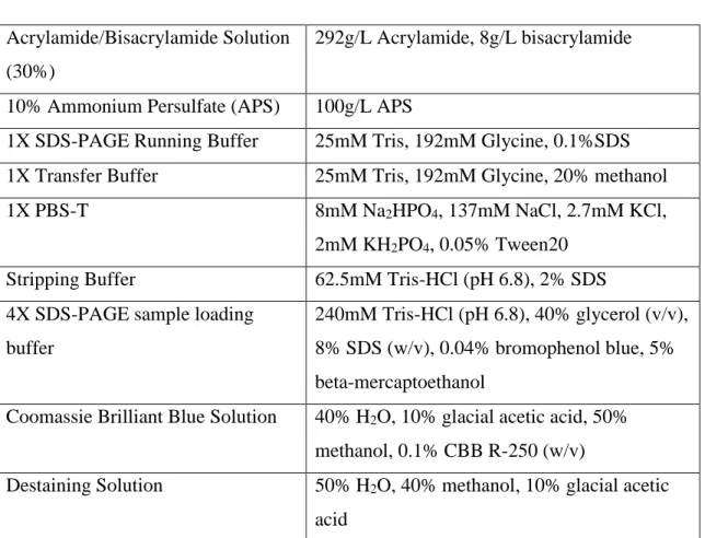
Figure 1.1. Mediator enables communication between enhancer-bound TFs and transcriptional complex at the promoter [62].
1.1.1.3 Mediator Complex & Nuclear Receptor Functions
Nuclear Receptors (NRs) are transcription factors which shows generally ligand-dependent activation to regulate sequence-specific gene expressions involved in development, homeostasis and cell differentiation. During translocating signals received from a cognate ligand to the promoter region of a target gene, NRs needs transcriptional coregulators that are mainly classified into two main categories namely coactivators and corepressors. Unlike corepressors operating with antagonist-bound NRs and block expression, coactivators help in the activation of gene expression. Many coactivator complexes are involved in chromatin remodeling, enzymatic reactions and the recruitment of transcriptional machinery. Steroid hormone receptors associate with steroid receptor coactivator (SRC) family members of the p160 class coregulators to control assembly of histone-modifying factors [19]. CREB-binding protein (CBP)-p300 is another coactivator that has histone acetyltransferase (HAT) activity regulating chromatin relaxation at the promoter region [20].
6
Mediator Complex acts directly on the RNA Pol II-complex and PIC formation. It is a general cofactor for ligand-dependent NRs and after purification process, many isolates of NRs are pulled down with Mediator. Med1, one of the subunits of Mediator, is most associated with nuclear receptors. It contains two leucine-rich amino acid motif "LXXLL" so-called "NR-box", which is frequently present in almost every nuclear receptor coactivators [21]. This motif is necessary for Mediator complex to provide direct interactions with NRs and regulate Med1-dependent activation of NRs such as estrogen receptor (ER), glucocorticoid receptor (GR), vitamin D receptor (VDR), peroxisome proliferator-activated receptor (PPAR), retinoic acid receptor (RAR), and retinoid X receptor (RXR). Furthermore, the importance of Med1 in the NR-dependent gene expressions is also confirmed with in vivo studies. For example, Med1 deletion in mouse embryonic fibroblasts (MEF) forms a deficiency in PPAR-mediated adipogenesis without affecting other NR-mediated developmental pathways [22, 23, 24].
Med25 is another subunit that contains LXXLL motif in its C-terminus and recently has been associated with the regulation of HNF4-mediated gene expressions. Direct binding of Med25 LXXLL motif to HNF4 organizes the assembly of the coactivators and induces the expressions of genes that are functional in lipid and drug metabolism [25].
Conclusion from these studies is that the Mediator Complex is a critical coactivator having a broad impact on NR-mediated transcription via its ability to interact with many proteins involved in transcriptional complex under favour of its multi-subunit structure.
7
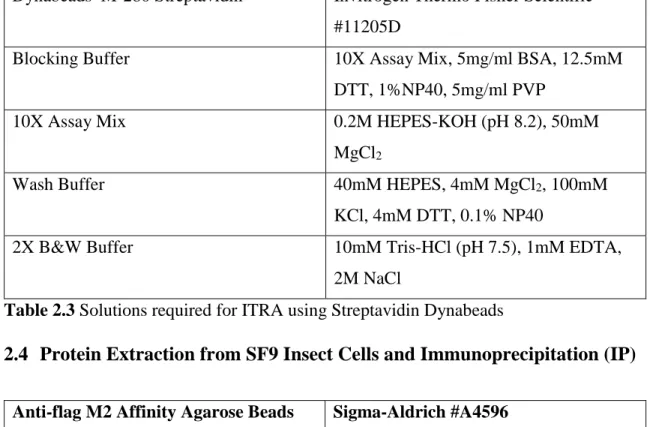
Figure 1.2. Modular structure of Mediator Complex and its relation with transcription factors [63].
1.2 Breast Cancer and Estrogen Receptor (ERα)
Breast Cancer is highly diagnosed cancer type after lung cancer in women worldwide. The incidence ratio observed in women to men is 125 to 1 [26]. This is because it depends on hormonal factors, especially steroid hormones whose endogenous level determines the risk of developing the disease. One of these steroid hormones is estrogen that is the immediate stimulus to mammary development coordinating the formation of ductal structures and enhancement of lobules from normal epithelium [27, 28]. Estrogen is synthesized from cholesterol mainly in ovaries, placenta and corpus luteum and stimulates estrogen receptor alpha (ERα).
ERα is a member of nuclear receptor family and its presence in a tumor is diagnostic marker. The variation in its expression and distribution is related to age and reproductive history [29]. Since breast cancer is a heterogeneous disease with respect to
clinical-8
pathological features and the status of receptor expressions, it is classified into distinct subtypes with different morphologies and clinical outcomes which are luminal A, luminal B, HER2 over-expression, basal and normal-like tumors. The expression of ERα in a tumor is accepted as a favorable prognostic biomarker and the large majority of ERα-positive breast tumors take place under luminal A and luminal B subtypes [30, 31]. 80% of breast cancer patients are ERα positive, which makes the receptor a good prognostic target for drug responsiveness [32].
1.2.1 Estrogen Receptor (ERα) Structure
Human ERα is located on the chromosome 6 and shares the common structure with all other nuclear receptors. It is composed of variety of regions named A/B, C, D, and E/F [78]. These regions forms different functional domains that are involved in transcriptional regulations. A/B region called the N terminal activation function domain (AF-1) and E/F region (AF-2) involved in ligand binding domain (LBD) works synergistically in the regulation of ligand-dependent transcriptional activity by recruiting and binding to a number of coactivators [79]. These two functional domains mostly work in a synergistic manner but AF-1 can also be activated in the absence of a ligand in certain cell and promoter specific contexts, depending on the phosphorylation status of ERα. For example, the phosphorylation site, Ser118 located on AF-1 region is an important target for MAPK signaling pathway involved in the ligand-independent transcriptional activation of the receptor. AF-2 domain consist of 12 α-helix forming a hydrophobic cleft which is masked until a ligand/ coactivator occupation and [80], is important for the interaction with Src-family of coactivators. Therefore, AF-2 is essential to regulate ligand-dependent activation by activating ERα through conformational changes. ERα binds to the ERE-sequence on DNA through its two zinc-finger domains which were designated as DNA-binding motifs. These protein structures organized by zinc ions are located in the DNA binding domain of the receptor (DBD, C region) [81, 82]. D region called hinge domain, on the other hand, has a role in binding to chaperone heat-shock proteins in the inactive state of ERα and in coordination of dimerization upon ligand stimulation [33, 34].
9
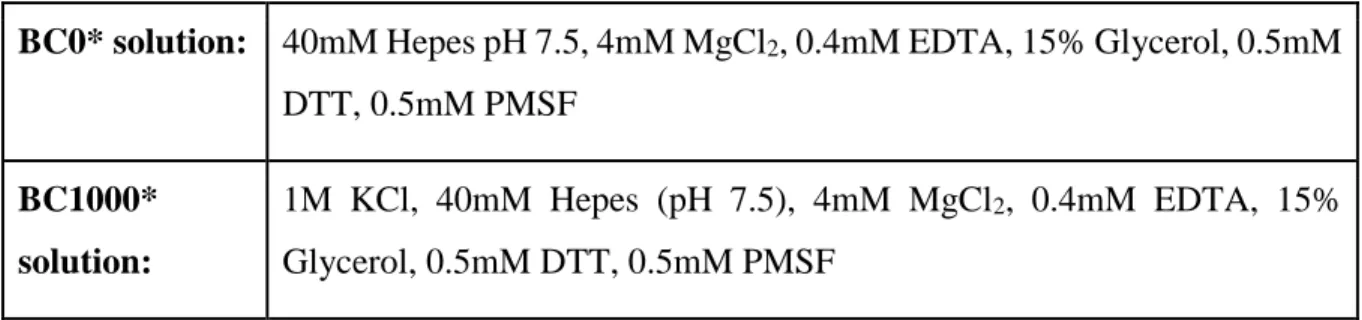
Figure 1.3. Functional and structural domains of ERα [64].
1.2.2 Estrogen Receptor-Mediated Transcription
The ERα-dependent transcriptional activities are divided into two distinct categories, depending on how the receptor initiates the formation of basal transcription machinery on the promoter of target genes. These two different signaling are called `genomic` and `non-genomic` pathways. Non-genomic signaling is appeared through rapid effect of estrogen binding to the membrane associated ERs (mER). mER regulates this activity either via cross-talk with other membrane-linked receptors or activation of a diversity of signaling cascades such as Raf/Ras/MAPKs, PKCs, PI3K/AKT, and cAMP protein kinase A (PKA). For example, ERK and PI3K/AKT activities increase when E2-bound ER activates EGF receptor. However, it is important to note that ER signaling through other receptors is cell type-specific. In addition to non-classical signaling, nuclear ERα regulates the genomic transcriptional activity in a canonical model which involves direct binding of the receptor to the specific sequences on promoter regions. This classical model of transcription mediated by nuclear ERα is the subject of this thesis and is elucidated more in detail in the following sections [34, 35, 36].
10
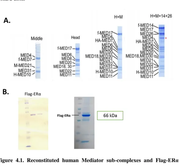
Figure 1.4. Estrogen and ERα-mediated transcription pathways [68].
1.2.3 Genomic Transcriptional Activity of Estrogen Receptor (ERα)
The ultimate goal of ERα, as a nuclear steroid hormone receptor, is to elicit the assembly of RNA Pol II transcriptional complex to the promoter regions of estrogen-responsive genes. In the canonical signaling pathway, the regulation of ERα-target gene expressions involves the direct binding of the ligand-bound receptor to the specific enhancer sequences on DNA. ERα transactivates as dimers, however, unliganded ERα is located in the cytoplasm as a monomer, bound to a heat shock (Hsp) chaperone protein complex. This chaperone Hsp competes with estrogenic ligands by interacting with the ligand binding domain (LBD) of ERα and keeps the receptor inactive state by preventing it from binding to DNA [37]. Upon binding of estrogen, ERα dissociates from chaperone protein and undergoes conformational changes leading to dimerization and activation of its transactivation domains. Active dimeric ERα translocates into the nucleus and binds to specific DNA sequences called estrogen response elements (EREs). This estrogen-responsive motif was first identified in the promoter of Xenopus laevis vitellogenin and shows properties of an enhancer such as acting in distance and orientation-independent manner. ERα binds directly to this cis-acting enhancers which are inverted six nucleotide palindromic repeats separated by a three nucleotide spacer [38]. Binding of dimeric ERα to ERE sequence causes allosteric effects on its structure unmasking interaction sites for
11
the recruitment of coactivators, thereby, the assembly of transcriptional machinery on the promoter regions of ERα-target genes. Therefore, in order to reveal the transcriptional regulations of estrogen-responsive genes, it is critical to elucidate intermediary coactivator proteins that bridges dimeric receptor in the enhancer region to the components of RNA PolII complex on the promoter.
1.3 Proposed Relations between Mediator Complex and ERα
Roeder group demonstrated the activity of Mediator Complex on ERα-mediated in vitro transcription by using ERE-containing promoter templates, purified transcription factors, purified Mediator and estrogen receptor expressed with baculovirus system. The cell-free system reveals that the presence of Mediator improves the ERα-dependent basal transcription [39, 40]. Additionally, Jiang group showed impaired development in the mammary gland by generating mutation in the LXXL motif on MED1 subunit in mice, suggesting the importance of MED1-interaction with the transcription factors [69].
1.3.1 Kinase Module and Breast Cancer & ERα
The kinase module of Mediator Complex that is composed of four subunits which are MED13, MED12, and CDK8-Cyclin C pairs binds to the core Mediator complex reversibly via its MED13 subunit. Therefore, Mediator purifications derived from nuclear extracts via different biochemical techniques were further distinguished according to whether it includes kinase module or not. Based on early biochemical studies, kinase module has been generally associated with the repression of transcription [41]. As purified Mediator containing kinase module restrained the activator-dependent transcription, the core Mediator lacking this module activated. EM-based studies on yeast Mediator explained this observation by the finding that when kinase module binds to the core Mediator, resulting conformation blocks the interaction surface between Pol II and core Mediator [42]. However, the notion that kinase module has only repressive role in transcription is inconsistent with new findings and it has been understood that kinase module can both repress and activate transcription depending on the cell-context.
12
To date, some of kinase module subunits are implicated in oncogenic alterations in many cancer types like colon cancer, melanoma, endometrial cancer (CDK8), osteosarcoma, T-ALL (CCNC) and prostate cancer (MED12). Recently, they are also implicated in the key role of gene transcriptions involving in the proliferation of breast cancer cells. It is revealed that kinase subunits are part of the enriched coregulators on the estrogen response element (ERE) site. By using streptavidin beads and biotinylated ERE-E4 templates, O’Malley group determined the enriched coregulators from nuclear extracts on E2 liganded ERα. Then mass spectrometry and immunoblotting analyses identified these 17 enriched proteins and revealed that kinase subunits; MED13, MED12, CDK8, and CCNC are some of them [43]. Consistent with the previous studies showing that high expressions of CDK8 and CCNC inversely correlates with shorter recurrence-free survival in breast cancer, CDK8 inhibitor (Senexin A) blocks the E2-induced mRNA transcription of ERα-responsive genes such as TFF1 and GREB1 in MCF7 cells. Also, both shRNA-based knockdown and CRSPR-Cas9-mediated knockout of CDK8 showed the same inhibitory effect on estrogen-stimulated transcription of GREB1 gene [44, 45]. All these studies suggest that kinase module of Mediator Complex might have a key role in the transcriptional regulation of estrogen-dependent genes in ER-positive breast cancer.
1.3.2 The Role of Mediator Complex in Tamoxifen Resistance
Tamoxifen is a selective ER modulator (SERM) that has been used for 30 years at all stages of breast cancer. Like other SERMs, its agonist or antagonist activity might be altered by coregulators recruited to the promoter in a cell-context manner. Whereas tamoxifen acts as an agonist in the uterus, it acts as an antagonist in the breast by competing with estrogen for ligand binding domain (LBD) of estrogen receptor and preventing transcriptional activity [47, 48, 49]. Although it has been an effective chemotherapeutic reagent, 50% ER positive breast cancer patients acquired resistance to tamoxifen.
Tamoxifen resistance is categorized into two groups which are de novo resistance that is unresponsive to tamoxifen treatment since the very beginning and acquired resistance. De novo resistance is modeled by the transfection of Her2 gene into MCF7 cell line that is then causing tumor growth in xenograft mice even in the presence of tamoxifen. On the
13
other hand, acquired resistant cell lines are obtained from initially sensitive cells after a long term therapy.
Although the molecular mechanism that causes tamoxifen resistance has not been fully understood, MED1 subunit of Mediator Complex is suggested to be the regulator of Her2-mediated tamoxifen resistance. Studies support that phosphorylation of MED1 at specific sites is important for its function and this is regulated by ERK activity mediated by HER2 signaling in tamoxifen resistant cells. Further, it is shown that impairment in its phosphorylation leads to recruitment of corepressors NCOR and SMRT to the ERα-target gene promoter, otherwise they are not assembled together with phosphorylated form of MED1. As in vivo analyses demonstrate that higher expression of MED1 in breast cancer patients is correlated with the decrease in recurrence-free survival, knockdown of MED1 in ERα positive cell lines results in the inhibition of ERα-mediated transcriptional activity [50, 51].
1.4 Baculovirus Expression System for Recombinant Protein Production in Insect Cells
In eukaryotic organisms, many proteins are not singular entity but multi-subunit complexes which are even part of a bigger cellular network. Therefore, it is very essential to reveal their structure, functional roles and interactions within the complex or with other protein assemblies in order to understand their necessities in many cellular events [52]. To investigate the role of these complexes at the molecular level, high amount and properly folded proteins are required to be used in structural studies like imaging of protein interactions in atomic resolution or biochemical techniques such as immunoprecipitation and DNA-pull down assays. Unfortunately, endogenous proteins are often synthesized in low amounts in their cell environment or hard to delete as some of them are essential [53]. Thus, recombinant protein production has been mandatory to obtain sufficient amount of functional proteins for experimental needs.
Prokaryotic expression system, especially the use of E.coli as an expression host, is one of the well-established methods in laboratories to produce recombinant proteins in cheap and easy way [54]. However, bacterial system does not allow the adjustment of expression
14
ratios in co-expressing of individual subunits when the vector containing multiple genes is delivered. Besides, eukaryotic protein complexes require post-translational modifications and proper folding which are not provided by a prokaryotic host to the all extend [55].
Baculovirus expression system has gained increased interest to obtain recombinant proteins in high yields and proper eukaryotic protein processing conditions. Baculovirus is a double stranded circular DNA that invades arthropods, especially insects. The most worked and manipulated baculovirus for the generation of recombinant expression vector is Autographa californica multicapsid nucleopolyhedrovirus (AcMNPV). The reason why AcMNPV is a good tool for more than 100 mg recombinant protein production per 1 liter of insect cells is because of to produce large amounts of polyhedrin production which appears in nuclei of insect cells during AcMNPV infection. This is turned into an advantage by using polyhedrin promoter (polh) to control the expression of a DNA sequence encoding a protein of interest. The generation of recombinant viral DNAs was first achieved in insect cells co-transfected with transfer plasmid containing gene of interest under the control of polh and purified AcMNPV genomic DNA. Homologous recombination (double crossover) provides the replacement of foreign DNA in transfer plasmid with polyhedrin gene in the baculovirus genome [56]. However, the low frequency of homologous recombination (estimated as 0.1%) and isolating low amounts of recombinant baculovirus vectors from co-transfected cells were required for a new modification. This problem was solved by using linearized baculovirus genome, which increases the frequency of homologous recombination up to 20% and decreases replication of non-recombinant viral genome replication [57]. Later, researchers improved another approach in which E.coli strain is generated to produce a plasmid (bacmid) that integrates with the gene of interest in transfer vector. When transfer vector is transformed into E.coli containing the viral bacmid, the gene of interest in transfer plasmid is integrated into the polyhedrin region of the bacmid via Tn7 transposition and deletes LacZ gene in the bacmid by permitting the blue/white selection. This approach enables scientists who are not familiar with virology but bacterial methods to make a viral progeny in insect cells in easier way.
15
Although modifications enabled researchers to obtain high amounts of recombinant individual proteins, the isolation of recombinant multi-subunit complexes from host cells co-infected with viruses expressing only one subunit was still an obstacle. Due to the difficulties in controlling expression ratios of individual subunits in infected-cells, resulting from incapability of titer-arrangements of viruses, co-infection was not an effective method for the complex formation. Therefore, it was more logical to eliminate co-infection and rather to use one baculovirus expressing all genes of interest. To achieve this, researchers created Multibac system by modifying transfer vectors allowing the integration of multiple genes in nonsequential way. They generated two vectors which are PFBDM and PUCDM. The expression of genes placed either side of the multiplication module via proper restriction sites in the expression cassettes is under the control of polh or p10 viral promoters. The multiplication module contains PmeI&AvrI and BstZ171&SpeI or Nru&SpeI restriction sites. Since these sites are compatible, the expression cassette containing two genes of a transfer vector can be cleaved and combined with another one and finally in this way, desired amounts of genes can be inserted into a baculovirus expression vector [58].
Multibac expression system has been widely used by scientists to purify functional and stoichiometric protein complexes in insect cells. Sf9 and High Five are two generally used cell lines. Since they can grow in either adherent or suspension, to be able to grow them in spinner flasks or stirred tanks provides researchers a chance to produce their proteins in an amount as large as they want. The largest protein complex that was produced by Dr. Cevher is active human core Mediator Complex. He used this multibac expression system to jointly express Mediator subunits and reconstituted functional 15-subunit Mediator.
16 1.5 Aim of the Study
The ultimate goal of our study is to find the estrogen receptor alpha (ERα) interactive form of Mediator Complex to prevent ERα-mediated tumorigenesis of breast cancer. By blocking ERα-Mediator interaction, we aimed to stop tumor formation specifically at transcriptional regulation by disrupting the expressions of only ERα-responsive genes but not the others in cell type specific manner. Our hypothesis is that Mediator Complex directs the signal transmission from enhancer-bound ERα to transcription machinery on promoters of ERα-responsive genes. To carry out investigation of our hypothesis, we first tried to identify the possible interaction subunits and recruited subunits on the estrogen response element site (ERE); then as a second study, aimed to clarify the role of Mediator on tamoxifen resistance in breast cancer by working on wild type and MCF-7 cells line acquired resistance to tamoxifen.
17
CHAPTER 2
Materials, Solutions & Buffers
2.1. Cell Culture Media, Supplements, Reagents, Buffers and Equipment
Product Name Brand & Catalog No
Grace`s Insect Media (TNM-FH) Lonza Biowhittaker #04-649F
Gentamicin Thermo Fisher #15750060
Poloxamer Sigma-Aldrich #16758
Fetal Bovine Serum (FBS) Biowest #181H-500
DMEM low glucose, without phenol red Thermo Scientific #11880028 Penicillin/Streptomycin Gibco #15140-122
Nonessential amino acid (NEAA) Lonza #BE13-114E Insulin solution from bovine pancreas Sigma #I0516
Cellfectin II Reagent Invitrogen #10362-100
β-Estradiol Sigma-Aldrich #E8875
Tamoxifen Sigma-Aldrich # T5648-1G
100mm and 6 well plates Corning Coster 10 ml, 250 ml and 500 ml spinner flasks
10X PBS pH 7.4 Home Made
Table 2.1 Solutions, reagents and equipment required for cell culture
2.1.1. Wild-type and Tamoxifen-resistant MCF7 Cell Culture Condition
• DMEM low glucose, without phenol red • 10% FBS-heat inactivated and filtered
• 1X Pen/Strep ( 100 units/ml penicillin and 100µg/ml streptomycin) • 1X NEAA
18 • 10 µg/ml insulin
2.1.2 Sf9 Insect Cell Culture Condition
• Grace`s Insect Media • 10% FBS-heat inactivated • 50 µg/ml gentamicin • 1X Poloxamer
2.2 SDS-PAGE, Western Blot and Coomassie Blue Staining Buffers
Acrylamide/Bisacrylamide Solution (30%)
292g/L Acrylamide, 8g/L bisacrylamide
10% Ammonium Persulfate (APS) 100g/L APS
1X SDS-PAGE Running Buffer 25mM Tris, 192mM Glycine, 0.1%SDS 1X Transfer Buffer 25mM Tris, 192mM Glycine, 20% methanol
1X PBS-T 8mM Na2HPO4, 137mM NaCl, 2.7mM KCl,
2mM KH2PO4, 0.05% Tween20 Stripping Buffer 62.5mM Tris-HCl (pH 6.8), 2% SDS 4X SDS-PAGE sample loading
buffer
240mM Tris-HCl (pH 6.8), 40% glycerol (v/v), 8% SDS (w/v), 0.04% bromophenol blue, 5% beta-mercaptoethanol
Coomassie Brilliant Blue Solution 40% H2O, 10% glacial acetic acid, 50% methanol, 0.1% CBB R-250 (w/v)
Destaining Solution 50% H2O, 40% methanol, 10% glacial acetic acid
19
2.3 Immobilized Template Recruitment Assay (ITRA)
Dynabeads M-280 Streptavidin Invitrogen Thermo Fisher Scientific #11205D
Blocking Buffer 10X Assay Mix, 5mg/ml BSA, 12.5mM
DTT, 1%NP40, 5mg/ml PVP
10X Assay Mix 0.2M HEPES-KOH (pH 8.2), 50mM
MgCl2
Wash Buffer 40mM HEPES, 4mM MgCl2, 100mM
KCl, 4mM DTT, 0.1% NP40
2X B&W Buffer 10mM Tris-HCl (pH 7.5), 1mM EDTA, 2M NaCl
Table 2.3 Solutions required for ITRA using Streptavidin Dynabeads
2.4 Protein Extraction from SF9 Insect Cells and Immunoprecipitation (IP)
Anti-flag M2 Affinity Agarose Beads Sigma-Aldrich #A4596
Flag Peptide Sigma #F3290
BC1000* (*salt concentration) BC0* (*salt concentration)
20mM Tris-HCl (pH: 7.9 at 4 oC) 20mM Tris-HCl (pH: 7.9 at 4 oC) 20% Glycerol 20% Glycerol 0.1 mM EDTA 0.1 mM EDTA 0.5 mM PMSF 0.5 mM PMSF 0.5 mM DTT 0.5 mM DTT 1M KCL
Table 2.4 Buffers for protein extractions from insect cells and IP 2.4.1 Antibodies Used in Immunoblotting and Immunoprecipitation
Product Name Brand & Catalog No Dilutions
Estrogen Receptor α (D8H8) rabbit mAb
Cell Signaling Tech. #8644 1:1000
20
Med15 rabbit pAb Proteintech #115661AP 1:1000 Med16 rabbit pAb Santa Cruz Biotech. 1:1000 Med25 (A-7) mouse mAb Santa Cruz Biotech.
#SC393759
1:1000
MEDX Home Made Rockefeller
University –R.G.R Lab
1:1000
MEDY rabbit pAb Home Made Rockefeller University –R.G.R Lab
1:1000
Med6 rabbit pAb Home Made Rockefeller University –R.G.R Lab
1:1000
Med12 rabbit pAb Home Made Rockefeller University –R.G.R Lab
1:1000
Med26 rabbit pAb Home Made Rockefeller University –R.G.R Lab
1:1000
MEDZ rabbit pAb Home Made Rockefeller University –R.G.R Lab
1:1000
Med30 rabbit pAb Home Made Rockefeller University –R.G.R Lab
1:1000
Rpb1 (8WG16) mouse pAb
Home Made Rockefeller University –R.G.R Lab
1:1000
Rpb5 rabbit pAb Home Made Rockefeller University –R.G.R Lab
1:1000
Rpb6 rabbit pAb Home Made Rockefeller University –R.G.R Lab
1:1000
P62 rabbit pAb Home Made Rockefeller University –R.G.R Lab
1:1000
P53 rabbit pAb Home Made Rockefeller University –R.G.R Lab
1:1000
Anti-mouse IgG, HRP-linked Antibody
21 Anti-rabbit IgG,
HRP-linked Antibody
Cell Signaling #7074S 1:10000
22
CHAPTER 3
Methods
3.1. Plasmid Construction
3.1.1. Primer Design
The DNA coding for flag tagged full-length human estrogen receptor alpha (hERα) was amplified by using the primers 5′- GC GAATTC ATG GAC TAC AAA GAC GAT GAC GAC AAG ACC ATG ACC CTC CAC-3′ as forward and 5′-GC GTCGAC TCA GAC CGT GGC AGG-3′ as reverse. EcoRI cleavage sequence was added to the forward primer and SalI cleavage sequence was inserted into the reverse primer. cDNA was used as a template in the PCR of full-length flag tagged hERα. The PCR product was cleaved with EcoRI and SalI, purified, and cloned into the PFBDM vector by ligation. The full-length hERα was fragmented according to its domains. The primers for residues 1-185 (N terminal region, transactivation domain), 185-355 (DNA binding domain and hinge) and 301-595 (ligand binding domain) of hERα are 5′- GC CTCGAG ATG CATCATCATCATCATCAT AAA ACC ATG ACC CTC CAC-3′, 5′-GC GCTAGC TCA ACA GTA GCG AGT CTC CTT-3′, 5′-GC CTCGAG ATG CATCATCATCATCATCAT AAA GCA GTG TGC AAT GAC-3′, 5′-GC GCTAGC TCA GGC CGT CAG GGA-3′, 5′-GC GAATTC ATG CATCATCATCATCATCAT AAA GAC CAG ATG GTC AGT-3′, 5′-GC GCGGCCGC TCA GAC CGT GGC AGG-3′ respectively. The hERα fragments were tagged with histidine residues. The PFBDM plasmid containing full-length flag tagged hERα was used as a template for the PCR
23
amplifications of fragments. Their PCR products were restricted with the enzymes corresponding to cleavage sites in their sequence, purified and inserted into PFBDM vector by restriction and ligation protocols as described below.
3.1.2 cDNA Synthesis
cDNA was synthesized from total mRNA of Hela cells using the protocol of Thermo Scientific Revert Aid First Strand cDNA synthesis kit #K1622, provided by the manufacturer.
3.1.3 Polymerase Chain Reaction (PCR) Protocol
Thermo Scientific Phusion High-Fidelity PCR Master Mix #F-5315 was used in reactions for the amplification of coding DNA of interests.
2x HF Phusion Master Mix 25 µl Forward Primer (10 µM ) 1.5 µl Reverse Primer (10 µM ) 1.5 µl cDNA (template DNA) 5 µl
ddH2O 17 µl
Total 50 µl
Cycle Step Time Temperature Cycle Initial Denaturation 30 sec 98°C 1 cycle
Denaturation 10 sec 98°C
35 cycles
24
Extension * 72°C
Final Extension 7 min 72°C 1 cycle *changes according to the length of amplified DNA
**changes according to the melting temperatures of primers
Table 3.1 Protocol and condition for PCR using Phusion High-Fidelity PCR Master Mix
0.8 % or 1% agarose gel was used to run PCR products with different sizes. Products were excised and purified by using Thermo Scientific GeneJet Gel Extraction Kit #K0691.
3.1.4 Digestion of PCR products with Restriction Enzymes
PFBDM vector and all PCR products were cleaved with restriction enzymes corresponding cleavage sites included in their primers. Thermo Scientific FastDigest Value Pack kit (#K1991) was used in all reactions.
Double digestion of PCR products 10X Fast Digest Buffer 2µl
Enzyme I 1µl Enzyme II 1µl PCR product 16µl
Double digestion of Pfbdm vector 10X Fast Digest Buffer 2µl Enzyme I 1µl
Enzyme II 1µl
PFBDM vector (300 ng/ul) 8µl ddh20 8µl
25
The reactions were incubated 2 hours @37°C in water bath. In the case of vector restriction, 1 µl of (Thermo Scientific FastAP Thermosensitive Alkaline Phosphatase #EF0652) alkaline phosphatase was added after 1hour incubation. Restricted products extracted and purified with GeneJet Extraction kit #K0691.
3.1.5 Ligation of inserts and PFBDM vector
Ligation reactions were set up based on the guidance of NEBligation calculator and occurred @16°C overnight.
3.2 DH5 α Competent Cells Preparation
50 µl of DH5α cells were grown in 2 ml of LB medium for 12-16 hours, then transferred into 100 ml of fresh LB medium. The DH5 α cells were incubated for 2-2.5 hours in 37°C shaker until the OD reached 0.6A. The cells were collected into a 50 ml conical tube and chilled on ice for 10 min. The cells were centrifuged at 2000 rpm for 10 min at 4°C and the supernatant was discarded. The pellet is dissolved in 10 ml of ice cold 0.1 M CaCl2. After centrifugation at 2000 rpm for 10 min at 4 °C, the supernatant was removed and the pellet was dissolved in ice cold 2 ml of 0.1M CaCl2. The suspension was incubated in cold room overnight. 1 ml of 50% glycerol was mixed and 100 µl of bacteria was aliquoted into centrifuge tubes. They were frozen in liquid nitrogen and stored at -80°C.
3.3 Transformation of ligation products to Dh5α competant cells
30 µl of Dh5α competant cells were mixed with the 20 µl of ligation product and incubated on ice for 30 min. They were heat shocked at 42°C in water bath for 45 sec and incubated for 2 min on ice. 800 µl of fresh LB medium without any antibiotic was added into the reaction mixture followed by the incubation for 2-3 hours at 37°C in shaker. The reaction tubes were centrifuged at 3000-4000 rpm for 5 min and excessive LB medium was discarded. The pellet was dissolved in 100 µl LB and spread on agar plate containing 100 µg/ml ampicillin. Bacteria colonies were grown at 37°C incubator overnight. Selected single colonies were inoculated in 2 ml of LB medium containing 100 µg/ml ampicillin
26
for bacteria amplification to purify large amount of plasmids containing inserts. They were incubated overnight at 37°C in shaker followed by plasmid extraction by using Thermo Scientific GeneJet Plasmid Miniprep Kit #K0503. The cloned product was checked by PCR.
3.4 Transformation of PFBDM vectors with the insert into DH10Bac
Same transformation protocol mentioned above was applied to DH10Bac, an E.coli strain that includes a baculovirus shuttle vector (Bacmid) and a helper plasmid. After transformation, a recombinant Bacmid was generated as a result of transposition of PFBDM expression construct. The agar plate contained 50 µg/ml kanamycin, 100 µg/ml ampicillin and 10 µg/ml tetracycline, 100 µg/ml X-Gal and 0.17mM IPTG for blue/white selection. Single colonies appeared after 36 hours and a white colony was selected inoculated in 4 ml of SOC medium containing three antibiotics for 14-16 hours and the Bacmid isolation was performed as mentioned below.
3.5 Isolation of Recombinant Bacmid DNA from DH10Bac Transformants A single transformed DH10Bac colony was grown in 4 ml of SOC medium containing 50 µg/ml kanamycin, 100 µg/ml ampicillin and 10 µg/ml tetracycline in a 37°C shaking incubator for 13-14 hours. 1.5 ml of bacterial culture was centrifuged at 14000 g for 1 min to get cell pellets. The supernatant was removed and the pellet was resuspended in 300 µl of Solution I, cell suspension buffer. After adding 300 µl of Solution II, cell lysis buffer, the mix was incubated for 5 min at room temperature until the suspension changed from turbid to translucent. 300 µl of 3M potassium acetate, pH 5.5, neutralization solution was added slowly and the suspension was kept on ice for 7 min. It was centrifuged for 10 min at 14000g in cold room. The supernatant was transferred into the tubes containing 800 µl of 100% isopropanol and was incubated for 7 min on ice. The sample was centrifuged at 13000 rpm for 15 min at room temperature. Without disturbing translucent DNA pellet at the bottom of the centrifuge tube, the supernatant was removed and 500 µl of 70% ethanol was added to wash the pellet. The sample was centrifuged at 13000 rpm for 5 min. This
27
washing step was repeated twice, then the centrifuge tube was kept in room temperature for 5-7 min to allow drying of ethanol. Finally, the Bacmid DNA was dissolved in 40 µl of 1X TE buffer, pH 8.0. Its concentration was measured by NanoDrop spectrophotometer and stored at +4°C to be used in transfection of Sf9 cells.
Solution I
(cell lysis buffer):
50mM Tris-HCl (pH 8.0), 10mM EDTA, containing RNAse A at 0.2 mg/ml
Solution II
(neutralization solution):
200mM NaOH, 1% SDS
TE Buffer: 10mM Tris-HCl (pH 8.0), 0.1mM EDTA
Table 3.2 Solutions used in Bacmid isolation
3.6 Cationic Liposome-mediated Transfection of Sf9 cells with Bacmid DNA
Approximately 8x10⁵ to 1x10⁶ cells per well was seeded into the 6 well plates. 6 µg of Bacmid DNA and 4 µl of Cellfectin Transfection Reagent were mixed with 200 µl of serum free Grace`s Insect Medium and incubated at room temperature for 15 min. The media on cells was aspirated and the Bacmid DNA-lipid mixture was added dropwise on top of cells. After a short incubation time, 800 µl of serum free Grace`s Insect media was added to make a final volume of 1 ml. After 5 hours post-transfection at 27°C, the transfection mixture was removed and 2 ml of full growth medium (Grace`s Insect medium, supplemented and 10% heat inactivated FBS) was added. The cells were incubated at 27°C for 5-7 days or until the morphology of Sf9 cells changed as a sign of viral infection. When the cells seemed enlarged and started to detach from the plate, they were collected into centrifuge tubes by scrapping. The media containing cells was centrifuged at 2000 rpm for 5 min. The supernatant media contained first generation of recombinant baculovirus particles and was stored in a cryovial at +4 °C as PO virus. To
28
check the existence of the recombinant protein, the cell pellet was dissolved in ddH20 and mixed with SDS-loading dye to be analyzed by Western Blotting.
Amplifying the Virus Stock: To amplify the virus titer, 100 µl of PO virus was added into 50 ml of Sf9 insect cells cultured in Grace`s insect media supplemented with 5% FBS and incubated for 5-6 days. Secreted viruses present in media were collected as P1 virus. By using 1 ml of P1 virus, same protocol was applied for the production of P2 virus stock.
3.7 Purification of Recombinant Flag-ERα Proteins from SF9 cells Infected with Flag-ERα-P2 virus stock
After incubation for 3 days with 1ml of P2 virus, 50x106 of Sf9 cells in spinner flask was transferred into a 50 ml falcon tube and centrifuged at 1500 rpm for 5 min. The cell pellet was dissolved in 4 ml of BC500 solution containing 0.5mM PMSF and 0.5mM DTT. The suspension was lysed three times with 10 minutes intervals on ice by using a dounce homogenizer and centrifuged at 12000 rpm for 25 min at 4°C in centrifuge. 66 µl of 10%NP40 and 2.6 ml of BC0 solution were added slowly by using an injector into the supernatant containing soluble recombinant proteins in order to make the salt-concentration of the solution 300mM and 0.1% NP40. The protein solution was mixed with 100 µl of anti-flag M2 agarose beads washed with 1 ml of BC300 solution containing 0.5mM PMSF, 0.5mM DTT, and 0.1% NP40 for five times. Beads are conjugated with Anti-flag M2 antibody that can recognize and bind to flag tagged ERα proteins. The mixture was incubated overnight and centrifuged at 1500 rpm for 3 min. Beads were washed with 1 ml of BC300 solution containing 0.5mM PMSF, 0.5mM DTT, and 0.05% NP40. By adding 75 µl of BC200 solution containing 0.5mM PMSF, 0.5mM DTT, and 0.05% NP40, the pulled-down proteins were then eluted from M2 agarose beads using flag peptide for 45 min (0.05 mg/ml). 10 µl of eluate was loaded into 7% of acrylamide gel to perform SDS page gel electrophoresis and the gel was then stained with Coomassie brilliant blue solution (50% methanol, 10% glacial acetic acid, 40% dH20) to check the recombinant protein.
29
BC0* solution: 40mM Hepes pH 7.5, 4mM MgCl2, 0.4mM EDTA, 15% Glycerol, 0.5mM
DTT, 0.5mM PMSF BC1000*
solution:
1M KCl, 40mM Hepes (pH 7.5), 4mM MgCl2, 0.4mM EDTA, 15% Glycerol, 0.5mM DTT, 0.5mM PMSF
Table 3.3 Salt Buffers used in recombinant protein extraction from insect cells
* The number near ‘BC’ refers to the salt (KCl) concentration in milimolar unit and concentrations were arranged by mixing both solutions in proper amounts. (e.g.: BC300 contains 300mM salt)
3.8 Immunoprecipitation Assay
M2 agarose beads (20 µl of the 50% slurry of beads (10 µl of packed beads) was used for each reaction) were washed with1 ml BC300 solution containing 0.1% NP40 for 5 times and centrifuged at 3000 rpm for 1 min. Recombinant flag-ERα nuclear extract in BC300 solution was added onto the M2 agarose beads with or without 30nM β-estradiol (Sigma-Aldrich, #E8875) or 4hydroxytamoxifen (4-OHT). The mixture was incubated for 3 hours at rotator in cold room. The nuclear extract was discarded after centrifuging at 3000 rpm, 1 min. The beads were washed with 1 ml of BC300 solution containing 0.1% NP40. The tubes were spinned quickly to make sure that any of excess buffer does not remain 200 µl of recombinant nuclear extracts in BC300 solution containing 0.1% NP40 (Med12, MEDZ, MEDX, MEDY, Med25) were added onto the beads separately. The mixture was incubated for 3 hours at rotator in cold room and, then, centrifuged at 3000 rpm for 1 min at +4°C to discard supernatant. The pull-down sample on beads was washed 5 times with 1ml of BC150 including 0.05% NP40. After removing all excess washing buffer from beads, 15 µl of SDS loading dye was added to be ready for Western Blotting.
3.9 Cell Culture
MCF7 cell line showing acquired resistance to tamoxifen (MCF7-TamR) was gifted from Sahin Lab (Bilkent University, Ankara). Both wild type and Tam-R MCF7 cells were
30
grown in low glucose DMEM without phenol red, supplemented with 10% heat-inactivated FBS, 1X non-essential amino acids, 1X Penicillin/Streptomycin and 10 µg/ml insulin. They were passaged by washing with 1X PBS (pH 7.4) followed by trypsinization for 3 min in 37°C incubator.
Sf9 insect cells were grown in Grace`s insect media supplemented with 10% heat-inactivated FBS and 50 µg/ml gentamicin. They were maintained in stirred flasks stably at 26-27°C room temperature.
3.10 Nuclear Extract (NE) Preparation from MCF7 and MCF7-TamR cells After discarding their medium, cells were washed twice with ice cold 1X PBS and detached in 10 ml of 1X PBS by using a scraper. To collect cell pellet, cells in 1X PBS were centrifuged at 1500 rpm for 10 min. The pellet was resuspended in 4 ml of 40mM Hepes pH 7.5, 1.5mM MgCl2, 10mM KCl, 0.5mM DTT (dithiothreitol), and 0.5mM PMSF (phenylmethylsulfonyl fluoride). The suspension was lysed three times with 10 min intervals in ice by using a douncer. The lysate was, then, centrifuged at 6000g for 10 min. The supernatant was kept in 25% glycerol at -80°C as a cytoplasmic fraction. Nuclear pellets were dissolved in an appropriate amount of 40mM Hepes pH 7.5, 1.5mM MgCl2 ,25% glycerol, 0.2mM EDTA, 0.5mM DTT, 0.5mM PMSF, and 0.3M NaCl. The suspension was incubated for 30 min in rotator at 4°C and centrifuged for 15 min at 10000g. The supernatant (NE) was snap-frozen in liquid nitrogen and stored at -80°C until usage. Protein concentrations were determined by Pierce BCA Protein Assay Kit #23227.
3.11 Immobilized Template Recruitment Assay Beads-immobilized DNA preparation
2XERE-ptata fragment (generously provided by Prof. Dr. Mesut Muyan) with length of ∼400 bp was obtained by PCR using primers biotinylated at their 5` ends and pERE as a template. 100 µl of Dynabeads M280 Streptavidin (Invitrogen) # 11205-D were washed twice with 300 µl of 1X binding and washing (B&W) buffer (5mM Tris-HCl pH 7.5, 0.5mM EDTA, 1M NaCl) containing 0.5 mg/ml BSA followed by three wash with 300 µl of 1X B&W buffer. 6 µg biotinylated 2XERE DNA was bound to beads in 1X B&W
31
buffer by incubation for 15 min at room temperature. Bead-immobilized DNAs were washed three times with 300 µl of 1X B&W buffer containing 0.5 mg/ml BSA followed by two 1X PBS washings and blocked with 200 µl of Blocking Buffer (10x Assay mix, 5 mg/ml BSA, 5mg/ml PVP, 12.5mM DTT, 1% NP40 ) at room temperature for 15 min. After two washings with 150 µl of Wash Buffer (40mM HEPES, 4mM MgCL2, 4mM DTT, 100mM KCl, 0.1% NP40), beads-DNAs immobilized on the beads were kept in 100 µl of wash buffer at 4°C until further use.
ERE-Coregulators Pull-down Assay
10 µl of bead-immobilized DNA was used for each reaction and washed three times with 1X assay mix (containing 0.25 mg/ml BSA and 0,025 %NP40) followed by incubation in 750 µg of 4-OHT sensitive and resistant MCF7 nuclear extracts at 30°C water bath for 50 min. Also 50 mg/ml BSA was added for each reaction to avoid nonspecific recruitments of coregulators on 2XERE fragments. After incubation, beads were washed three times with 300 µl of 1X assay mix. Supernatant was completely removed and beads were resuspended in 15 µl of 2X SDS loading dye. After 5 min boiling, samples were loaded into 7-10% acrylamide gel for SDS-page gel electrophoresis followed by immunoblotting.
3.12 Immunoblotting
Samples were transferred from SDS-Page polyacrylamide gel to the PVDF membrane activated with 100% methanol in 1X Tris-glycine and 20% methanol for 2.5-3 hours at 300mA on ice. The membrane in which the proteins are stuck was blocked at room temperature for 2 hours in 5% nonfat milk-1X PBS. The blocked membrane was cut into strips of different size ranges followed by addition of primary antibody and incubated at 4°C overnight. After six washings with 1X PBS-0.1% Tween, the membrane was incubated in secondary antibody-HRP conjugates (horse mouse #7076S, goat anti-rabbit #7074S Cell Signaling Technology) in 0.1% nonfat milk-1XPBS for 2 hours at RT, followed by 1X PBS-0.1% Tween washings. Signal development was performed either on X-ray film or with the Amersham Imager 680 by using Pierce ECL Western Blotting, after an appropriate exposure time.
![Figure 1.1. Mediator enables communication between enhancer-bound TFs and transcriptional complex at the promoter [62]](https://thumb-eu.123doks.com/thumbv2/9libnet/5841532.119744/20.918.172.652.107.445/figure-mediator-enables-communication-enhancer-transcriptional-complex-promoter.webp)
![Figure 1.2. Modular structure of Mediator Complex and its relation with transcription factors [63]](https://thumb-eu.123doks.com/thumbv2/9libnet/5841532.119744/22.918.177.577.105.518/figure-modular-structure-mediator-complex-relation-transcription-factors.webp)
![Figure 1.3. Functional and structural domains of ERα [64].](https://thumb-eu.123doks.com/thumbv2/9libnet/5841532.119744/24.918.171.857.126.201/figure-functional-structural-domains-erα.webp)
![Figure 1.4. Estrogen and ERα-mediated transcription pathways [68].](https://thumb-eu.123doks.com/thumbv2/9libnet/5841532.119744/25.918.158.803.102.456/figure-estrogen-erα-mediated-transcription-pathways.webp)