Molecular Identification and Phylogeny of Some
Hypocreales Members Isolated from Agricultural Soils
Rasime DEMİREL
Department of Biology, Faculty of Science, Anadolu University, TR26470 Eskişehir, Turkey
Abstract: Hypocreanlean fungi are include important plant pathogens around the
world. Head blight and crown rot disease of cereals caused by these species are responsible for large economic losses due to reduction in seed quality and contamination of grain with their mycotoxins. Although morphological and biochemical tests are still fundamental there is an increasing more towards molecular diagnostics of these fungi. This paper reviews to PCR identification of Hypocreanlean fungi isolated from agricultural soil from Eskisehir City. Five Hypocreanlean fungi belong to 4 different genera as Bionectria, Fusarium, Gibberella and Nectria were isolated from 56 soil samples. DNA of these strains were isolated by glass beads and vortexing extraction method and used for PCR amplification with universal fungal specific primers. The internal transcribed spacer (ITS) regions of fungal ribosomal DNA (rDNA) were sequenced by CEQ 8000 Genetic Analysis System. The ITS-5.8S sequences obtained in this study were compared with those deposited in the GenBank Database. Phylogenetic position of investigated closely related Hypocreanlean fungi was determined.
Key words: Hypocreanlean; PCR; ITS; Phylogeny; Eskisehir
Tarımsal Topraklardan İzole Ed len Bazı Hypocreales Üyeler n n
Moleküler Teşh s ve F logen s
Öz: Hypocreanlean mantarları Dünya'dak öneml b tk patojenler d r. Bu türler n
sebep olduğu baş tahr bat ve tahıl hasar hastalığı, tohum kal tes nde azalma ve tahılın m kotoks nler le bulaşması neden yle büyük ekonom k kayıplardan sorumludur. Morfoloj k ve b yok myasal testler hala temel olsa da, bu mantarların moleküler teşh s ne yönel m g derek artmaktadır. Bu makale, Esk şeh r'dek tarım topraklarından zole ed lm ş olan Hypocreanlean mantarlarının PCR le dent f kasyonunu ncelemekted r. B onectr a, Fusar um, G bberella ve Nectr a olmak üzere 4 farklı c nse a t 5 Hypocreanlean mantarı 56 farklı toprak numunes nden zole ed lm şt r. Bu suşların DNA'sı, cam boncuklar ve vorteks ekstraks yon yöntem le zole ed lm ş ve ün versal fungal spes f k pr merler le PCR ampl f kasyonu ç n kullanılmıştır. Fungal r bozomal DNA'nın (rDNA) ç transkr ps yonlu ayırıcı (ITS) bölgeler , CEQ 8000 Genet k Anal z S stem le d z lenm şt r. Bu çalışmada elde ed len ITS-5.8S d z ler , GenBank ver tabanında depolanan d z lerle karşılaştırılmıştır. İncelenen b rb r ne yakın akraba Hypocreanlean mantarlarının f logenet k konumu bel rlenm şt r.
Anahtar kel meler: Hypocreanlean; PCR; ITS; F logen ; Esk seh r
Introduction
The best-known Hypocreanlean fungi are a broad order that are include members of Fusarium and Acremonium genera. They are anamorphs of teleomorph genera, such as
Gibberella, Nectria and Bionectria that are mainly seen in agricultural, ecological, or biodiversity studies (Howard, 2002; Stone et al., 2004).
These genera are widely distributed in soil and on organic substrates and have been isolated from permafrost in the arctic and from the sand of Sahara. They are amongst the fungi most frequently isolated by the plant pathologist. The predominant interest the genus has been and still is in their role as plant pathogens (Booth, 1971; Ismail et al., 2015) as well as there are species which are highly mycotoxigenic, producing a range of toxins affecting wildlife, livestock and humans (Antonissen et al., 2014). The current fungal taxonomic systems have been still identified by macroconidia and m i c r o c o n i d i a i n t h e a s e x u a l s t a g e , morphological character of chlamydospore, host range, and secondary metabolites. However, the plasticity and intergradations of the phenotypic traits offered difficulty in identifying the filamentous fungi (Ismail et al., 2015; Young et al., 2000). In addition, because of their capacity for rapid change, species identification presents certain problems (Booth, 1971; Hsuan et al., 2011). For these reasons, the molecular biological method has been recently introduced in Hypocreanlean fungi systematic and the molecular variation at the DNA level has been studied in many works (Young et al., 2000). In addition to DNA sequencing, phylogenetic a n a l y s e s h a v e b e e n s u p p o r t e d s t r o n g information about genetic relationship of closely related Hypocreanlean fungi (Hsuan et al., 2011). This paper evaluates the use of ITS sequences for identification and phylogenetic analysis of closely related Hypocreanlean fungi isolated from agricultural soils in Eskisehir province.
Material and Method Fungal Strains
All of the strains used in this study were obtained from agricultural soils in Eskisehir province and identified using traditional methods according to the Booth (1971), Gerlach & Nirenberg (1982) and Nelson et al. (1983). Additional information on these and related strains can also be found elsewhere (Demirel et
al., 2005). All strains were stored in suitable conditions at the Culture Collections of KUKENS (WDCM101), the Centre for Research and A p p l i c a t i o n o f C u l t u r e C o l l e c t i o n s o f Microorganisms. Cultures were maintained at 4°C on potato dextrose agar (PDA) for use in the present study.
DNA Extraction, PCR Amplification and Sequencing
Genomic DNA extraction were conducted with strains grown on PDA for 7 days at 25ºC using a modified method of Van Burik et al. (1998). DNA concentration were estimated visually in 1% agarose gels containing 5 µg/mL ethidium bromide by comparing band intensity with known quantities of DNA high range markers and the extracted DNA was stored at −20ºC. To examine the phylogenetic relationship among the test strains of Fusarium, the nuclear ribosomal ITS1-5.8S-ITS2 region was amplified w i t h p r i m e r s I T S 1 ( 5 ´ -TCCGTAGGTGAACCTGCGG-3´) and ITS4 (5´- TCCTCCGCTTATTGATATGC-3´) (White et al., 1990). PCRs were performed using Techne Thermal Cycler (Techgene, Techne, UK) in 25-mL solution containing 1 µL of genomic DNA, 2.5 µL of 2.5-µM forward and reverse primers, 2.5 µL of Taq buffer + KCl-MgCl (Fermentas), 2.5 µL of 2
25-mM MgCl (Fermentas), 2 µL of 2.5-mM 2
d N T P m i x , 0 . 2 5 µ L o f 5 - U / µ L Ta q D N A polymerase (Fermentas) and 11.75 µL of sterile deionised water. The amplification conditions consisted of initial denaturation at 95 ºC for 2 min, followed by 35 cycles of denaturation at 95 ºC for 1 min, annealing at 55 ºC for 45 s, extension at 72 ºC for 1.30 s and final extension at 72 ºC for 5 min. To confirm the amplification, 5 µl of the PCR product together with marker (GeneRuler DNA Lader 50 bp Fermentas) was resolved by gel electrophoresis on 1% agarose gel containing 5 µg/mL ethidium bromide in 1X TAE buffer. Gel were photographed by Gel Documentation system (Uvitec M02 4611) (Demirel, 2016).
After agarose gel blocks containing DNA fragment were cut out and purified with Promega
®
Wizard SV Gel and PCR Clean-Up System, Cycle sequencing products were purified with Dynabeads® Sequencing Clean-Up to remove unincorporated dye-labeled nucleotides. Then, all the sequencing reactions were performed using CEQ™ DTCS Quick Start Kit (Beckman Coulter) by CEQ 8000 Genetic Analysis System.
Data Analysis
The ITS sequences were blasted with GenBank sequences (Altschul et al., 1990) to verify their identity; the closest Blast results are reported for each taxon (Table 1). The alignments were performed using the Muscle in MEGA 6.0 software package, together with the other sequences of morphologically and phylogenetically related species that were obtained from NCBI GenBank (Tamura et al., 2013). The aligned data sets were investigated using ML analysis based on the Tamura–Nei model (Tamura and Nei, 1993) as implemented i n t h e M E G A 6 . 0 w i t h 1 0 0 0 b o o t s t r a p replications. All the positions containing gaps and missing data were eliminated. Fusarium oxysporum (KT794176) was used as the out group. The obtained sequence data have been deposited in GenBank with accession numbers.
Results and Discussion
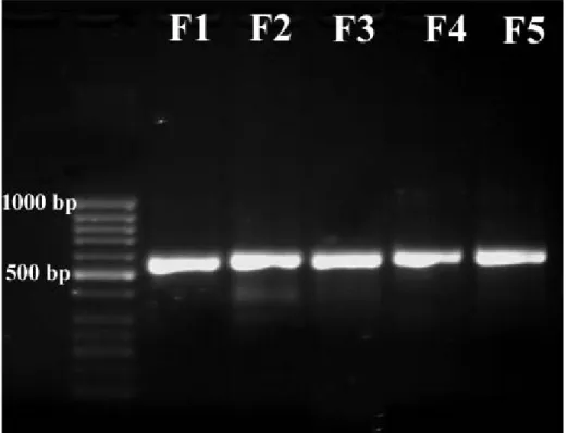
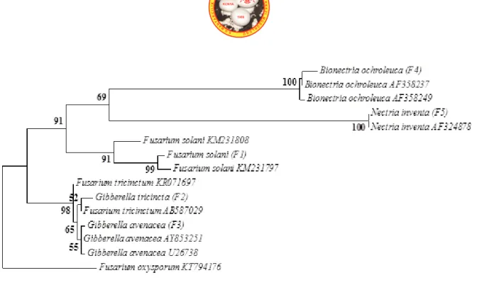
The PCR products (570 bp) were obtained from all of the species by using the universal fungal primers (ITS1/ITS4), Figure 1 shows that the sizes obtained for the full ITS region amplified of all of the strains. The rDNA base sequences belong to investigated strains are presented in Table 1 together with closest Blast results. When each of this sequences were investigated by Blast, identity and coverage values were found between 98-100% and 97-100%, respectively (Table 1). The phylogenetic trees were obtained by comparison to all sequences with Genbank nucleotide sequence database that have ITS1-5.8S rRNA-ITS2 sequences (Figure 2). Figure 2 shows that the members of genera Bionectria, Fusarium, Gibberella and Nectria have almost identical
phylogenetic tree based on ITS region was structured at higher divergence levels. For investigated mainly closely related members, identical positions and four sections for specific clades such as Bionectria, Fusarium, Gibberella and Nectria genera were found.
The genus Fusarium is the anamorph stage of Gibberella genus (Samuels et al., 2001). The members of these genera are known as main and wide plant pathogens (Howard, 2002; Stone et al., 2004; Dragich and Nelson, 2014; Chehri, 2016). These two genera have been distinguished with especially teleomorph structures of Gibberella genus. The complexity about the their morphologic and microscopic identification has been related with varies problems such as depending on the host, loosing of stock cultures, limitations associated with morphological characters (Summerell et al., 2003; Hsuan et al., 2011; Antonissen et al., 2014). The findings of this study demonstrated the efficiency of ITS region and phylogenetic analysis of belong to these two genera. Figure 2 shows that Fusarium and Gibberella genera have considerably identical topology with the ITS locus. The genus Gibberella occurred in two main clades and two clear divergences, namely Gibberella avenacea and Gibberella tricincta, were noted. Furthermore, investigated members of Fusarium and Gibberella genera are polyphyletic.
The genus Nectria is a big genus with about 650 members and many species of Nectria genus are known as plant pathogens, and some of them are toxigenic to animals and humans (Schroers and Samuels, 1997). The genus Bionectria is one of the other plant pathogenic Hypocreanlean fungi and very similar to Nectria member (Schroers, 2001; Samaga et al., 2014; Melo et al., 2014). These two genera have some differences about their morphologic and chemical structure. However, Bionectria and Nectra genera have very similar morphologic and microscopic properties and distinguish of them has been very problematic for mycologist (Schroers and Samuels, 1997; Schroers, 2001).
Phylogeny based on the ITS region in this study showed a successfully topology for these genera and indicated main phylogenetic position of them as closely related but distinctly different members (Figure 2).
Figure 1. Full ITS PCR products amplified from all of the strains with ITS1/ITS4
primers. M, molecular-weight markers (50 bp GeneRuler DNA Lader, Fermentas)
Table 1. Newly generated ITS sequences with their closest GenBank sequences (according to Blast
Figure 2. Best-scoring maximum likelihood tree based on the Tamura–Nei model calculated using MEGA
6.0 based on ITS sequences showing the relationships of the newly generated sequences in this study with previously known taxa in the NCBI GenBank. The scale bar denotes 0.02 substitutions per position. The tree with the highest log likelihood (-1499.8212) is shown. Initial tree for the heuristic search were obtained by applying the neighborjoining method to a matrix of pairwise distances estimated using the maximum composite likelihood approach. The tree is drawn to scale, with branch lengths measured in the number of substitutions per site. The analysis involved 15 nucleotide sequences. All positions with less than 50% site coverage, containing gaps, or missing data were eliminated. There were a total of 374 positions in the final dataset. The tree is rooted with Fusarium oxysporum (KT794176) (bootstrap 1000).
Conclusions
The results of this study demonstrated the efficiency of rDNA region and phylogenetic analysis in taxonomic studies of closely related members of Hypocreanlean fungi. In particular, ITS region was found to be success because of its high performance with regard to easy
application, topology, identification and clearly discrimination. In addition, high quality sequences of the ITS locus obtained in the present study have been deposed in the NCBI database for bridge over to other taxonomic studies of Hypocreanlean fungi.
References
Altschul S.F., Gish W., Miller W., Myers E.W., Lipman D.J., Basic Local Alignment Search Tool, J Mol Bio, 215: 403-410 (1990).
Antonissen G., Martel A., Pasmans F., Ducatelle R., Verbrugghe E., Vandenbroucke V., Li S., Haesebrouck F., Immerseel F.V., Croubels S., The Impact of Fusarium Mycotoxins on Human and Animal Host Susceptibility to Infectious Diseases, Toxins (Basel), 6(2): 430–452 (2014).
Booth C., The genus Fusarium, CAB, Kew, UK; 237 p. (1971).
Chehri K., Molecular Identification of Pathogenic Fusarium Species, The Causal Agents of Tomato Wilt in Western Iran, J Plant Prot Res, 56(2): 143-148 (2016)
Demirel R., Ilhan S., Asan A., Kınacı E., Oner S., Microfungi in Cultivated Fields in Eskişehir Provience (Turkey), J Basic Microbiol, 45: 279-293 (2005).
Demirel R., Sariozlu N.Y., İlhan S., Polymerase Chain Reaction (PCR) Identification of Terverticillate Penicillium Species Isolated from Agricultural Soils in Eskişehir Province,. Braz Arch Biol Technol, 56 (6): 980-984 (2013).
Dragich M., Nelson S., Gibberella and Fusarium Ear Rots of Maize in Hawai'I, Plant Dis, 102: 1-8 (2014). Gerlach W., Nirenberg H., The Genus Fusarium a Pictorial Atlas, Biologische Bundesanstalt für Land-und
Forstwirtschaft Institut für Microbiologie, 406 p. (1982).
Howard D.H., Pathogenic fungi in Humans and Animals, In: Mycology Series, Volume 16, and Second Edition, CRC Press, 800 p., New York (2002).
Hsuan H.M., Salleh B., Zakaria L., Molecular Identification of Fusarium Species in Gibberella Fujikuroi Species Complex from Rice, Sugarcane and Maize from Peninsular Malaysia, Int J Mol Sci, 12: 6722-6732 (2011).
Ismail M.A., Abdel-Hafez S.I.I., Hussein N.A., Abdel-Hameed N.A., Contributions to the Genus Fusarium in Egypt with Dichotomous Keys for Identification of Species, Tomasz M. Karpiński Publisher, 175 pp., Poland (2015).
Melo I.S., Valente A.M.M.P., Kavamura V.N., Vilela E.S.D., Faull J.L., Mycoparasitic Nature of Bionectria sp. Strain 6.21, J Plant Prot Res, 54(4): 327-333 (2014).
Nelson P.E., Toussoun T.A., Marasas W.F.O., Fusarium Species: An Illustrated Manual for Identification, The Pennsylvania State University Press, 193 p. (1983).
Samaga P.V., Rai V.R., Rai K.M.L., Bionectria ochroleuca NOTL33—An Endophytic Fungus from Nothapodytes Foetida Producing Antimicrobial and Free Radical Scavenging Metabolites, Ann Microbiol, 64: 275–285 (2014).
Samuels G.J., Nirenberg H.I., Seifert K.A., Perithecial Species of Fusarium, In: Summerell B.A., Leslie J.F., Backhouse D., Bryden WL., Fusarium: Paul E. Nelson Memorial Symposium, APS Press, St. Paul, MN, USA, 1-14 (2001).
Schroers H.J., Samuels G.J., Bionectria: A Genus for Species of the Nectria Ochroleuca Group, Zeitschrift Für Mycologia, 63(2): 149-154 (1997).
Schroers H.J., A monograph of Bionectria (Ascomycota, Hypocreales, Bionectriaceae) and Its Clonostachys anamorphs, Stud Mycol, 46: 1-96 (2001).
Stone J.K., Polishook J.D., White J.F., Endophytic Fungi in Biodiversity of Fungi: Inventory and Monitoring Methods, Academic Press, p. 241-270, Burlington (2004).
Summerell B.A., Salleh B., Leslie J.F., A Utilitarian Approach to Fusarium Identification, Plant Dis, 87(2): 117-128 (2003).
Tamura K., Stecher G., Peterson D., Filipski A., Kumar S., MEGA6: Molecular Evolutionary Genetics Analysis Version 6.0. Molecular Biology and Evolution, 30: 2725-2729 (2013).
Tamura K., Nei M., Estimation of the Number of Nucleotide Substitutions in the Control Region of Mitochondrial DNA in Humans and Chimpanzees, Mol Biol Evol, 10: 512-526 (1993).
Van Burik J.A., Schreckhise R.W., White T.C., Bowden R.A., Myerson D., Comparison of Six Extraction Techniques for Isolation of DNA from Filamentous Fungi, Med Mycol, 36(5): 299–303 (1998). White T.J., Bruns T., Lee S., Taylor J., Amplification and Direct Sequencing of Fungal Ribosomal RNA
Genes for Phylogenetics. In: Innis M.A., Gelfand D.H., Sninsky J.J., White T.J., editors. PCR Protocols: A Guide to Methods and Applications. Academic Press, New York, 315–322 (1990). Young-Mi L., Choi Y., Min B., PCR-RFLP and Sequence Analysis of the rDNA ITS Region in the Fusarium