REGULATION OF HUMAN MONOCYTE
DIFFERENTIATION INTO M1- AND M2-LIKE
MACROPHAGES
A DISSERTATION SUBMITTED TO
THE GRADUATE SCHOOL OF ENGINEERING AND SCIENCE
OF BILKENT UNIVERSITY
IN PARTIAL FULFILLMENT OF THE REQUIREMENTS
FOR THE DEGREE OF
DOCTOR OF PHILOSOPHY
IN
MOLECULAR BIOLOGY AND GENETICS
By
Defne Bayık
REGULATION OF HUMAN MONOCYTE DIFFERENTIATION INTO M1-
AND M2-LIKE MACROPHAGES
By Defne Bayık May, 2016
We certify that we have read this dissertation and that in our opinion it is fully adequate, in scope and in quality, as a thesis for the degree of Doctor of Philosophy.
________________________ ________________________
İhsan Gürsel Dennis M. Klinman
(Advisor) (Co-Advisor) ________________________ Mayda Gürsel ________________________ Özlen Konu ________________________ Ali Güre ________________________ Mesut Muyan
Approved for the Graduate School of Engineering and Science
__________________ Levent Onural
Abstract
REGULATION OF HUMAN MONOCYTE
DIFFERENTIATION INTO M1- AND M2-LIKE MACROPHAGES
Defne Bayık
Ph.D. in Molecular Biology and Genetics Supervisor: İhsan Gürsel
Co-supervisor: Dennis M. Klinman May, 2016
Myeloid-derived suppressor cells (MDSC) play a key role in down-regulating activated T and NK cells. MDSC are emerging as targets for cancer immunotherapy since they protect tumor cells from immune elimination. We previously showed that the TLR7/8 agonist R848 and the TLR2/1 dual agonist PAM3 had opposite effect on the maturation of human monocytic MDSC (mMDSC). While the former triggered them to differentiation in M1-like macrophages with pro-inflammatory/anti-tumoricidal capacity, the latter generated immunosuppressive M2-like macrophages. This work seeks to identify the soluble factors that regulate the differentiation of mMDSC into macrophages. Our studies reveal that TNFα and M-CSF are essential for mMDSC to mature into functional M1- and M2-like macrophages, respectively. IL-6 and IL-10 play secondary roles but when used in combination with TNFα or M-CSF exceed the effects of TLR agonists. Understanding the response of mMDSC to cytokines should help efforts to direct the mMDSC maturation to therapeutic benefit.
The finding that PAM3 could induce human mMDSC to mature into M2-like macrophage triggered us to study the effect of this TLR agonist on other monocyte populations. Our findings reveal that PAM3 was unique among TLR agonists in generating M2-like macrophages. We compared the polarizing activity of PAM3 to that of M-CSF. PAM3 was slightly less efficient than M-CSF in driving maturation of HLA-DR+ monocytes based on phenotypic characterization and phagocytic ability. Yet macrophages generated by PAM3 or M-CSF were equally capable of suppressing T cell proliferation. Analysis of gene regulatory networks by microarray and subsequent validation of the pathways identified by using specific inhibitors defined the NF-κB – COX-2 axis as playing a primary role. However, PAM3 also induced
driven cultures. Our findings clarified the pathways by which immunosuppressive M2-like macrophage arise from human monocytes and identify PAM3 as a potential therapeutic modulator of monocyte differentiation in patients with autoimmune disease.
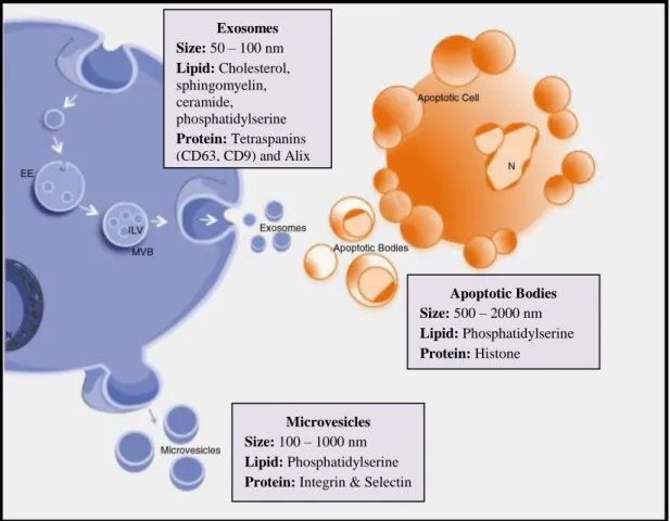
Extracellular vesicles (EV) are a heterogeneous population of biological nanoscaled particles that serve as vectors to enhance intercellular communication. In addition to this physiological role evidence indicates that EV can be harnessed as therapeutic agents for cancer. The major limitation to EV-based therapeutics is their rapid clearance by the reticuloendothelial system (RES). To overcome this problem, we sought to reduce macrophage uptake of EV by blocking scavenger receptors. In vitro results using human and murine cells suggests that inhibiting class A scavenger receptors selectively impairs EV uptake by monocytes and macrophages. In vivo studies document reduced liver accumulation and enhanced plasma circulation of i.v. injected EV after such blockade. These findings provide a strategy for reducing EV uptake by the RES thereby increasing their targeting and activity.
Keywords: Myeloid-derived suppressor cells, HLA-DR+ human monocytes, M1-like macrophages, M2-like macrophages, TLR agonists, cytokines, extracellular vesicles, scavenger receptors
Özet
İNSAN MONOSİTLERİNİN
M1- VE M2-BENZERİ MAKROFAJLARA
DÖNÜŞÜMÜNÜN DÜZENLENMESİ
Defne Bayık
Moleküler Biyoloji ve Genetik Bölümü, Doktora Tez Danışmanı: İhsan Gürsel
Eş Tez Danışmanı: Dennis M. Klinman Mayıs, 2016
Miyeloid türevi baskılayıcı hücreler (MDSC) aktive olmuş T ve NK hücrelerini susturulmasında önemli rol oynarlar. Tümör hücrelerinin bağışıklık sistemi tarafından elemesini engellediklerinden dolayı, MDSC kanser immünoterapi için yeni bir hedef olarak ortaya çıkmaktadır. Önceki sonuçlarımız TLR7/8 agonisti R848 ve TLR2/1 ikili agonisti PAM3’ün insan monositik MDSC (mMDSC) olgunlaşması üzerinde ters bir etkiye sahip olduğunu göstermişti. İlk agonist mMDSClerin pro-inflamatuar/anti-tümorisidal kapasiteli M1 benzeri makrofajlara farklılaşmasını tetiklerken, ikinci agonist immunsupresif M2 benzeri makrofajlar oluşturmuştur. Bu çalışma mMDSClerin makrofajlara farklılaşmasını düzenleyen çözünür faktörler tespit etmeyi amaçlamaktadır. Araştırmalarımız mMDSClerin fonksiyonel M1 ve M2 benzeri makrofajlara olgunlaşması için sırasıyla TNFα ve M-CSF’in önemli olduğunu ortaya koymaktadır. IL-6 ve IL-10 ikincil bir rol oynamakla birlikte TNFα veya M-CSF ile kombinasyon halinde kullanıldığında TLR agonistlerinden daha etkilidir. mMDSC sitokinlere nasıl tepki verdiğini anlamak bu hücrelerin terapötik amaçla olgunlaşmasını yönlendirmek açısından yardımcı olacaktır.
PAM3’ün insan mMDSClerini M2-benzeri makrofajlara dönüştürdüğüne dair bulgular, bu TLR agonistinin diğer monosit popülasyonları üzerinde etkisini araştırmaya yönlendirmiştir. Bulgularımız PAM3’ün M2-benzeri makrofajlar oluşturabilme kapasitesinin TLR agonistleri arasında benzersiz olduğunu ortaya koymaktadır. PAM3 ve M-CSF’in kutuplaştırıcı aktivitesinin fenotipik nitelendirilme ve fagositik yetenek açısından karşılaştırılması PAM3’ ün M-CSF’e oranla HLA-DR+ monositleri dönüştürmede daha az etkili olduğunu göstermiştir. Yine de PAM3 veya M-CSF tarafından oluşturulan makrofajlar T hücresi çoğalmasını eşit derecede bastırabilmektedirler. Mikrodizi analizi yöntemi ile düzenleyici gen şebekelerinin
sonucunda NF-κB–COX-2 ekseninin makrofaj dönüşümünde birincil rol oynadığı tespit edilmiştir. PAM3 aynı zamanda IL-6 bağımlı bir yol üzerinden monosit farklılaşmasını neden olurken, bu şebeke M-CSF tarafından kullanılmamaktadır. Bulgularımız, insan monositlerinin bağışıklık baskılayıcı M2 makrofajlara kutuplaşmasını yönlendiren yollara açıklık getirmiş ve PAM3’ü otoimmün hastalarda monosit farklılaşmasını kontrol edebilecek potansiyel bir terapötik modülatör olarak tanımlamıştır.
Hücre dışı kesecikler (EV), hücreler arası iletişimi arttırmak için hizmet eden biyolojik kökenli, nano büyüklükte heterojen parçacıklar topluluğudur. Fizyolojik rolüne ek olarak EVlerin kanser tedavisinde kullanılabileceğine dair kanıtlar bulunmaktadır. EV-bazlı tedavilerdeki başlıca sınırlama EVlerin retikülo-endotelyal sistem (RES) tarafından hızlı bir şekilde temizlenmeleridir. Bu sorunu aşmak için, çöpçü reseptörlerini bloke ederek EVlerin makrofajlar tarafından alımını azaltmak hedeflenmiştir. İnsan ve fare hücrelerini kullanarak elde edilen in vitro sonuçlar A sınıfı çöpçü reseptörlerinin bloke edilmesinin seçici olarak monosit ve makrofajlar tarafından EV alımını engellediğini göstermektedir. İn vivo çalışmalar A sınıfı çöpçü reseptörleri bloklanmasının damar içi enjekte edilerin EVlerin karaciğer birikimi azaltırken plazma dolaşımını arttırdığını belgelemiştir. Bu çalışma RES alımını engelleyerek EVlerin hedef ve aktivitelerini geliştirmeye yönelik bir strateji tanımlamıştır.
Anahtar Kelimeler: Myeloid türevi baskılayıcı hücreler , HLA-DR+ insan monositler, M1-benzeri makrofajlar , M2-M1-benzeri makrofajlar, TLR agonistleri , sitokinler , hücre dışı kesecikler, çöpçü reseptörleri
Acknowledgements
First and foremost I would like to express my deepest gratitude to my supervisors Prof. Dr İhsan Gürsel and Dr. Dennis M. Klinman for their care, trust, patience and guidance. It has been a privilege to work with them. I am grateful for all the knowledge they shared and the values they taught. I cannot thank them enough for helping me to become a researcher and to develop as a person. I hope that I can make them proud.
I would also like to express my heartfelt thanks to Assoc. Prof. Dr. Mayda Gürsel for broadening my perspective on life and science. She has been a true inspiration for me. I am extremely thankful to Assoc. Prof. Dr. Işık G. Yuluğ for her support throughout my graduate life. I would like to express my gratitude to Assoc. Prof. Dr. Özlen Konu for being part of my Ph.D. progress committee and for her feedbacks. I also would like to thank Assoc. Prof. Dr. Ali Güre and Assoc. Prof. Dr. Mesut Muyan for accepting to become members of my thesis jury, and sparing time to evaluate and improve my thesis.
I would like to express my gratitude to Debra Tross for her help, especially with the microarray experiments. I am grateful to Roberta Matthai and Kathleen Noer for all the sorting that they have done and to the NIH Blood Bank staff for the samples they provided. Special thanks to Dr. Dionysios C. Watson and Dr. Avinash Srivatsan for collaborating on studies with extracellular vesicles.
I am really grateful to past and present members of the Gürsel group: Fuat Cem Yağcı, Tamer Kahraman, Arda Gürsel, Mehmet Şahin, İhsan Dereli, Yusuf İsmail Ertuna, Merve Deniz Abdüsselamoğlu, Kübra Almacıoğlu, Begüm Han Horuluoğlu, Gözde Güçlüler, particularly to Banu Bayyurt and Gizem Tinçer-König for their support and friendship. I had to privilege to work alongside great people. I am thankful to the Klinman Group members Emilie Goguet, Julia Scheiermann, Kathy Parker and Neslihan Turan and Debra Tross for their collegiality, discussions and support.
I feel lucky to be a part of the MBG family, where I met amazing people with whom I will remain lifetime friends, especially Gülşah Dal-Kılınç, Özlem Tufanlı, Büşra Yağbasan, Şeyma Demirsoy, Füsun Doldur-Ballı, Merve Mutlu and Dilan Çelebi-Birand. I would like to dedicate special thanks to Gözde Güçlüler and Begüm Han Horuluoğlu for their invaluable friendship and support.
I am especially grateful to Verda Bitirim for her incredible friendship. Through good times and bad, she will remain as my best friend.
Last but not least, I want to thank all my family for their life-long support and encouragement. I would like to express my deepest love to my parent Sedef and Nezihi; and my grandmothers Gül and Göksel. Thank you for being awesome parents and grandparents. I would like to express my heartfelt gratitude Dennis Watson. Without his constant love and patience, I couldn’t have completed this thesis. We will remain united in work as in life. Finally I would like to dedicate this thesis to my grandfather, whom will be deeply missed.
I was financially supported by The Scientific and Technological Research Council of Turkey (TÜBİTAK) BİDEB 2211 scholarship during my Ph.D. studies. My work at the NIH as part of the NIH-Bilkent University individual Graduate Partnership Program (GPP) was supported by the NIH intramural funds.
Table of contents
Abstract ... ... iii Özet ... ... v Acknowledgements ... viii Table of contents ... x List of figures ... xvList of tables ... xix
Abbreviations ... xx
Chapter 1 .... Introduction ... 1
1.1 The Immune System ... 1
1.2 Myeloid Cells ... 2
1.2.1 Monocytes ... 2
1.2.2 Macrophages ... 3
1.2.2.1 M1 Macrophages ... 3
1.2.2.2 M2 Macrophages ... 6
1.2.2.3 Macrophages Generated by M-CSF and GM-CSF ... 9
1.2.2.4 Tumor-associated Macrophages ... 10
1.2.3 Myeloid Derived Suppressor Cells ... 12
1.2.3.1 MDSC under disease conditions ... 13
1.2.3.2 MDSC-Mediated Immune Suppression ... 14
1.2.3.3 Response of MDSC to Soluble Factors... 16
1.2.3.4 Therapeutic strategies for MDSC targeting ... 19
1.2.3.4.1 Response of MDSC to TLR Agonists ... 22
1.3.2 Problems associated with EV-based therapeutics ... 27
1.3.3 EV Uptake Receptors ... 29
1.3.4 Scavenger Receptors Class A and B Families ... 31
1.4 Aim and outline of the thesis ... 33
Chapter 2 .... Materials and Methods ... 34
2.1 Materials ... 34
2.1.1 TLR Ligands ... 34
2.1.2 Recombinant Cytokines ... 35
2.1.3 Cytokine Neutralizing Antibody ... 36
2.1.4 Flow Cytometry Antibody ... 37
2.1.5 ELISA Antibodies ... 39
2.1.6 Cell Culture Media and Standard Solutions Components... 39
2.2 Methods ... 40
2.2.1 Collection and isolation of human blood samples ... 40
2.2.1.1 Sorting of mMDSC and HLA-DR+ monocytes ... 40
2.2.1.2 Sorting of naïve CD4+ T Cells ... 41
2.2.2 Stimulation of mMDSC and HLA-DR+ monocytes ... 42
2.2.3 Effect of cytokines on re-polarization of HLA-DR+ monocytes... 42
2.2.4 Microscopy image of HLA-DR+ monocytes ... 42
2.2.5 Analysis of surface and intracellular markers with flow cytometry ... 43
2.2.6 Functional analysis of differentiated cells ... 43
2.2.6.1 Cytotoxicity functional assay ... 43
2.2.6.2 T Cell proliferation assay ... 44
2.2.6.2.1 Staining of naïve CD4+ T cells with CFSE ... 44
2.2.6.2.2 Preparation of Dynabeads® Human T Actiator CD3/CD28 ... 44
2.2.6.3 Phagocytosis Assay ... 45
2.2.7 ELISA for cytokines ... 45
2.2.8 Analysis of gene expression with microarray ... 46
2.2.8.1 RNA isolation ... 46
2.2.8.2 Expression analyses ... 46
2.2.9 Generation of bone marrow-derived macrophages ... 47
2.2.9.1 Isolation of bone marrow cells ... 47
2.2.9.2 Sorting for mouse mMDSC ... 47
2.2.9.3 Sorting of mouse monocytes ... 47
2.2.9.4 Stimulation of bone marrow-derived cells ... 48
2.2.10 Cell lines ... 48
2.2.11 Isolation of extracellular vesicles ... 49
2.2.12 Staining of extracellular vesicles ... 49
2.2.12.1 Staining with SYTO® RNASelect™ Green Fluorescent Stain ... 49
2.2.12.2 Staining with DiR ... 50
2.2.13 Uptake of EV by mouse cell lines ... 50
2.2.14 Uptake of EV by primary murine macrophages ... 51
2.2.15 Uptake of EV by human peripheral blood mononuclear cells ... 51
2.2.16 In vivo biodistribution of EV ... 52
2.2.17 Statistical analyses ... 52
Chapter 3 .... Results ... 53
3.1 Efforts to Regulate Macrophage Differentiation ... 53
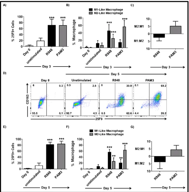
3.1.1 PAM3 and R848 induce mMDSC to differentiate into M1- and M2-like macrophages ... 53
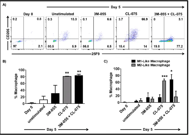
3.1.2 Co-stimulation of TLR7 and TLR8 is required for effective M1-like macrophage polarization of mMDSC ... 54
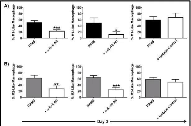
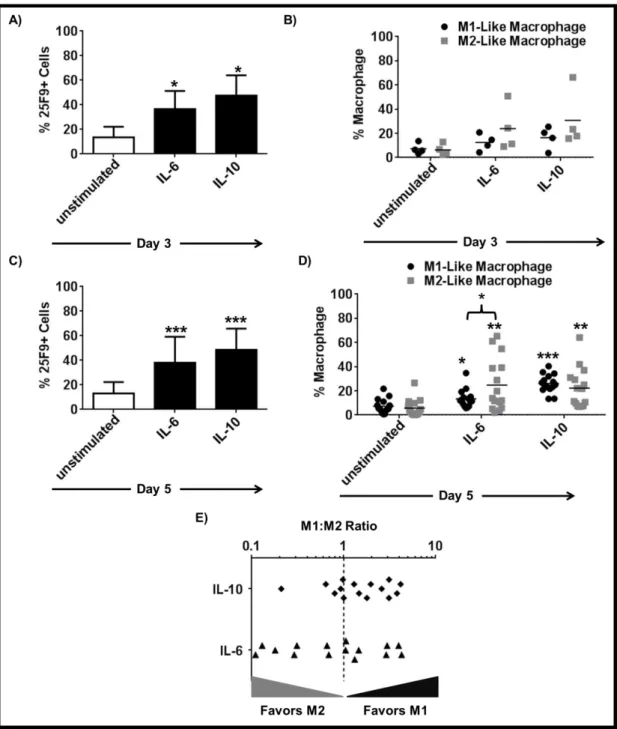
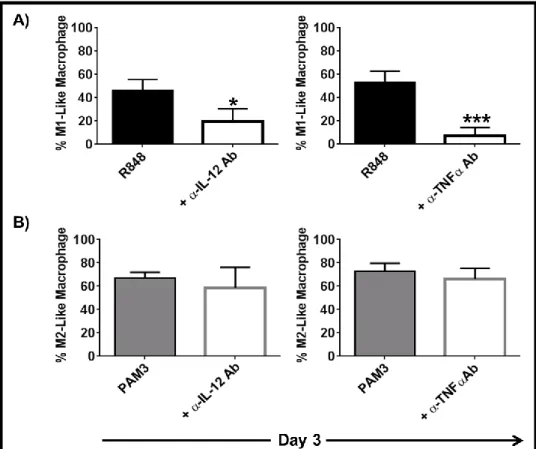
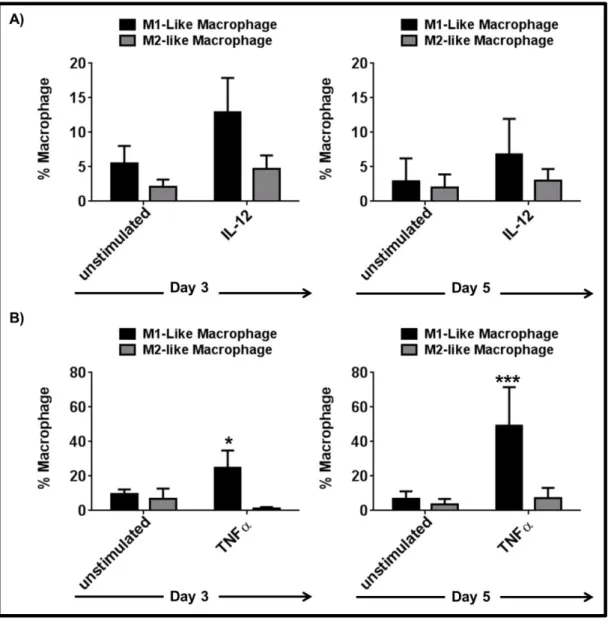
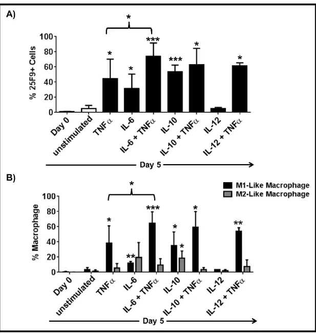
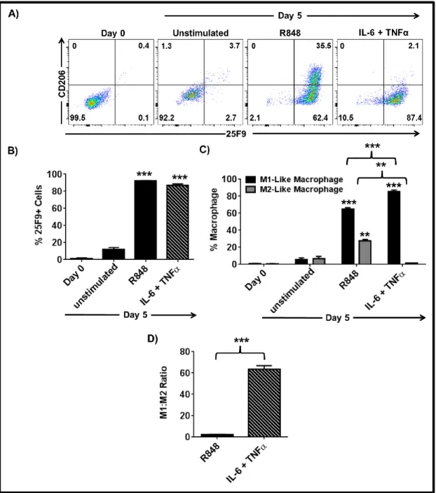
3.1.3 IL-6 and IL-10 are important but not sufficient to induce the complete differentiation of mMDSC ... 55 3.1.4 TNFα but not IL-12 supports the differentiation of mMDSCs into M1-like macrophages ... 59 3.1.5 Combining TNFα with IL-6 recapitulates the effect of R848 on mMDSC ... 62 3.1.6 M1-like macrophages generated by R848 vs IL-6 and TNFα combination differ in morphology ... 65 3.1.7 Macrophages generated by stimulation of mMDSC with IL-6 and TNFα lyse tumor cells effectively ... 65 3.1.8 IFNγ is efficient driver of M1-like macrophage differentiation from mMDSC . 66 3.1.9 Regulatory networks of R848, IFNγ and IL-6 plus TNFα-dependent differentiation of mMDSC ... 67 3.1.10 GM-CSF does not induce differentiation of the mMDSC into macrophages ... 70 3.1.11 PGE2 alters the pattern of macrophage differentiation from mMDSC ... 72
3.1.12 IL-4, IL-8, IL-13 and TGF-β are not essential for M2-like macrophage differentiation from mMDSC ... 73 3.1.13 IL-1β partially supports M2-like macrophage differentiation from mMDSC .... 74 3.1.14 High dose M-CSF supports M2-like macrophage differentiation from mMDSC as an independent signal ... 77 3.1.15 Low dose M-CSF in combination with IL-6 and IL-10 recapitulates the effect of PAM3 ... 80 3.1.16 mMDSC stimulated with PAM3 or combination of IL-6, IL-10 and low dose M-CSF remain functionally suppressive ... 83 3.1.17 mMDSC preferentially differentiate into M2- rather than M1-like macrophages ..
... 84 3.1.18 Macrophages differentiated from HLA-DR+ monocytes are plastic ... 85 3.1.19 TLR2/1 signaling polarizes HLA-DR+ monocytes into M2-like macrophages .. 86 3.1.20 PAM3 response of mMDSC and HLA-DR+ monocytes are indistinguishable... 90 3.1.21 Variation in PAM3 response of human and mouse monocytes ... 90
3.1.22 PAM3 and M-CSF generate M2-like macrophages from HLA-DR+ monocytes 92 3.1.23 PAM3 and M-CSF differ in their ability to induce HLA-DR+ monocytes to
differentiate into M2-like macrophage ... 94
3.1.24 Functional activity of PAM3 and M-CSF induced M2-like macrophages ... 94
3.1.25 Morphology of PAM3 and M-CSF induced M2-like macrophages ... 95
3.1.26 PAM3 and M-CSF-induced M2-like macrophages originated from HLA-DR+ monocytes differ in lineage and functional marker expression ... 96
3.1.27 PAM3 and M-CSF generate M2-like macrophages from HLA-DR+ monocytes that differ in phagocytic activity ... 98
3.1.28 Regulatory network underlying PAM3 and M-CSF-induced M2-like macrophage differentiation of HLA-DR+ monocytes ... 100
3.1.29 Validation of selected targets identified with microarray ... 103
3.1.30 Mouse macrophage cell line uptakes EV in Scavenger Receptor Class A-dependent manner ... 104
3.1.31 Primary mouse macrophages differentially uptake EV depending on the level of scavenger receptor expression ... 106
3.1.32 Human monocytes uptake EV in scavenger receptor-dependent manner ... 107
3.1.33 Administration of scavenger-receptor inhibitor alters the biodistribution of EV ... ... 108
Chapter 4 .... Discussion ... 110
Chapter 5 .... Future Perspectives ... 130
Bibliography ... 133
Appendices ... 172
Appendix A - Gene Lists ... 172
Appendix B - Figures ... 181
List of figures
Figure 1.1: Subsets of myeloid-derived suppressor cells ... 12 Figure 1.2: Biogenesis and properties of three main classes of extracellular vesicles. ... 25 Figure 3.1: TLR agonists selectively drive mMDSC differentiation into M1- and M2-like macrophages. ... 54 Figure 3.2: Stimulation through both TLR7 and TLR8 is required for differentiation of mMDSC into M1-like macrophages. ... 55 Figure 3.3: PAM3 and R848 induce mMDSC to secrete IL-6 and IL-10. ... 56 Figure 3.4: IL-6 and IL-10 have regulatory roles in R848- and PAM3-induced differentiation of mMDSC. ... 57 Figure 3.5: IL-6 and IL-10 partially support macrophage differentiation from mMDSC. ... 58 Figure 3.6: Addition of IL-6 plus IL-10 has the same effect as each cytokine separately on mMDSC differentiation. ... 59 Figure 3.7: TNFα is secreted in the course of R848-induced mMDSC differentiation. ... 60 Figure 3.8: IL-12 and TNFα regulate R848- but not PAM3-induced macrophage differentiation from mMDSC. ... 61 Figure 3.9: TNFα but not IL-12 supports the differentiation of mMDSC into M1-like macrophage. ... 62 Figure 3.10: TNFα and IL-6 cocktail is the most effective cytokine combination inducing M1-like macrophage differentiation. ... 63 Figure 3.11: Combination of IL-6 with TNFα is superior to R848 in generating M1-like macrophages. ... 64 Figure 3.12: Distinct morphology of unstimulated, R848 or IL-6 + TNFα stimulated mMDSC at Day 5. ... 65 Figure 3.13: mMDSC stimulated with R848 or the combination of IL-6 plus TNFα effectively lyse tumor cells. ... 66 Figure 3.14: IFNγ polarizes mMDSC towards M1-like macrophages. ... 67
Figure 3.15: R848, IL-6 plus TNFα and IFNγ-induced M1-like macrophage differentiation is regulated by a NF-κB-dependent network. ... 69 Figure 3.16: GM-CSF does not induce mMDSC to become macrophage. ... 71 Figure 3.17: PGE2 regulates differentiation status of mMDSC. ... 73
Figure 3.18: IL-4, IL-8, IL-13 and TGF-β do not support the differentiation of mMDSC into M2-like macrophages. ... 74 Figure 3.19: IL-1β partially supports M2-like macrophage differentiation by mMDSC. ... 76 Figure 3.20: High dose M-CSF is effective inducer of M2-like macrophages from mMDSC. .. 79 Figure 3.21: Combination of 1 ng/ml M-CSF with IL-6 and IL-10 is not sufficient to recapitulate the effect of PAM3. ... 81 Figure 3.22: Combination of 5ng/ml M-CSF with IL-6 and IL-10 drives M2-like macrophage polarization as effectively as PAM3. ... 82 Figure 3.23: mMDSC stimulated with PAM3 or IL-6, IL-10 and low dose M-CSF combination retain the suppressive activity. ... 84 Figure 3.24: mMDSC preferentially differentiate into M2-like macrophages when exposed to TNFα and M-CSF simultaneously. ... 85 Figure 3.25: M1-like and M2-like macrophages generated from HLA-DR+ monocytes can change their phenotype when exposed to opposing stimuli. ... 86 Figure 3.26: PAM3 preferentially generates M2-like macrophages from HLA-DR+ monocytes. ... 88 Figure 3.27: Differences in HLA-DR+ monocyte differentiation after stimulation with PAM3 vs FSL-1. ... 89 Figure 3.28: mMDSC and HLA-DR+ monocytes respond similarly to PAM3. ... 90 Figure 3.29: Mouse mMDSC and monocytes do not primarily differentiate into M2 macrophages in response to PAM3... 92 Figure 3.30: M-CSF and PAM3 polarized HLA-DR+ monocytes towards M2-like macrophage phenotype. ... 93 Figure 3.31: PAM3-induced M2-like macrophage differentiation of HLA-DR+ monocytes is independent of M-CSF. ... 94
Figure 3.32: Macrophages generated from HLA-DR+ monocytes in the presence of PAM3 and M-CSF suppress proliferation of T cells. ... 95 Figure 3.33: Distinct morphology of unstimulated, PAM3 or M-CSF stimulated HLA-DR+ monocytes at Day 5... 96 Figure 3.34: CD16 is differentially up-regulated in PAM3 and M-CSF macrophages. ... 97 Figure 3.35: PAM3 and M-CSF induced macrophages slightly differ in marker expression. .... 98 Figure 3.36: PAM3 vs M-CSF-induced macrophages differ in their ability to phagocytose. .... 99 Figure 3.37: Shared network of genes up-regulated during the course of PAM3- and M-CSF-induced macrophage differentiation. ... 100 Figure 3.38: Interaction of distinct set of genes up-regulated by PAM3 vs M-CSF with shared regulatory network. ... 102 Figure 3.39: NF-κB is the main regulator of PAM3- and M-CSF-induced M2-like macrophage differentiation... 104 Figure 3.40: Scavenger receptor class A blockade with DS interferes with EV uptake of murine macrophage cell line. ... 105 Figure 3.41: Scavenger receptor class A blockade with DS interferes reduces EV uptake of primary murine macrophages. ... 106 Figure 3.42: Human monocytes take up EV in a scavenger receptor class A dependent manner. ... 108 Figure 3.43: Scavenger receptor blockade decreases the uptake of EV by the liver. ... 109 Figure 4.1: Schematic representation of differentiation of mMDSC into M1-like macrophages. ... 115 Figure 4.2: Schematic representation of differentiation of mMDSC HLA-DR+ monocytes into M2-like macrophages. ... 126 Figure B1: Confirmation of IL-10 and TNFα neutralization with ELISA ... 181 Figure B2: Expression pattern of additional surface markers in PAM3 vs M-CSF stimulated HLA-DR+ monocytes. ... 181 Figure B3: Secreted IL-6 levels are higher in PAM3 treated cultures as compared to M-CSF treated cultures. ... 182
Figure B5: FCS concentration determines the level of spontaneous macrophage differentiation from human monocytes. ... 183
List of tables
Table 1.1: Ligands of TLR ... 3
Table 1.2: Therapeutic agents for MDSC targeting ... 19
Table 1.3: EV uptake receptors ... 29
Table 2.1: Ligands for TLR ... 34
Table 2.2: Recombinant Cytokines ... 35
Table 2.3: Neutralizing antibodies against secreted cytokines. ... 36
Table 2.4: Fluorescence-conjugated antibodies ... 37
Table 2.5: Staining volume and antibody amounts ... 40
Table 2.6: Cell lines and culture conditions ... 48
Table 2.7: EV concentrations ... 50
Table 3.1: Comparison of M2:M1 Ratio and actual M2-like macrophage numbers of PAM3 or IL-6 + IL-10 + IL-1β stimulated mMDSC cultures. ... 77
Appendix Table 1: Symbol of genes upregulated more 2-fold in R848, IL-6 plus TNFα or IFNγ stimulated HLA-DR+ monocytes (alphabetically ordered)………172
Appendix Table 2: Symbol of genes upregulated more than 2-fold in PAM3 and M-CSF stimulated HLA-DR+ monocytes (alphabetically ordered). ... 174
Abbreviations
Ab Antibody
APC Antigen presenting cell
asRNA Antisense RNA
ARG1 Arginase 1
ATCC American Type Culture Collection
BSA Bovine serum albumin
BMDC Bone marrow-derived dendritic cell
BMDM Bone marrow-derived macrophage
CCL/CXCL Chemokine ligand
cDNA Complementary deoxyribonucelaic acid
COX-1 (PTGS1) Cyclooxygenase 2/Prostaglandin-endoperoxide synthase 1 COX-2 (PTGS2) Cyclooxygenase 2/Prostaglandin-endoperoxide synthase 2
CS Chondroitin sulfate
ddH2O Double-distilled water
DC Dendritic cell
DMEM Dulbecco’s modified eagle medium
DMSO Dimethyl sulfoxide
DS Dextran sulfate
ELISA Enzyme linked-immunosorbent assay
EV Extracellular vesicles
GvHD Graft-versus-host disease
gMDSC Granulocytic myeloid-derived suppressor cells
FLA Flagellin
GM-CSF (CSF-2) Granulocyte macrophage colony-stimulating factor
Gr-1 Granulocyte receptor 1
HLA-DR Human leukocyte antigen – antigen D related
IFNα Interferon alpha
IFNγ Interferon gamma
IL Interleukin
iNOS Inducible nitric oxide synthase
IPA Ingenuity Pathway Analysis
I.P. Intraperitoneal
IRF Interferon regulatory factor
I.V. Intravenous
KO Knockout
L-Arg L-Arginine
LPS Lipopolysaccharide
MAPK Mitogen-activated protein kinase M-CSF (CSF-1) Macrophage colony stimulating factor MDSC Myeloid-derived suppressor cells
MFI Mean fluorescence intensity
MHC Major histocompatibility complex
mMDSC Monocytic myeloid-derived suppressor cell
MPLA Monophosphoryl Lipid A
MSC Mesenchymal stem cell
MSR1 (SR-A) Macrophage Scavenger Receptor 1 MyD88 Myeloid differentiation factor 88 NAMPT Nicotinamide phosphoribosyltransferase
NCI National Cancer Institute NF-κB Nuclear factor-kappa B
NIBIB National Institute of Biomedical Imaging and Bioengineering NIH National Institutes of Health
NK Natural Killer
NO Nitric oxide
OMV Outer membrane vesicle
O/N Overnight
PAM3 Pam3CysSerLys4
PAMP Pathogen-associated molecular patterns PBMC Peripheral blood mononuclear cells
PBS Phosphate buffered saline
PGN Peptidoglycan
PRR Pathogen recognition receptor
PS Phosphatidylserine
R848 Resiquimod
RES Reticuloendothelial system
RNA Ribonucleic acid
RPMI Roswell Park Memorial Institute
RT Room temperature
SR-BI Scavenger receptor class B I
STAT Signal transducers and activators of transcription TGF-β Transforming growth factor beta
TLR Toll-like Receptor
TNFα Tumor necrosis factor alpha VEGF Vascular endothelial growth factor
Chapter 1
Introduction
1.1
The Immune System
The mammalian immune system is a network of physical barriers, leukocytes and soluble factors that provide host resistance to non-self or altered/missing-self [1]. The immune system has been broadly divided into two arms - innate and adaptive - based on the specificity, type and rapidity of the response, [2].
Monocytes/macrophages, dendritic cells (DC), neutrophils, natural killer cells (NK cells), mast cells, eosinophils and basophils comprise the cellular elements of the innate immune system that provides the first line of host defense against pathogenic microorganisms. These cells express germline-encoded receptors, so called pattern recognition receptors (PRR), specialized to recognize pathogen and danger-associated molecules and to discriminate between self, altered-self and non-altered-self [3]. PRR include several receptor families: AIM2-like receptors (ALR), C-type lectin receptors (CLR), nucleotide-binding oligomerization domain (Nod)-, leucine-rich repeat-containing receptors (NLR), RIG-I-like receptors Toll-like receptors (TLR) and cytosolic DNA sensors, all of which are primarily classified based on structural similarities and target molecule specificity [4-6]. PRR recognize common microbial pathogen/danger-associated molecular patterns (PAMPs or DAMPs) including nucleic acids (double- or single-stranded DNA and RNA) protein components, lipids, lipoproteins, glycolipids and polysaccharides expressed by bacteria, viruses, fungi and parasites and to a lesser extent the cellular contents of dying host cells [3, 4, 7, 8]. Detection of PAMPs or DAMPs initiates an inflammatory response that involves pathogen killing, elimination of debris via phagocytosis, release of pro-inflammatory mediators, and the recruitment/activation of innate and/or adaptive immune cells [9]. This process is followed by a resolution phase which involves different set of mediators and cells specialized to restore the homeostasis [10].
The adaptive immune system, also known as the acquired immune system, is the second arm of immune defense. T cells and B cells belong to this arm, mount pathogen-specific immune responses, and confer long-lasting protection against the same pathogen [2]. The type of response generated by these cell types is different. T cells are important for cell-mediated immunity, whereas B cells secrete antibodies as part of the humoral immune response [11].
Activation of both T and B cells is regulated by cellular interactions and factors released by the innate immune cells [12].
1.2
Myeloid Cells
1.2.1 Monocytes
Monocytes are elements of the mononuclear phagocytose system (MPS) which also includes macrophages and dendritic cells. These cells help maintain immune homeostasis and support the induction of immunity through their ability to phagocytose a large spectrum of particles [13]. Monocytes originate from myeloid precursors in the bone marrow or fetal liver from which they are released into the systemic circulation [14, 15]. Colony stimulating factors (CSFs) regulate the generation of monocytes and their maturation into tissue-resident macrophages. Severe monocytopenia develops in mice deficient in M-CSF or the M-CSF receptor CSF-1R [16, 17]. Conversely, i.v. injection of M-CSF for several days enhances the frequency of peripheral monocytes by 10-fold [18].
Monocytes constitute 5-10% of human and 2-5% of mice circulating leukocytes. Most of these monocytes survive for only 2-3 days. Yet microbial products, pro-inflammatory mediators and growth factors can trigger monocytes to migrate into tissues where they replenish tissue-resident macrophages and/or give rise to DC and inflammatory macrophages [19, 20].
Human monocytes are broadly categorized in three subclasses depending on their expression of CD14 (co-receptor for TLR4) and CD16 (FcγRIII). Classical monocytes represent the largest subset (85%) and have a CD14highCD16low expression profile. Non-classical monocytes represent 10% of the population and are CD14dimCD16high. The remaining 5% are composed of intermediate monocytes that are CD14highCD16med [21, 22]. These subsets express different levels of HLA-DR, which can be used to discriminate the CD16 expressing subset from NK cells [23]. These chemokine receptors similarly define monocyte subsets in mice. The classical monocyte population is CD11bhighLy6ChighCCR2high while the non-classical and intermediate monocytes are represented by Ly6Clow and CX3CR1high cells, respectively [24, 25].
Transcriptome profiling of monocytes shows that intermediate and non-classical monocytes are closely related and might have a developmental relationship [26]. Although the in vivo roles of the monocyte subsets are not well defined, several behavioral and functional differences have been noted. CCR2 is expressed by classical monocytes, aiding their recruitment to sites of infection/inflammation by CCL2 (MCP-1) and CCL7 (MCP-3), where they differentiate into phagocytes [27-29]. In contrast, trafficking of non-classical monocytes is regulated through
CX3CR1 [30]. Unlike classical monocytes, intermediate and non-classical monocytes engage in patrolling behavior by circulating through vessels and entering uninflamed tissues [31, 32].
1.2.2 Macrophages
Inflammation occurs when the host responds to infection or tissue damage [33]. This response includes the maturation of monocytes into macrophages that play a critical role in limiting the infection and subsequently resolving the inflammation [34]. Macrophages also contribute to the regulation of metabolism, wound healing, organogenesis and tissue remodeling [35, 36]. Based on differences in function, macrophages are broadly categorized into two sub-groups: M1 and M2 (in humans M1-like and M2-like macrophages). This nomenclature is linked to the Th1-Th2 paradigm, which reflects the activation status of functionally distinct types of T cells [37]. When stimulated by bacteria or viruses, macrophages undergo “classical” activation towards and M1 phenotype [37, 38]. In contrast, parasitic infections and allergens trigger “alternative” activation into M2 macrophages. These help downregulate the immune response during the resolution phase of inflammation and promote tissue remodeling and repair [34, 39].
1.2.2.1 M1 Macrophages
M1 macrophages are primarily activated by bacterial or viral products that are recognized by PRRs in particular TLRs [7]. Following recognition of PAMPs or endogenous danger molecules these receptors initiate a highly conserved signaling cascade to generate an immune response that involves the release of pro-inflammatory mediators, pathogen killing and attraction of cytotoxic T lymphocytes [40, 41]. Each TLR has evolved to recognize a different set of ligands and their specificity is influenced by the subcellular localization of the receptors [42]. A list of well characterized TLRs expressed by monocytes is provided in Error! Not a valid bookmark
self-reference.. More recently TLR10, TLR11, TLR12 and TLR13 were identified [43].
Table 1.1: Ligands of TLR
Receptor Ligand Origin Subcellular Localization
TLR2/1 Triacetylated
Receptor Ligand Origin Subcellular Localization TLR2 Lipoprotein, peptidoglycan, glycolipid, lipoteichoic acid, zymosan, heat-shock protein Bacteria, Mycobacteria, Fungi, Host Plasma membrane TLR2/6 Diacetylated lipoprotein, zymosan Mycoplasma, gram-positive bacteria, fungi Plasma membrane TLR3 Double-stranded
RNA Viruses Endolysosome
TLR4 Lipopolysaccharide, heat-shock protein
Gram-negative
bacteria, Host Plasma membrane
TLR5 Flagellin Bacteria Plasma membrane
TLR7/8 Single-stranded RNA Viruses Endolysosome
TLR9 Unmethylated CpG
ODN Bacterial DNA Endolysosome
Modified from Akira and Takeda [44] .
Several studies indicate that the M1 macrophages generated by TLR signaling (tested with LPS) do not exhibit a full pro-inflammatory response and that co-stimulation with IFNγ might be required for complete activity. This conclusion is based on experiments examining the expression pattern of IL-12 which is an important cytokine that regulates early events in the anti-microbial response and activates NK cells and CTL [45]. IL-12 is a heterodimer formed by p35 and p40 subunits expressed from different promoters [46]. Although constitutively expressed at low levels, expression of the p35 subunit is augmented with stimulation with IFNγ [47, 48]. In contrast, p40 subunit expression is induced by LPS and levels are increased further with IFNγ [47, 49]. Thus, IFNγ primes monocytes to more efficiently produce IL-12 in response
to TLR agonists. Macrophage polarization and function is regulated by a combination of transcription factors. NF-κB and AP-1 are up-regulated through the TLR signaling pathway [50]. For M1 macrophages, STAT1 and interferon regulator factors (IRF) are crucial for the expression of pro-inflammatory mediators [51, 52].
The anti-microbial function of M1 macrophages has two arms. These cells can directly kill pathogens by producing nitric oxide (NO) and reactive oxygen intermediates (ROI) [53]. In addition, they generate a Th1 biased immune response characterized by the release of factors that activate and recruit other cells. Polarization of the type I T cell response is mediated by production of pro-inflammatory molecules and expression of the co-stimulatory molecules CD80 and CD86 along with MHC Class II to present antigens to T cells [54]. These mediators include are IL-1β, IL-6, IL-12, IFNγ and TNFα, which are required for activation of Th1 immune responses [55-58]. Moreover, secretion of chemoattractants (such as CCL15, CCL20, CXCL9, CXCL10, CXCL11 and CXCL13) drives recruitment of NK and Th1 T cells at the site of infection [39, 59].
Unrestrained activation of M1 macrophages has been linked to the pathogenesis of chronic inflammatory conditions and autoimmune diseases [60]. Chronic inflammation and infection can increase cancer incidence [61, 62]. For example, patinets with Crohn’s disease and other inflammatory bowel abnormalities have an increased prevalence of colorectal cancer [63]. Approximately 25% of cirrhotic patients develop hepatocellular carcinoma within 12 years [64]. Hepatitis B and Helicobacter pylori are linked to liver and stomach cancers, respectively [65]. Mechanistically, there is a relationship in which pro-inflammatory macrophages produce reactive oxygen species that can induce spontaneous cancer driving mutations in surrounding cells [66]. Furthermore, unresolved inflammation drives stromal cell accumulation and activation which creates a pro-tumorigenic niche [67]. Elevated TNFα or IL-12 levels have been linked to development and progression of inflammatory and autoimmune diseases including ankylosing spondylitis, Crohn’s disease, multiple sclerosis, psoriasis, rheumatoid arthiritis, Sjögren’s syndrome and ulcerative colitis [68-74].
Considering the role of macrophages in different disease and conditions, therapeutic strategies were developed in preclinical and clinical setting to target maturation, migration and effector functions of the macrophages [75]. Monoclonal antibodies against TNFα, IL-1 or IL-12 family are the mostly studied treatment applications for autoimmune diseases [76, 77]. Due to side effects of these systemic therapies, new approaches were investigated to increase the specificity of the antibodies. In murine model of LPS/D-Galactosamine-induced hepatotoxicity, anti-TNFα antibody conjugated to anti-F4/80 antibody was tested to increase targeting of active macrophages. Targeted TNFα antibody significantly more effective than untargeted antibody
and completely abolished the serum TNFα levels [78]. Still, human trials with monoclonal antibody against TNFα infliximab were unsuccessful, exacerbating the symptoms [79]. These results suggest that alternative treatment options are required.
1.2.2.2 M2 Macrophages
The designation M2 encompasses a heterogeneous population of immunomodulatory macrophages. Three main sub-groups have been defined based on the polarizing stimuli (IL-4, IL-13, immunoglobulin complexes, IL-10 and glucocorticoids). These have somewhat different phenotypes and primary functions. However it is unclear whether these subgroups are distinct or whether they represent different parts of a shared spectrum of activation [80].
IL-4 and IL-13 produced by mast cells, Th2 cells, eosinophils, basophils or macrophages during parasite infection or allergen exposure are the main drivers of “M2a” type macrophages [81]. In conditions of tissue damage and helminthic infection, IL-4 induces the expansion of tissue-resident macrophages and the recruit of additional macrophages from distinct sites [82-84]. IL-4 and IFNγ work in an antagonistic manner to regulate expression of markers and macrophage function. FIZZ1, Ym1, macrophage mannose receptor (MMR, CD206), C-type lectin receptor DC-SIGN (CD209), scavenger receptors SR-A (CD204) and CD163, are markers of M2 macrophages and are significantly enhanced in macrophages following IL-4 and/or IL-13 but not IFNγ stimulation [85-90]. Their expression is regulated by the cooperation of transcription factors STAT6 and KLF4, which are activated by IL-4R and IL-13R-mediated signaling cascades [91-93]. Stimulation of peritoneal macrophages with IL-4 or IL-13 also up-regulates MHC Class II, although the level of expression was modest when compared to IFNγ-treated macrophages [87, 94]. In addition to surface marker expression, IL-4 and IL-13 regulate the cytokine profile of macrophages, most notably by blocking LPS-induced IL-1β, IL-10, IL-12 and TNFα production from monocytes [95-98]. Instead, IL-4 stimulation induces the production of anti-inflammatory cytokines such as TGF-β, IL-10, CCL18 (AMAC-1) and IL-1Ra [96, 99, 100]. Unlike IFNγ and LPS stimulated murine BMDM, which produce iNOS, IL-4 and IL-13, IL-4 treatment favors expression of arginase [101]. The most important difference between M1 and M2 macrophages is their functional activity. M2a macrophages have impaired killing function, associated with a lack of superoxide production [102]. Rather, macrophages activated by a combination of IL-4 plus glucocorticoids secrete growth factors which promote the proliferation of epithelial cells in vitro [100]. These macrophages also have pro-fibrotic function and produce TGFβ and PDGF to modulate collagen formation [103]. Thus, M2a macrophages have pro-angiogenic and remodeling capacity like tissue-resident macrophages [34]. Of note, the primary role of macrophages generated by IL-4 or IL-13 is to mediate Th2 type immune responses [83]. Alternatively activated macrophages are generated in response to parasites
including protozoa, fungi and helmiths, such as Trypanosoma cruzi, Heligmosomoides polygyru and Nippostrongylus brasiliensis to eliminate parasites and generate Th2 memory CD4+ T cells [83, 104, 105]. Although their roles are slightly different both IL-4 and IL-13 are required for a anti-parasite response [106].
Uncontrolled alternatively activated macrophage have been linked to recurrent infections and asthma. Francisella tularensis is an intracellular bacterium that causes tulameria. Although initial recognition of the bacteria triggers pro-inflammatory macrophages, at later stages this pathogen restrains classical activation and triggers an alternative macrophage response characterized by IL-4 and IL-13 production. This mechanism enables immune escape of the bacteria [107]. A similar response is induced by Mycobacterium tuberculosis and Leishmania major [108, 109]. IL-4 and IL-13 levels as well as M2 macrophage frequency are elevated in patients with asthma and allergen-induced asthma in animal models [110-113]. In this setting targeting M2 macrophages or reducing IL-13 provides protection [114, 115]. A phase II clinical trial demonstrated that administration of recombinant IL-4 variant that recognizes IL-4Rα subunit ameliorates asthma symptoms [116].
TLR desensitization is a negative feedback loop that protects the host from a dysregulated TLR response. This mechanism includes the release of receptor antagonists and expression of genes that downregulate various components of the TLR signaling pathway [117]. One such desensitization mechanism includes the promotion of “M2b” macrophages. Analysis of gene regulatory networks of LPS tolerant murine BMDM indicate that there are two distinct sets of genes that are differentially regulated by TLR4. The first genes activated after LPS exposure mainly contains pro-inflammatory mediators. Repeated LPS exposure generates tolerant macrophages that upregulate a second set of genes specifically involved in pathogen recognition and clearance but not in pro-inflammatory responses. p38 MAPK signaling was important for the regulation of both sets of genes [118]. In this context, desensitized macrophages expressed higher levels of the p50 subunit of the NF-κB complex. Homodimerization of p50 impairs p65/p50-dependent signaling events such as the production of IFNβ and phosphorylation of STAT1 [119]. These studies suggest that prolonged exposure to LPS can generate tolerant macrophages by altering the gene signature. However several other groups concluded that treatment with LPS alone was not sufficient to drive polarization of immunoregulatory macrophages and additional signaling through the FcγRI or complement receptor was required [120-123]. For example, ligation of the immunoglobulin complex (IgG) with FcγRI on BMDM reversed the pro-inflammatory response by blocking LPS-dependent transcription of IL-12 while enhancing IL-10 production [121, 124]. This change in macrophage phenotype affected the T cell response: stimulation of T cells with antigens in the presence of FcγRI-ligated macrophages resulted in high IL-4 and low IFNγ whereas in the absence of macrophages high
IFNγ secreting Th1 cells arose [125, 126]. The in vivo implication of this interaction was tested in an LPS-induced endotoxin shock model, in which FcγR-ligated macrophages were adoptively transferred into mice injected with a lethal dose of LPS. Mice receiving ligated macrophages were protected against this lethal challenge in a response that involved the up-regulation of IL-10 [127]. The effect of complement engagement was slightly dissimilar as C5a or anti-C3R antibodies interfered with IL-12 production by IFNγ-primed, LPS-treated human and mouse macrophages but did not influence IL-10 secretion [122, 123]. Neither FcγR nor complement activation reduced TNFα production suggesting that these macrophages are different from M2a macrophages in their immunosuppressive capacity [120, 122]. Transcriptome analysis demonstrated that M2b macrophages uniquely secreted CCL1 to attract Treg and eosinophils, which express CCR8 [128-130]. Functional and phenotypical comparisons revealed that this subset of macrophage more closely resemble M1 than M2a macrophages, the only difference being reduced IL-12 and elevated IL-10 levels [54]. Similar to M2a macrophages, M2b macrophages increase susceptibility in leishmaniasis [131].
“M2c” macrophages are a heterogeneous population defined by the stimulus used to induce them: IL-10, glucocorticoids (GC) or TGFβ [132]. IL-10 induced macrophage are considered to be deactivated [133]. IL-10 treatment prevents the production of pro-inflammatory mediators such as NO, TNFα, IL-12, IL-1β and IFNγ, and interferes with macrophage function such as oxidative burst and cytotoxicity [133-135]. Similarly, TGFβ antagonizes the production of TNFα, IL-1β and IL-18 [136]. GC induces anti-inflammatory macrophages by elevating IL-10, IL-1RII decoy receptor and CD163 expression [137]. Unlike IL-4/IL-13, IL-10 interferes with the antigen presenting capacity of macrophages by reducing the expression of MHC Class II [138]. 10 macrophages primarily rely on phophorylation of STAT3 [139]. Studies using IL-10 KO mice established that macrophages generated by IL-IL-10 enhance susceptibility to intracellular infections (L. major, L. monocytogenes, C. trachomatis, M. bovis bacille Calmette-Guérin (BCG)) due to reduced pro-inflammatory mediator production, such as IL-6, IL-12, TNFα, NO, prostaglandins [140-144]. Still these macrophages play a protective role in reducing pathogenic immune responses with colonic macrophages generated by exposure to IL-10 helping to suppress the response against gut flora [145].
The categories M2a, M2b and M2c do not include the full spectrum of M2 macrophages such as tumor-associated macrophages (TAMs) that are generated by combinations of various polarizing agents. It is also debatable whether tissue-resident macrophages generated by M-CSF, which share a resemblance to M2 macrophages should be categorized separately [146]. These subsets of M2 macrophages are discussed under different topics. Due to the enormous heterogeneity and ongoing debates about M2 subsets, a consortium of scientists has
recommended that the stimulant(s) used to generate monocytes and macrophages should be clearly described when reporting results [147].
1.2.2.3 Macrophages Generated by M-CSF and GM-CSF
Tissue-resident macrophages include Kupffer cells, splenic and alveolar macrophages, histiocytes and microglia [148]. While these cells are heterogeneous and have specialized functions associated with their anatomic location, they share the same primary role of clearing cellular debris and apoptotic/necrotic cells [149].
Experiments in mouse models indicate that tissue-resident macrophages originate from hematopoietic organs prior to birth as well as blood monocytes and can proliferate to replenish their numbers under homeostatic conditions or following an inflammatory response [150-155]. M-CSF is responsible for maintaining microglia and osteoclasts whereas GM-CSF is critical for the generation of alveolar and peritoneal macrophages [156]. M-CSF binds to the c-fms receptor (also known as CSF-1R or CD115) expressed on myeloid cells [157].
Treating mice repeatedly with anti-M-CSF Ab resulted in a depletion of tissue-resident macrophages particularly in the liver, gut, kidney and testis whereas thioglycollate-elicited peritoneal and LPS-driven lung inflammatory macrophages were unaffected [158]. One of the properties of tissue-resident macrophages is believed to involve maintaining organogenesis by regulating the tissue microenvironment. In line with this hypothesis, M-CSF or CSF-1R KO mice exhibit defects in osteopetrosis and were infertile [16, 159]. Conversely, daily injection of recombinant M-CSF conjugated to Fc increased the absolute number of osteoclasts in the epiphyseal plate by 2.5-fold and significantly increased the frequency of testicular macrophages [160]. Another property of these cells is to trigger tissue regeneration following injury. This function was tested in a dextran sodium sulfate-induced colitis model. The proliferation of epithelial cells was impaired in M-CSF deficient mice, suggesting that regenerating colonic epithelium from progenitors required the presence of macrophage [161].
For these reasons, culturing with M-CSF and GM-CSF was established as the standard protocol for inducing the maturation of murine bone marrow and human peripheral blood monocytes into macrophages in vitro. Such cells can then be further stimulated with TLR agonists, cytokines and soluble factors to induce differentiation into M1 and M2 subsets [147]. In this context, macrophages generated in the presence of CSFs exhibit characteristics of both M1 and M2 macrophages that differ in transcriptome profile. Analysis of murine BMDM revealed that macrophages generated with GM-CSF can stimulate T cells 30-fold more effective than those produced by M-CSF. Consistent with pro-inflammatory properties, GM-CSF macrophages expressed 60-fold higher levels of TNFα when compared to M-CSF macrophages, which
primarily produced IL-10 and CCL2. Additional differences were observed after LPS stimulation. GM-CSF but not M-CSF generated macrophages up-regulated IL-12 in response to LPS [162]. Culturing human CD14+ monocytes with GM-CSF for 6 days resulted in a IL-12 producing macrophages with the ability to present antigens to T cells. In contrast, macrophages generated in the presence of M-CSF secreted high levels of IL-10 and failed to activate Th1 cells [55]. Gene expression analysis of human monocytes stimulated with M-CSF showed that further activation with IL-4 did not significantly alter the gene expression pattern [163]. These results suggest that the default polarization pathway of monocytes under physiological conditions could be M2-like and that M-CSF-driven macrophages should be categorized as M2 macrophages.
1.2.2.4 Tumor-associated Macrophages
M2 macrophages can contribute during the latter stages of cancer progression [132]. Tumor-associated macrophages (TAM) originate from bone marrow-derived monocytes that are recruited to tumor sites by the chemoattractants CCL2, VEGF and M-CSF produced by tumor cells [164-167]. The activation state of these macrophages is heterogeneous as determined by local stimuli [168, 169]. During the late stages of the tumorigenesis, TAM are characterized by an inability to produce IL-12 or NO due to altered classical NF-κB signaling [170-172]. Instead, they secrete very low levels of TNFα, high levels of IL-6, and the anti-inflammatory cytokines IL-10 and TGF-β which inhibit cytotoxic T cell activation, induce generation/recruitment of Tregs, and support tumor cell survival [37, 39, 170, 173, 174]. Furthermore, TAM release pro-angiogenic factors and growth hormones (particularly VEGF and MMP-9) to promote vascular development and regulate the proliferation of tumor cells [175-177]. Enzymes (including metalloproteases, plasmin and cathepsins) produced by TAM are essential for remodeling of extracellular matrix, and activity that triggers migration of tumor cells to distant sites [178-180]. Thus, TAM collectively protect established tumors by enabling their escape from immune recognition, supporting angiogenesis, tumor growth and metastasis [181]. These macrophages are phenotypically characterized by high levels of CD68 and CD163 expression [182, 183]. TAM have been detected in the solid tumors of patients with breast, pancreatic, and non-small cell lung cancer, endometrial and ovarian carcinoma, colon adenocarcinoma, and the lymph nodes of patients with Hodgkin’s or angioimmunoblastic T cell lymphoma [182-189]. Several studies demonstrated a correlation between high numbers of TAM and poor disease outcome in breast cancer, melanoma, lymphoma and pancreatic cancer [185, 189-192]. For these reasons, efforts have concentrated on understanding the mechanisms by which TAM are generated and in developing approaches to target them therapeutically.
Following identification of TAM, therapeutic approaches to prevent the recruitment and action of these cells were recently tested [178]. Most of these strategies focuses on preventing TAM infiltration [193]. Among multiple soluble targets, M-CSF is the one studied extensively. High M-CSF is detected in the microenvironment many types of cancer and linked to poor prognosis [181, 194]. Indeed, M-CSF was shown to support both accumulation and maintenance of TAM in the tumor microenvironment [167, 195, 196]. The importance of M-CSF was tested in several pre-clinical setting using monoclonal antibodies, kinase inhibitors and antisense oligonucleotides [197, 198]. Systemic treatment of breast cancers with tyrosine kinase inhibitor PLX3397, which block CSF-1R signaling, depleted >70% of the TAM and decreased the metastatic rate >85%. This reduction in TAM frequency associated with improved response to chemotherapeutic agent PTX as measured by the degree of tumor shrinkage and enhanced cytotoxic T cell influx [196]. Daily oral delivery of another CSF-1R inhibitor JNJ-28312141 also significantly reduced the tumor growth. Injection of siRNA against M-CSF significantly reduced the growth rate of neuroblastoma cells prolonging the survival period [199]. Limited evidence indicates that these preclinical findings can be translated into human. One such example is diffuse-type giant cell tumor (Dt-GCT), in which chromosal translocation is leads to overproduction of M-CSF [200]. In patients with Dt-GCT (n=7), a phase I trial with anti-CSF-1R antibody named RG7155 reduced the percentage of TAMs in tumor biopsies and resulted in significant symptomatic improvement [201]. Similarly anti-CCL2 antibodies were tested in patient with various cancers either alone or in combination with conventional chemotherapy in phase I and phase II studies [202-204]. Based on clinical criteria, all these studies observed better outcome. Yet, they have not checked for the changes in the TAM percentages in the tumor microenvironment. However, more recent studies indicated that interrupted anti-CCL2 may associate with increased metastasis due to enhanced angiogenesis [205]. Another approach is to reeducate TAM towards anti-tumoricidal phenotype. IFNγ and anti-CD40 agonist antibody was tested for this purpose [206, 207]. In mouse models of pancreas cancer, administration of CD40 agonist resulted in up-regulation of CD86 and MHC Class II indicating that TAM acquire M1 macrophage properties [208]. A phase I clinical trial combining CD40 agonist with gemcatibine in patients with pancreatic adenocarcinoma (n=21) detected an increase in serum cytokine levels of IL-6, IL-8 and IL-10; and reduced tumor mass [209]. TLR receptors a
1.2.3 Myeloid Derived Suppressor Cells
Myeloid-derived suppressor cells (MDSCs) are a heterogenous population of immature myeloid cells that arise in the bone marrow. Under normal physiological conditions, these cells rapidly differentiate into macrophages, DCs or neutrophils [210, 211]. C/EBPβ, HIF-1α and STAT3 maintain the MDSC survival and activity [212-218].In certain disease and inflammatory states MDSCs can proliferate and migrate into affected tissues where they suppress ongoing immunity [219-221]. Several studies found that the differentiation of MDSC into antigen-presenting cells is abrogated under these conditions [210, 217, 222]. There are two major types of MDSC: monocytic (mMDSC) and granulocytic (gMDSC, also known by polymorphonuclear MDSC) (Figure 1.1).
Figure 1.1: Subsets of myeloid-derived suppressor cells
Monocytic and granulocytic are the two main sub-types of MDSC. These cells differ in their frequency, marker expression, suppressive mechanism and capacity to mature into macrophages. Adapted from Gabrilovich and Nagaraj [210].
These subsets differ in their expression of markers, mechanism by which they exert suppressive activity, and biological function [210]. Murine MDSCs are CD11b positive cells defined by the differential expression of granulocyte receptor-1 (Gr-1), which consists of two epitopes Ly6C
Ly6G high and Ly6C low [223]. gMDSC constitute 70-80% of the total MDSC population with the remaining being mMDSC [223, 224]. In humans, MDSCs are characterized by the expression of the myeloid cell markers CD11b and CD33 and by lack of lineage specific markers (CD3/CD19/CD56) and MHC Class II (HL-DR) [210, 220]. Discrimination between subgroups is mainly accomplished based on their expression of CD14 and CD15. Cells expressing high levels of CD15 but negative for CD14 are gMDSC [225-228] while CD14highHLA-DRlow/- cells are mMDSC [229-235].
1.2.3.1 MDSC under disease conditions
MDSC frequency is enhanced in many diseases including cancer, sepsis, allergy, traumatic stress, infection and autoimmunity. In healthy individuals MDSC constitute <0.5% of the total PBMC, while the percentage of MDSC in the peripheral blood can increase up to 10-fold in patients with HNSCC, multiple myeloma, non-Hodgkin’s lymphoma, prostate, renal, thyroid, bladder, pancreatic, breast, colorectal, esophageal, gastrointestinal, hepatocellular and non-small cell lung carcinoma, [225, 226, 229, 231-253]. In patients with renal cell carcinoma, the increase in the gMDSC population (almost 25-fold) was more profound than in the mMDSC population (7-8-fold) and a direct correlation between the absolute number and percentage of circulatory mMDSC with clinical stage was detected [241, 254]. MDSC constitute 1-15% of the total tumor mass in a number of different cancers [236, 237]. Several murine models yielded the same observations. The accumulation of MDSC in the tumor microenvironment correlated with enhanced growth, angiogenesis, and metastasis [236, 241, 255-257].
It is also noteworthy that MDSCs can dampen immunological responses against parasitic, bacterial and viral infections and interfere with the efficacy of vaccines. A number of studies reported that MDSC expand in patients with toxoplasmosis or trypanosomiasis [258, 259]. By suppressing T cell activation, MDSC reduce responsiveness against these parasites [260]. Exposure to vaccinia or influenza A virus can also trigger the accumulation of MDSC at the site of infection [261, 262]. Although the frequency of hepatic MDSCs in HBV-infected mice was twice as the frequency in uninfected mice [263], no correlation between the frequency of mMDSC and the stage of hepatitis B/C virus infection was found in humans [234]. In this context, human myeloid cells co-cultured with HCV-infected hepatocytes acquired the ability to suppress T and NK cells by up-regulating ARG1 and ROS, suggesting a potential link between the HCV infection and MDSC generation [264, 265]. Both mMDSC and gMDSC frequencies are significantly increased in patients with human immunodeficiency virus type 1 (HIV-1) [266, 267]. In vitro findings demonstrate that the HIV-1 transactivator protein and gp120 are directly responsible for the expansion of mMDSC by 2-3-fold [266, 268]. This increase in MDSC inhibits HIV-specific CD8+ T cell responses such that MDSC accumulation positively correlates
with viral load and disease progression [266, 267]. In line with these finding, vaccination of macaques against Simian immunodeficiency virus (SIV) resulted in a 2-fold increase in the MDSC frequency [269]. Priming mice with Complete Freud’s Adjuvant (CFA) containing heat-killed Mycobacteria triggered expansion of splenic gMDSC by 10-fold over the course of 10 days [270, 271]. Similarly, vaccination with Mycobacterium bovis bacillus Calmette-Guérin (BCG) led to accumulation of mMDSC at the site of injection [272]. For these reasons, MDSC are considered a targetable population that can be manipulated to boost immunity.
Studies using murine models support the observation that MDSC frequencies change as a function of disease state and inflammation. In a murine model of polymicrobial sepsis, splenic MDSC numbers increased by 50-fold [273]. Traumatic stress was examined in mice by performing midline incision under anesthesia. 12 hours after surgery, the number of splenic MDSCs was increased by 6-fold and remained at high levels for >3 days [274]. It should be recognized that increasing MDSC frequency can be beneficial in certain disease states. For example in murine models of experimental autoimmune encephalomyelitis (EAE) and inflammatory bowel disease (IBD), MDSC were expanded in the spleen and blood and served to inhibit autoreactive T cells and reduce disease burden [221, 275]. Adoptive transfer of MDSC attenuated the symptoms of EAE, IBD, uveitis and rheumatoid arthritis [276-280]. Similarly, following transplantation, MDSC percentages were enhanced to suppress alloreactive T cells and induce generation of Tregs [281, 282]. In a murine model of graft-versus-host disease, adoptive transfer MDSCs significantly increased the number of surviving mice [283].
1.2.3.2 MDSC-Mediated Immune Suppression
The primary function of an MDSC is to suppress cytotoxic CD8+ T and NK cell responses [222, 284]. This role is of particular relevance in the tumor microenvironment where MDSC down-regulate anti-tumor immunity via several mechanisms [285]. MDSC interfere with activation of T cells by altering availability of amino acids. L-Arg is a nonessential amino acid important to immune metabolism [286]. L-Arg starvation of T cells triggers cell-cycle arrest at the Go-G1
phase by preventing up-regulation of cyclinD3 and Cdk4 [287]. Additionally, depletion of L-Arg downregulates (by approximately 3-fold) the expression of the T cell receptor ζ chain, which is crucial for the formation of the T cell antigen receptor [288, 289]. L-Arg is metabolized into L-ornithine and urea by arginase (ARG1) or into citrulline by inducible nitric oxide synthase (iNOS) [286]. In line with these observations both enzymes are highly but differentially expressed by tumor-infiltrating MDSC [288, 290, 291]. The inhibitory activity of mMDSC is mediated by iNOS expression, whereas gMDSC has higher levels of ARG1 [223, 251, 292]. MDSC also deplete L-Arg by internalization through the cationic amino-acid transporter 2B [293, 294]. Expression of iNOS supports the production of nitric oxide (NO) by
MDSC. NO suppresses effector T cell proliferation by interfering with the downstream of IL-2R signaling pathway [291].
T cells also depend on exogenous cystine for proliferation and protein synthesis. Cystine is generated by joining two molecules of cysteine, a function performed by macrophages and DC [295, 296]. MDSCs complete with macrophages and DC for the uptake of cysteine, thereby reducing the availability of cystine to T cells [297]. Another mechanism of MDSC-dependent suppression involves the generation of reactive oxygen species (ROS). gMDSC are the main source of ROS and produce 3-fold more ROS per cell than mMDSC [224, 270]. Constitutive STAT3 signaling results in up-regulation of NADPH oxidase subunits, which generates superoxide [298]. Superoxide spontaneously reacts with a variety of molecules to induce ROS including H2O2 and hydroxyl radicals. H2O2 contributes to the maintenance of MDSC and at the
same time suppresses the activity of cytotoxic T cells [299, 300]. Superoxide produced by gMDSC also reacts with NO secreted by mMDSC and forms peroxynitrite (PNT) [292]. By preventing formation MHC Class I-tumor peptide complex, PNT protects tumor cells from recognition by cytotoxic T cells [301]. Finally, MDSCs can inhibit the migration of CD4+ and CD8+ T cells by expressing ADAM17; an enzyme that cleaves L-selectin on the T cells [302-304].
Suppression of NK cells is mediated by production of TGF-β and/or contact inhibition. IL-2 activated NK cells co-cultured with murine MDSC fail to phosphorylate STAT5, activate the JAK3 pathway, or produce perforin (which is crucial to their cytotoxic activity) [305]. Murine MDSC induce NK cells to down-regulate expression of the activation markers NKG2D, NKp46 and NKp44 and the production of IFNγ in vitro and in vivo by producing TGF-β1 [306, 307]. TGF-β produced by MDSC also suppresses the activation of human NK cells [235].
Additionally, MDSC support immunosuppressive responses by directing the differentiation of myeloid and T cells into suppressive subpopulations. The only study investigating the interaction between MDSC and myeloid cells revealed that contact-dependent cross-talk between MDSCs and tumor infiltrating macrophages drove the latter population towards M2 macrophages by preventing IL-12 secretion [308]. In humans and in murine models, the frequency of Tregs correlated with MDSC levels [254, 309]. MDSC can induce Tregs by three mechanisms. In vitro and in vivo studies confirm that IL-10 and IL-10-dependent TGF-β production by MDSC can increase the frequency of Tregs by 2-fold by converting non-dividing CD4+ T cells or Th17 cells into Foxp3 expressing Tregs [234, 310, 311]. MDSC also induce expansion of Tregs in a CD40-dependent manner such that CD40-deficient MDSC fail to support Treg expansion [312]. Lastly, ARG1 can double the size of pre-existing Treg