The existence of Listeria monocytogenes in a cattle slaughterhouse and
identification of serotypes by mPCR
Özgür ÇADIRCI, Ali GÜCÜKOĞLU, Göknur TERZİ GÜLEL, Tolga UYANIK, Mustafa ALİŞARLI
Ondokuz Mayıs University, Faculty of Veterinary Medicine, Department of Food Hygiene and Technology, Samsun, Turkey.Summary: This study was carried out to investigate the presence of Listeria monocytogenes in samples obtained from a slaughterhouse in Samsun, Turkey. A total of 300 samples (25 samples per month) analyzed by Immunomagnetic Separation (IMS) based on conventional culture method. Then isolates were verified and serotyped by PCR using specific primers. According to the analysis, a total of 18 isolates were identified as L. monocytogenes. Distribution of these isolates were as; 5 isolates (serotype 1/2b or 3b) from 1 cattle carcass, 5 isolates (serotype 1/2b or 3b) from 1 knife, 2 isolates (serotype 4b or 4d, 4e) from 1 knife sharpener, 5 isolates (serotype 1/2a or 3a) from 1 floor sample, 1 isolate (serotype 1/2a or 3a) from 1 cowhide, respectively. As a result, carcasses are considered to pose a hazard to public health due to L. monocytogenes contamination. The development of slaughterhouse conditions and production technology, improving the education level of employees in food safety issues, prevention of contamination of foodborne pathogens to carcasses in slaughterhouses and implementation of effective sanitation methods should be provided.
Keywords: IMS, Listeria monocytogenes, serotyping, slaughterhouse.
Sığır kesimhanelerinde Listeria monocytogenes varlığı ve serotiplerinin mPCR tekniğiyle belirlenmesi
Özet: Samsun ilindeki mezbahada Listeria monocytogenes varlığının araştırılması amacıyla yapılan bu çalışma 12 aylık bir dönemi kapsamaktadır. Ayda bir kez olmak üzere her ay 25 örnek alınarak toplam 300 örnek L. monocytogenes varlığı bakımından analiz edildi. Örneklerden L. monocytogenes varlığının araştırılmasında Immunomagnetic Separation (IMS) bazlı klasik kültür tekniği, identifikasyonda biyokimyasal testler kullanıldı. İdentifiye edilen izolatlar PCR ile doğrulandı ve spesifik primerler ile serotiplendirildi. Yapılan biyokimyasal doğrulama testleri ve PCR analizleri sonucunda; 1 karkas örneğinden 5 izolat, 1 bıçak örneğinden 5 izolat; 1 bıçak bileyicisi örneğinden 2 izolat; 1 zemin örneğinden 5 izolat; 1 deri örneğinden 1 izolat olmak üzere toplam 18 izolat L.
monocytogenes olarak belirlendi. Yapılan serotiplendirme sonucunda 1 karkas örneğinden elde edilen 5 izolat, 1 bıçak örneğinden elde
edilen 5 izolat L. monocytogenes 1/2b (3b); 1 bıçak bileyicisi örneğinden elde edilen 2 izolat L. monocytogenes 4b (4d, 4e); 1 zemin örneğinden elde edilen 5 izolat ve 1 deri örneğinden elde edilen 1 izolat L. monocytogenes 1/2a (3a) olarak tespit edildi. Sonuç olarak karkaslardaki L. monocytogenes kontaminasyonunun halk sağlığı açısından tehlike oluşturabileceği değerlendirilmiştir. Mezbaha şartlarının ve üretim teknolojisinin geliştirilmesi, çalışan personelin gıda güvenliği hususunda eğitim düzeyinin arttırılması ve mezbahalarda etkin sanitasyon yöntemlerinin uygulanması ile gıda kaynaklı patojenlerin karkaslara bulaşmasının önlenmesi sağlanmalıdır.
Anahtar sözcükler: IMS, Listeria monocytogenes, mezbaha, serotiplendirme.
Introduction
There are no pathogens on the carcass of healthy animals initially which have been slaughtered in compliance with hygiene rules. It is known that pathogenic factors are infecting carcass surfaces due to environmental contamination sources most of the times. Pathogens reproduce in carcass surfaces, and hence both infection and intoxication are formed. It is stated that, besides having Listeria species as the most common foodborne pathogen group in raw beef products, the existence of L. monocytogenes in processed meat products pose food safety risk (2, 3, 20, 30).
L. monocytogenes is a Gram-positive, intracellular
bacterium and one of the most important food-borne
pathogen all over the world (21). It is especially influential with a mortality rate of 20%-30% among pregnants, newborns, elderly and immunosuppressed individuals. The disease is seen in two different forms known as invasive (especially pregnant uterus, central nervous system) and non-invasive (referred to as febrile listerial gastroenteritis) listeriosis. Although the incidence of listeriosis is lower than other foodborne pathogens, it is stated that rates of death from listeriosis are quite high (approximately 30%) (24).
Virulence of L. monocytogenes is dependent on virulence-associated genes such as actA, hlyA, iap, inlA,
inlC, mpl, plcA, plcB, opuCA, prfA, and clpC.
most important virulence factor and allows the induction of “a pore-forming toxin” by L. monocytogenes serotypes (10, 21, 24).
There are 13 serotypes of L. monocytogenes and serotypes of 1/2a, 1/2b and 4b which are responsible for 98% of listeriosis cases in human beings. L.
monocytogenes serotypes have diversity in terms of
virulence. 4b serotype is the dominant cause in food-borne listeriosis outbreak (11).
In this research, i) isolation of L. monocytogenes from the floors, tools, hands of staff and beef carcasses, ii) identification of L. monocytogenes by IMS based conventional culture method and confirmation by PCR, iii) determination of L. monocytogenes serotypes by mPCR are aimed.
Materials and Methods
In this study, 300 samples were collected in a slaughterhouse between June 2012 and May 2013. Checkpoints where samples were taken: inner wall (2 samples), floor (2 samples), hands of staff (3 samples), knifes (3 samples), knife sharpeners (3 samples), carcass hooks (3 samples), cowhide (3 samples), rectal swap (3 samples) and carcass surfaces (3 samples). Samples were periodically collected as 25 per month during one year and were brought to the laboratory under cold chain as soon as possible.
Wet-dry swab method was used in sample collection. While collecting samples from carcass surface, swab samples from 3 different areas (neck, legs and abdomen) were considered as a single sample (12).
IMS (immunomagnetic separation) based culture technique suggested by ISO 11290-1 (The International Standards Organization) (19) and Anon. (14) were used for L. monocytogenes isolation.
Swab samples were put in sterile tubes containing 10 ml Half Fraser Broth (Oxoid, CM0895; SR0166) and were incubated for 24 hours at 30°C. Following the pre-enrichment procedure, IMS process was conducted according to the manufacturer’s instructions.
50 µl Dynabeads-Listeria complex that was obtained from the IMS process was plated onto Listeria Selective Agar (Oxford Formulation, Oxoid, CM0856; SR0140) and petri dishes were incubated for 24-48 hours at 35°C. Five typical colonies (middle part collapsed dark brown or green color with a diameter of 2-3 mm surrounded by a black halo) were picked and plated onto TSA-YE (Tryptic Soy Agar - Yeast Extract, Fluka, 22091, Oxoid LP0021) in order to perform biochemical tests and then plaques were incubated for 24 hours at 30°C. As a result of gram stain, catalase, oxidase, mobility, indole, mobility, CAMP, β-hemolysis and rhamnose, xylose and mannitol fermentation tests, the isolates were identified as L.
monocytogenes. L. monocytogenes ATCC 7644 (serotype
1/2c), L. monocytogenes RSKK 472 (serotype 1/2b), L.
monocytogenes RSKK 471 (serotype 1/2a), L. monocytogenes RSKK 475 (serotype 4b), S. aureus ATCC
6923 and R. equi ATCC 3370 were used as positive control strains.
Confirmation of Listeria monocytogenes with PCR:
In order to verify obtained isolates with PCR, hlyA primers designed by Bohnert et al. (9) and iap primer pairs designed by Köhler et al. (22) were used (Table 1).
Genomic DNA extraction: Genomic DNA extraction
has been done in accordance with the method suggested by Amills et al. (4) by using Proteinaz K (Sigma, P2308) and Chelex 100 (Sigma C7901, Chelating Ion Exchange Resin).
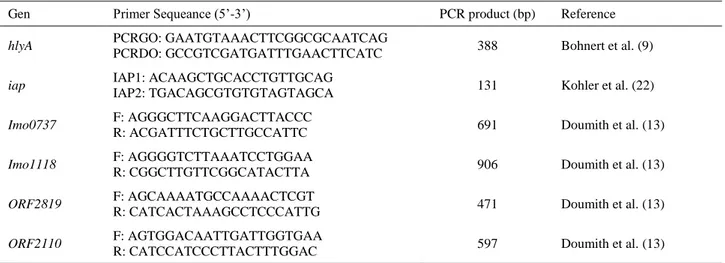
Table 1. List of L. monocytogenes genes and primers used in the PCR assay (13). Tablo 1. PCR analizinde kullanılan L. monocytogenes genleri ve primer dizilimleri (13).
Gen Primer Sequeance (5’-3’) PCR product (bp) Reference
hlyA PCRGO: GAATGTAAACTTCGGCGCAATCAG
PCRDO: GCCGTCGATGATTTGAACTTCATC 388 Bohnert et al. (9)
iap IAP1: ACAAGCTGCACCTGTTGCAG
IAP2: TGACAGCGTGTGTAGTAGCA 131 Kohler et al. (22)
Imo0737 F: AGGGCTTCAAGGACTTACCC
R: ACGATTTCTGCTTGCCATTC 691 Doumith et al. (13)
Imo1118 F: AGGGGTCTTAAATCCTGGAA
R: CGGCTTGTTCGGCATACTTA 906 Doumith et al. (13)
ORF2819 F: AGCAAAATGCCAAAACTCGT
R: CATCACTAAAGCCTCCCATTG 471 Doumith et al. (13)
ORF2110 F: AGTGGACAATTGATTGGTGAA
PCR amplification and electrophoresis: For hlyA
gene, PCR mix was prepared as such; 1X PCR Buffer in total 50 μl volume, 1.5 mM MgCl2, 0.1 mM dNTP, 0.5 U
Taq-Polymerase (Sigma D4545), 1 μM from each primer and 5 μl template DNA. Amplification of hlyA gene was carried out in Thermal Cycler (Bio-Rad MJ mini Gradient CA - ABD) at 94°C with 5 minutes initial denaturation and 35 cycles, at 94°C with 30 seconds denaturation, at 65°C primer annealing for 45 seconds, at 72°C primer extension and at 72°C last extension (9).
For iap gene, PCR mix was prepared as such; 1X PCR Buffer in total 50 μl volume, 1.5 mM MgCl2, 0.1 mM
dNTP, 0.5 U Taq-Polymerase 1 μM and 5 μl DNA from each primer. Amplification of iap gene was programmed as 5 minutes initial denaturation at 94°C; 35 cycles of 30 seconds denaturation at 94°C, 45 seconds annealing at 55°C, 45 seconds extension at 72°C; 5 minutes of final extension at 72°C (22).
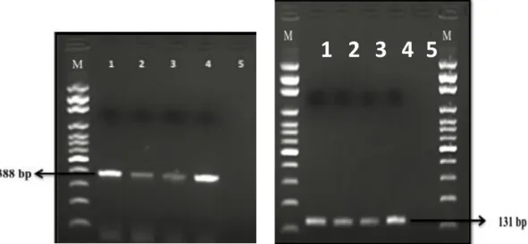
Amplicons were carried out in 2% agarose at 80 volt current for electrophoresis process (Bio-Rad PowerPac Basic Power Supply, Bio-Rad Wide Mini Sub-Cell GT Cell). hlyA and iap genes were visualized at UV-transilluminator at 388 bp and 131 bp, respectively.
Serotyping: In order to serotype L. monocytogenes
isolates, primer sequences designed by Doumith et al. (13) were used (Table 1).
PCR amplification and electrophoresis: For
serotyping, PCR mix was prepared as such: 1X PCR Buffer in total 50 μl volume, 1.5 mM MgCl2, 0.1 mM
dNTP, 0.5 U Taq-Polymerase, 1 μM from each primer and 5 μl template DNA. Serotyping conditions in Thermal Cycler were programmed as such: first denaturation at 94°C for 3 minutes; 35 cycles of denaturation at 94°C for
40 seconds, annealing at 53°C for 75 seconds, primer extension at 72°C for 45 seconds and final extension at 72°C for 5 minutes (13). Amplicons were carried out in 2% agarose at 80 volt current for electrophoresis process (Figure 2). For defining the current serotype the evaluation was as such: If an isolate was Imo0737 positive: serotype 1/2a or 3a. If an isolate was both Imo0737 and Imo1118 positive: serotype 1/2c or 3c. If an isolate was ORF2819 positive: serotype 1/2b or 3b. If an isolate was both
ORF2819 and ORF2110 positive: serotype 4b or 4e, 4d.
Results
It was detected that, 5 of the samples (5/300, 1.6%) taken from slaughterhouse have been contaminated with
L. monocytogenes. Amongst the samples contaminated
with L. monocytogenes, one of them was obtained from cattle carcass (2.7%), one from blade (2.7%), one from blade sharpener (2.7%), one from floor (2.7%) and one from cowhide (2.7%). A total of 18 isolates (Table 2), which obtained from 5 positive samples, were determined to have hlyA and iap genes and confirmed as L.
monocytogenes (Figure 1).
Three different serotypes (1/2a or 3a, 1/2b or 3b, and 4b or 4d, 4e) were obtained from 18 L. monocytogenes isolates. It has been detected that 10 of the isolates obtained from carcass and blade were L. monocytogenes 1/2b or 3b; 2 of the isolates obtained from blade sharpener were L. monocytogenes 4b or 4d, 4e; 6 of the isolates obtained from floor and skin sample were L.
monocytogenes 1/2a or 3a. Serotypes of 1/2b and 1/2a
were detected as dominant from the samples obtained from slaughterhouse whilst serotype 1/2c was not detected (Table 2).
Figure 1. Electrophorese image of hlyA (388 bp) and iap (131 bp) gene L. monocytogenes isolates by PCR. M: 100 bp DNA marker, lane 1: positive control for hlyA and iap gene (L. monocytogenes ATCC7644); lanes 2–4: hlyA and iap gene positive L. monocytogenes isolate; lane 5: negative control.
Şekil 1. hlyA (388 bp) ve iap (131 bp) genlerinin elektroforez görüntüsü. M: 100 bp DNA marker, sütun 1: hlyA ve iap genleri pozitif kontrol (L. monocytogenes ATCC7644); sütun 2–4: hlyA ve iap pozitif L. monocytogenes izolatları; sütun 5: negatif kontrol.
Figure 2. Electrophorese image of positive and negative control. M: 100 bp DNA marker, lane 1: positive control of L. monocytogenes 4b (RSKK 475); lane 2: positive control of L. monocytogenes 1/2a (RSKK 471); lane 3: positive control of L. monocytogenes 1/2b (RSKK 472); lane 4: positive control of L. monocytogenes 1/2c (ATCC 7644); lane 5: negative control.
Şekil 2. Pozitif ve negatif kontrollerin elektroforez görüntüsü. M: 100 bp DNA marker, sütun 1: L. monocytogenes 4b pozitif kontrol (RSKK 475); sütun 2: L. monocytogenes 1/2a pozitif kontrol (RSKK 471); sütun 3: L. monocytogenes 1/2b pozitif kontrol (RSKK 472); sütun 4: L. monocytogenes 1/2c pozitif kontrol (ATCC 7644); sütun 5: negatif kontrol.
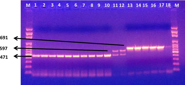
Figure 3. Serotype identification of Listeria monocytogenes isolates by multiplex PCR assay. M: 100 bp DNA marker, lanes 1-10: serotype 1/2b isolates; lanes 11-12: serotype 4b isolates; lanes 13-18: serotype 1/2a isolates.
Şekil 3. Listeria monocytogenes izolatlarının multiplex PZR ile serotiplerinin belirlenmesi. M: 100 bp DNA marker, sütun 1-10: serotip 1/2b; sütun 11-12: serotip 4b; sütun 13-18: serotip 1/2a.
M
1
2
3
4
5
6
7
8
9
10
11
12
13
14
15
16
17
18
M
471
597
691
M
1
2
3
4
5
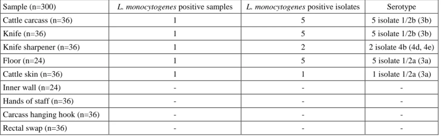
906 691 597 471Table 2. Occurrence of L. monocytogenes isolates and serotypes diversity in slaughterhouse samples. Tablo 2. Mezbaha örneklerinde L. monocytogenes varlığı ve serotiplerinin dağılımı.
Sample (n=300) L. monocytogenes positive samples L. monocytogenes positive isolates Serotype
Cattle carcass (n=36) 1 5 5 isolate 1/2b (3b)
Knife (n=36) 1 5 5 isolate 1/2b (3b)
Knife sharpener (n=36) 1 2 2 isolate 4b (4d, 4e)
Floor (n=24) 1 5 5 isolate 1/2a (3a)
Cattle skin (n=36) 1 1 1 isolate 1/2a (3a)
Inner wall (n=24) - - -
Hands of staff (n=36) - - -
Carcass hanging hook (n=36) - - -
Rectal swap (n=36) - - -
Discussion and Conclusion
From a total of 300 samples obtained from slaughterhouse, 5 of them (1.66%) were detected as L.
monocytogenes positive. In various studies conducted in
Turkey, the incidence of L. monocytogenes was reported to be between 1% and 5.17% in slaughterhouses (2, 20).
In this study, L. monocytogenes was isolated from cattle carcass, knife, knife sharpener, floor and cattle skin. During slaughtering, extraction of the cowhide and shredding, knives, knife sharpeners and also floors have been thought to be the primary contamination source of microorganism to cattle carcass. Since no microorganism have been isolated from staff hands, it can be suggested that contamination was not human originated.
In our study, 1 of 48 (2.08%) samples obtained from slaughterhouse wall and floor was detected L.
monocytogenes positive. In contrast with the findings of
our study, Yeşilyurt (32) detected the incidence of L.
monocytogenes 4.2% (2/48) from the samples of
slaughterhouse wall and floor.
In the study of Akkaya et al. (2), L. monocytogenes was not detected in the samples obtained from the wall of cold storage and the floor of meat transport vehicle. On the other hand, 10 of 100 (%10) samples obtained from slaughterhouse floor, 2 of 65 samples (3.07%) obtained from slaughterhouse inner wall, 1 of 65 samples (1.53%) obtained from cold storage floor and 1 of 20 (5%) samples obtained from the sides of the meat section of the transport vehicle were L. monocytogenes positive. In a study of Alişarlı et al. (3), L. monocytogenes was detected from slaughterhouse floor by the level of 6.7%. The findings of Akkaya et al. (2) and Alişarlı et al. (3) were consistent with the findings of our study. Similar to our findings, Sammarco et al. (29) reported that slaughterhouse surfaces and materials, equipments were the source of contamination.
On the other hand, in a study of Kahraman et al. (20), it has been stated that in 30 samples (6.97%) amongst 430
samples obtained from meat processing plants and 7 samples (7%) amongst 100 samples obtained from surfaces were L. monocytogenes positive. Researchers indicated that surfaces and equipments in food processing plants were the focal points for the microorganism.
In a study regarding the existence of L.
monocytogenes at tools and materials used in
slaughterhouse, the agent was detected in 10 of 120 (8.33%) samples obtained from cutting blades, choppers, shredding saws, hooks and cutting boards. Amongst 24 samples obtained from each tools and materials, the composition for positive L. monocytogenes was as such; 2 from cutting blades (8.33%), 2 from choppers (8.33%), 1 (4.16%) from meat saws, 3 from hooks (12.50%) and 2 from cutting boards (8.33%) (3). On the other hand, different from our study Akkaya et al. (2) detected L.
monocytogenes from the samples collected from
slaughterhouses (4 from shredding countertops (40%), 3 from knives (8.57%), 2 from choppers (20%), 6 from hooks (13.33%). Whilst the results of knife samples were very close, the other results were quite different. This may be because of sampling time, sampling technique, analysis method and the structure of the materials used. It was further considered that the lack of workers in implementation of hygiene rules was related with the sanitation quality of the slaughterhouses.
In a study of Kahraman et al. (20), L. monocytogenes was detected in 12 samples (9.23%) obtained from equipments. The percentages were also similar (10%, 8.8%, 11.9%) in the studies of Beumer and Giedfel (8), Alişarlı et al. (3) and Gudbjornsdottir et al. (17) respectively. On the other hand, Legnani et al. (23) reported the level of L. monocytogenes as 0.71% in their study.
Yeşilyurt (32) reported that he could not detect L.
monocytogenes from any of the materials including 36
blades, 36 grinder and 72 hooks. One of 36 (2.78%) samples obtained from the hands of the workers and
gloves was declared to be detected L. monocytogenes positive. In the study of Akkaya et al. (2), 35 samples were obtained from the hands of the slaughterhouse workers and L. monocytogenes was detected as positive only in 1 sample (2.85%). Gudbjornsdottir et al. (17) also reported
L. monocytogenes positive samples from the hands of
workers from red meat and poultry plants. Alişarlı et al. (3), Aarnisalo et al. (1) and Kahraman et al. (20) could not detect L. monocytogenes from workers hands. The results of our study were similar to those by Alişarlı et al. (3), Aarnisalo et al. (1) and Kahraman et al. (20). Different from our findings, however, Yeşilyurt (32) and Akkaya et al. (2) had positive results. Considering those contradictory results, one may assume that results differ from each other due to lack of attention and hygiene of workers which plays a key role for food safety.
In this study, L. monocytogenes was detected in 1 of 36 samples (2.77%) obtained from cattle carcass surface. In parallel with our findings, the ratio was 6.8% in the study of Akkaya et al. (2) and 6.2% in the study of Alişarlı et al. (3). In the study of Fenlon et al. (16) in UK, L.
monocytogenes was detected from 7% of carcasses. These
datas seem to support the findings of our study.
In a study of Barros et al. (7), L. monocytogenes was detected from 25.9% of bovine carcass samples whilst McNamara (26) detected only 4.1% from 2098 carcass samples. Vanderline et al. (31) reported the contamination level as 0.77% from 130 imported carcasses samples in Australia. Antoniollo et al. (5) identified L. monocytogenes with a level of 4.3% from 69 lamb carcass
samples. In the study of Nicolas et al. (27) in France, L.
monocytogenes was identified in 4 samples (20%)
amongst 20 samples obtained from cattle carcass and 1 sample (14.28%) amongst 7 samples obtained from lamb carcass. It is seen that the results of the studies differ a lot. It is considered that the results of the studies for each country may differ because the terms of sanitation applied in slaughterhouses, different sampling techniques, method of analysis, hygiene conditions.
L. monocytogenes has 13 serotypes according to
somatic (O) and flagella (H) antigenic structures. Serotype 4b, 1/2a, 1/2b and 1/c have been reported to cause 98% of foodborne listeriosis cases worldwide (21, 24). As a result of serotyping of isolates obtained in our study, we studied 18 isolates and detected that 55.6% was L. monocytogenes 1/2b, 33.3% was L. monocytogenes 1/2a and 11.1% was
L. monocytogenes 4b. It was found in our study that 1/2b,
1/2a and 4b serotypes were dominant serotypes.
Hofer et al. (18) serotyped a total of 3122 L.
monocytogenes isolates by using antiserum, 70 of which
had milk product origin, that were obtained from different sources like food, human, animal and environment from Brazil between 1971-1997. In accordance with their
findings, it was reported that, amongst milk product derived isolates 58% were L. monocytogenes 1/2a, 26.6% were L. monocytogenes 4/b, 14% were L. monocytogenes 1/2b and 1.4% were L. monocytogenes 1/2c. Within the total 3122 samples, L. monocytogenes 4b was the dominant serotype with the ratio of 45.4% which was followed by L. monocytogenes 1/2a as 20% and L.
monocytogenes 1/2b as 20%. Makino et al. (25) also
reported L. monocytogenes 1/2b was the dominant serotype from cheese samples (18).
In Turkey, Özkiraz (28) detected L. monocytogenes in 8 of 100 modified atmosphere packaged beef meat products sold in Samsun. It was detected that; from a total of 11 isolates, 3 of them were L. monocytogenes 1/2a, 4 of them were 1/2b, 1 was 1/2c and 3 were 4b. In studies on poultries, Erol and Şireli (15) isolated L. monocytogenes in frozen chicken carcasses that were offered for sale in Ankara with a method recommended by USDA/FSIS at the ratio of 30%. As a result of their serotyping, they detected L. monocytogenes 1/2a was the dominant serotype (73%) which followed by 1/2b, 1/2c and 4b respectively. Besides, Ayaz and Erol (6) reported that, as a result of serotyping L. monocytogenes isolates obtained from turkey meat, they detected 4b (51.4%) as dominant serotype which followed by 1/2a (27%) and 1/2b (21%) respectively. In our study, unlike the researchers, 1/2b was the most common serotype.
As a result, it was observed that L. monocytogenes could contaminate the carcasses during slaughtering because of slaughterhouse conditions, equipments and environment. Healthy meat can be obtained by transporting the cattles from farm to the slaughterhouse carefully, having necessary examinations and controls before slaughtering, having hygienic cutting in order to have both good flow of blood and animal welfare. Designing all departments in the slaughterhouse in accordance with hygiene rules and taking necessary measures in transition from clean areas to polluted areas, using tools, equipments and materials easy to clean and disinfect at all step, having well trained staff in food safety, applying quality management systems in slaughterhouses and having good hygiene practice procedures will help to prevent the contamination to carcasses.
Acknowledgements
This study was supported by Ondokuz Mayıs University, Samsun, Turkey, Scientific Research Project Programs (Project No: PYO.VET -1901.12.005). This research was presented as a poster at the 6th National Veterinary Food Hygiene Congress, held on 7-11 October 2015 in Van, Turkey.
References
1. Aarnisalo K, Tallavaara K, Wirtanen G, et al. (2006):
The hygienic working practices of maintenance personnel and equipment hygiene in the Finnish food industry. Food
Control, 17, 1001-11.
2. Akkaya L, Alişarlı M, Çetinkaya Z, et al. (2008):
Occurrence of Escherichia coli O157:H7/O157, Listeria monocytogenes and Salmonella spp. in beef slaughterhouse environments, equipment and workers. J Muscle Foods, 19,
261-274.
3. Alişarlı M, Akkaya L, Alemdar S (2003): An investigation
on the prevalence of Listeria monocytogenes and Salmonella spp. in a cattle slaughterhouse.
Fleischwirtschaft Int, 3, 41-44.
4. Amills M, Francino O, Jansa M, et al. (1997): Isolation of
genomic DNA from milk samples by using Chelex® resin. J
Dairy Res, 64, 231-238.
5. Antoniollo PC, Bandeira-Fda S, Jantzen MM, et al. (2003): Prevalance of Listeria spp. in Brazil. J Food Prot, 66, 328-330.
6. Ayaz ND, Erol İ (2011): Serotype distribution of Listeria
monocytogenes isolated from turkey meat by multiplex PCR in Turkey. J Food Safety, 31, 149-153.
7. Barros MAF, Nero LA, Silva LC, et al. (2007): Listeria
monocytogenes: Occurrence in beef and identification of the main contamination points in processing plants. Meat
Sci, 76, 591-596.
8. Beumer RE, Giefel MC (1999): Pathogens in domestic
kitchens: facts and fiction. Food microbiology and food safety into the next millennium: Proceedings of the 17th
international conference of the international committee on food microbiology and hygiene (ICFMH). Veldhoven, Netherland, 345-347.
9. Bohnert M, Dilasser F, Dalet C, et al. (1992): Use of
specific oligonucleotides for direct enumeration of Listeria monocytogenes in food samples by colony hybridization and rapid detection by PCR. Res Microbiol, 143, 271-280.
10. Chakraborty T, Hain T, Domann E (2000): Genome
organization and the evolution of the virulence gene locus in Listeria species. Int J Med Microbiol, 290, 167-174.
11. Churchill RLT, Lee H, Hall JC (2006): Detection of
Listeria monocytogenes and the toxin listeriolysin O in food.
J Microbiol Methods, 64, 141-170.
12. Commission of the European Communities (1987): Code
of good hygienic practices. EG-Document, VI/5938/87
(PVET/2140).
13. Doumith M, Buchrieser C, Glaser P, et al. (2004):
Differentiation of the major Listeria monocytogenes Serovars by Multiplex PCR. J Clin Microbiol, 42, 3819-22.
14. Anon. (1996): Cell separation and protein purification.
Technical handbook. 2nd Ed. Dynal A.S., Norway Printed: 02 96.
15. Erol İ, Şireli UT (1999): Donmuş broyler karkaslarında
Listeria monocytogenes’in varlığı ve serotip dağılımı. Turk
J Vet Anim Sci, 23, 765-770.
16. Fenlon DR, Wilson J, Donachie W (1996): The incidence
and level of Listeria monocytogenes contamination of food sources at primary production and initial processing. J
Appl Bacteriol, 81, 641-650.
17. Gudbjornsdottir B, Suihko ML, Gustavsson P (2004):
The incidence of Listeria monocytogenes in meat, poultry and seafood plants in Nordic countries. Food Microbiol, 21,
217-225.
18. Hofer E, Ribeiro R, Feitosa DP (2000): Species and
serovars of the genus Listeria isolated from different sources in Brazil from 1971 to 1997. Mem Inst Oswaldo
Cruz, 95, 615-620.
19. International Organization for Standardization (1997): EN ISO 11290- 1. Microbiology of Food and Animal
Feeding Stuffs – Horizontal Method for the Detection and Enumeration of Listeria monocytogenes. Part 1: Detection Method. Geneva, Switzerland: International Organization for Standardization.
20. Kahraman T, Çetin Ö, Dümen E, et al. (2010): Incidence
of Salmonella spp. and Listeria monocytogenes on equipment surfaces and personnel hands in meat plants.
Revue Med Vet, 161, 108-113.
21. Koçan D, Halkman AK (2006): Listeria monocytogenes ve
listeriozis. Gıda, 31, 133-140.
22. Köhler S, Leimeister-Wachter M, Chakraborty T, et al. (1990): The gene coding for protein p60 of Listeria
monocytogenes and its use as a specific probe for Listeria monocytogenes. Infect Immun, 58, 1943-50.
23. Legnani P, Leoni E, Berveglieri M, et al. (2004): Hygienic
control of mass catering establishments, microbiological monitoring of food and equipment. Food Control, 15,
205-211.
24. Liu D (2006): Identification, subtyping and virulence
determination of Listeria monocytogenes, an important foodborne pathogen. J Med Microbiol, 55, 645-659.
25. Makino SI, Kawamato K, Takeshi K, et al. (2005): An
outbreak of foodborne listeriosis due to cheese in Japan during 2001. Int J Food Microbiol, 104, 189-196.
26. McNamara AM (1995): Establishment of baseline data on
the microbiota of meats. J Food Safety, 15, 113-119.
27. Nicolas JA, Espaze EP, Catimel B, et al. (1989): Isolation
of Listeria from French meat products. Zbl Bakt, 272,
242-247.
28. Özkiraz A (2016): Modifiye atmosfer paketli sığır kıyma ve
kuşbaşı örneklerinde Listeria monocytogenes ve serotiplerinin belirlenmesi. MSc, Ondokuz Mayıs
University, Samsun, Turkey.
29. Sammarco ML, Ripabelli G, Ruberto A, et al. (1997):
Prevalance of Salmonella, Listeria and Yersinia in the slaughterhouses environment and on work surfaces, equipment and workers. J Food Prot, 60, 367-371.
30. Sofos JN (2008): Challenges to Meat Safety in the 21st
Century. Meat Sci, 78, 3-13.
31. Vanderlinde PB, Shay B, Murray J (1998):
Microbiological quality of Australian beef carcasses meat and frozen bulk packed beef. J Food Protect, 61, 437-44.
32. Yeşilyurt C (2010): Aydın ili mezbahalarında E. coli
O157:H7 ve Listeria monocytogenes varlığının araştırılması. PhD, Adnan Menderes University, Aydın,
Turkey.
Geliş tarihi: 05.12.2016 / Kabul tarihi: 02.03.2017
Address for correspondence: Doç. Dr. Özgür ÇADIRCI
Ondokuz Mayıs University, Faculty of Veterinary Medicine, Department of Food Hygiene and Technology,
Kurupelit, Samsun,Turkey. Telephone: +90 362 3121919-2812 Fax: +90 362 4576622