Case Report
Abstract
Bee pollen is given to children by mothers in order to strengthen their immune systems. There are no studies related with the side effects of bee polen in the literature. In this article, the literature was reviewed by presenting a case of allergic eosinophilic gastropathy related with bee polen. A 5-year old child was admitted due to abdominal pain. Edema was detected on the eyelids and pretibial region. In laboratory investigations, pathology was not detected in terms of hepatic and renal causes that would explain the protein loss of the patient diagnosed with hypoproteinemia and hypoalbuminemia. Urticaria was detected during the follow-up visit. When the history of the patient was deepened, it was learned that bee pollen was given to the patient every day. The total eosinophil count was found to be 1 800/mm3. Allergic gastroenteropathy was considered because of hypereosinophilia and severe abdominal pain and endoscopy was performed. Biopsy revealed abundant eosinophils in the whole gastric mucosa. A diagnosis of allergic eosinophilic gastropathy was made. Bee polen was discontinued. Abdominal pain and edema disappeared in five days. Four weeks later, the levels of serum albumin and total eosinophil returned to normal. (Turk Pediatri Ars 2015; 50: 189-92)
Keywords: Bee polen, eosinophilic gastroenteropathy, good allergy
189
Does bee pollen cause to eosinophilic
gastroenteropathy?
Belgin Usta Güç1, Suna Asilsoy2, Oğuz Canan3, Fazilet Kayaselçuk4
1Division of Pediatric Allergy and Immunology, Adana Maternity and Children Hospital, Adana, Turkey
2Division of Pediatric Allergy and Immunology, Başkent University Adana Research and Application Center, Adana, Turkey 3Division of Pediatric Gastroenterology, Başkent University Adana Research and Application Center, Adana, Turkey 4Department of Pathology, Başkent University Adana Research and Application Center, Adana, Turkey
Introduction
Food allergy (FA) is an important problem which is ob-served commonly in the childhood and manifested by different clinical findings (1). The majority of the clinical findings which generally develop in the early childhood are IgE-mediated. IgE-mediated reactions may involve the skin, gastrointestinal (GI) system and respiratory system. Signs appear in a short time after exposure to allergen. However, mixed type food allergies include both IgE-mediated and cell mediated mechanisms and clinical findings (eosinophilic esophagitis, eosinophilic gastritis, esosinophilic gastroenteritis) generally occur within a certain time after consumption of food (1, 2). Currently, substances including bee pollen, honey and royal jelly are given to children by their mothers with the objectives of protection from infectious and aller-gic diseases, increasing appetite and strengthening the body. Use of these types of substances with the above mentioned objectives is gradually increasing, but there
are no studies related with this subject in the literature. In contrast, there are many case reports reporting that these types of substances lead to anaphylaxis, urticar-ia, asthma, abdominal pain, diarrhea and pruritus (3-8). The relation between bee pollen and allergic eosino-philic gastropathy has been reported in the literature, though rarely (6). In this article, a case of allergic eo-sinophilic gastropathy causing to a picture of protein losing enteropathy related with bee pollen is presented and the literature is reviewed.
Case
A five-year old male patient presented with complaints of abdominal pain which had been lasting for one week, vomiting and swelling in the eyelids and legs which started three days ago. In the history, it was learned that he had constipation since infancy and he defecated every day but with difficulty. His personal and familial history were insignificant.
Address for Correspondence: Belgin Usta Güç, Adana Kadın Doğum ve Çocuk Hastanesi, Çocuk Allerji ve İmmunoloji Bilim Dalı, Adana, Türkiye. E-mail: [email protected]
Received: 01.04.2013 Accepted: 18.03.2014
©Copyright 2015 by Turkish Pediatric Association - Available online at www.turkpediatriarsivi.com DOI: 10.5152/TurkPediatriArs.2015.1105
This study was presented at the 30th European Academy of Allergy and Clinical Immunology Congress, 11-15 June 2011, İstanbul.
T
URKISHA
RCHIVESofP
EDIATRICS TÜRK PEDİATRİ ARŞİVİPhysical examination findings were as follows: body weight: 17 kg (50 p), height: 107 cm (50 p), apical heart beat: 82/min, blood pressure: 100-80 mmHg, general status: well, consciousness: open, cooperat-ed, orientcooperat-ed, mild periorbital and pretibial edema. Examination of the other systems was found to be normal.
Laboratory findings were as follows: Complete blood count: hemoglobin: 14.1 g/dL, WBC: 14 800/mm3, platelets: 510 000/mm3, total eosinophil count: 1 800 / mm3. The percentage of eosinophils was found to be 12% on peripheral smear. Renal function tests, sodium, potassium, liver enzymes and complement 3 and 4 lev-els were found to be normal. The other laboratory tests were as follows: total protein: 2.97g/dL (low), albumin: 1.94 g/dL (low), calcium: 8.34 mg/dL, IgG: 213 mg/dL (345-1236), Ig A: 60 mg/dL (14-139), IgM: 114 mg/dL (43-207), IgE: 335 IU/mL.
Nephrotic syndrome was considered, since total pro-tein and albumin was found to be low, however, lack of proteinuria in complete urinalysis and in 24-hour urine excluded this diagnosis. Parasites or occult blood were not found on repeated stool examination. Considering that decreased albumin level might be caused by hepat-ic pathology, liver functions tests were performed and the hepatic parenchyma was evaluated by abdominal ultrasonography and observed to be normal. There was no diarrhea in the history and the patient had chronic constipation in contrast. Therefore, protein losing en-teropathy was not considered initially.
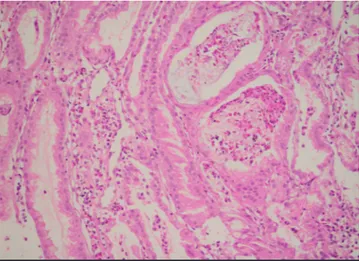
Short-term urticarial rash occured while the patient was being followed up. When nutritional history was taken from the mother once again, it was learned that the patient was given a product called bee pollen each day by adding into a spoon of milk. In the follow-up, it was observed that the abdominal circumference in-creased. On repeated ultrasonography, moderate asci-tes was observed. Pediatric gastroenterology was con-sulted considering allergic gastroenteropathy because of a neutrophil count of 1 800/mm3 and severe abdom-inal pain. Pedaitric gastroenterology performed upper gastrointestinal endoscopy. Upper gastrointestinal en-doscopy revealed edema and hyperemia prominent in the whole gastric mucosa, fragility especially in the an-trum, prepyloric antrum and corpus, marked hypertro-phy in the corpus pili (thicker than 5 mm), old and new blood residues in the lumen, nodular appearance in the antrum and hyperemia in the antrum and duedonum. On histopathological examination, edema and conges-tion was found in the gastric mucosa of the fundus and abundant eosinophils were found in the tunica propria and on the surface (Figure 1). A diagnosis of allergic gastropathy was made as a result of clinical, biochemi-cal and histopathologibiochemi-cal examinations.
Skin prick tests were performed with food allergens (milk, egg, egg white, cacao, wheat, nut, peanut, banana, orange, tomato, peach, fish, chicken meat, soya) and found to be negative. Patch test with bee pollen was performed in terms of possible late type reaction and found to be negative. Ingestion of pollen was discon-tinued and abdominal pain and edema which started to regress in five days disappeared completely after two weeks. Serum albumin, calcium, total eosinophil lev-els returned to normal on the follow-up visit after four weeks. On follow-up upper gastrointestinal endoscopy, sparsely dotted hyperemia was observed in the whole gastric mucosa and the general appearance was found to be normal. On histopathological examination, it was observed that the congestion in the gastric mucosa of Turk Pediatri Ars 2015; 50: 189-92 Usta Güç et al. Bee pollen and eosinophilic gastroenteropathy
190
Figure 1. Edema and congestion in the gastric mucosa, abundant eosinophils in the tunica propria and on the surface
Figure 2. Marked regression in extensive eosinophilia and con-gestion in the gastric mucosa
the fundus and intensive eosinophilia in the tunica pro-pria were markedly regressed (Figure 2).
Discussion
Consumption of substances like bee pollen has in-creased gradually in recent years. There are no stud-ies related with the adverse effects of these substanc-es. When the literature is reviewed, it is observed that there are case reports indicating that these substances lead to side effects including urticaria, bronchospasm, severe abdominal pain, anaphylaxis, hypereosinophilia, eosinophilic gastropathy and neurological findings and increase atopic sensitivity (3-8). We think that edema, hypoproteinemia and allergic eosinophilic gastropathy developed in our patient as a result of consumption of bee pollen.
Food allergies are divided into three classes includ-ing IgE-mediated, non-IgE mediated and mixed type in which both mechanisms are involved. Food aller-gies may lead to a wide spectrum of symptoms. The clinical picture may range from chronic nonspecific GI system findings to severe anaphylaxis picture. The common type of food allergy is in the form of type 1 reaction which is IgE mediated. IgE mediated reactions may cause to symptoms including urticaria, oral allergy syndrome, anaphylaxis, angioedema, acute rhinocon-juntivitis, acute bronchospasm and usually develop in minutes (1, 2).
Mixed type food allergies in which both IgE mediat-ed and cell mmediat-ediatmediat-ed mechanisms are involvmediat-ed usually cause late-onset reactions and may become symptom-atic days after exposure to the responsible agent. Atopic dermatitis, contact dermatitis, eosinophilic gastropathy and eosinophilic esophagitis may develop due to mixed type food allergies (1). The symptoms of mixed type food allergies (eosinophilic esophagitis, eosinophilic gasstritis, eosinophilic gastroenteritis) start late and not right after exposure. Therefore, it is difficult to associate the symptoms with diet (9).
Allergic eosinophilic gastropathy is a disease charac-terized with eosinophilic infiltration of the different parts and layers of the gastrointestinal tract (9). It is ob-served substantially rarely in the childhood compared to adults. It causes problems, including abdominal pain, diarrhea, melena, vomiting, weight loss, iron deficien-cy anemia and protein losing enteropathy. Eosinophilic infiltration is prominent histopathologically. The main complaints of our patient included vomiting and
ab-dominal pain. However, our patient had constipation in contrast to diarrhea which is generally observed in this clinical picture. Since eosinophilic infiltration may be observed in many conditions including gastro-esophageal reflux disease, parasitic contamination and inflammatory bowel disease, the diagnosis of eosino-philic gastropathy should be considered after the other possibilities are excluded (9, 10). The diagnostic criteria of eosinophilic gastroenteritis include gastrointestinal symptoms, eosinophilic infiltration in one or more ar-eas which can be demonstrated by biopsy, absence of any involvement outside the gastrointestinal tract and absence of parasitic contamination (9, 10). There is no atopic history in approximately half of the patients (10). Frequently, there is a strong familial history of food al-lergy. Although eosinophilia in the peripheral blood is an important variable suggesting the diagnosis, it is not found in approximately 20% of the patients. Therefore, it is not considered as a diagnostic criterion (11). Since a history of food intolerance and atopy can not be proved objectively in most of the patients, they are not manda-tory for the diagnosis. Hence, skin prick tests and food specific IgE levels are found to be normal in approxi-mately half of the patients (10). In our patient, hypo-proteinemia and eosinophilia were found in addition to the complaints including abdominal pain, vomiting and constipation. A diagnosis of eosinophilic gastrop-athy was made as a result of GI endoscopy and biopsy examination.
Double-blind placebo controlled food provocation test which is gold standard in IgE mediated food allergy is not used in the diagnosis of allergic eosinophilic gast-ropathy. Skin prick tests, atopy patch tests, serum food specific IgE levels may be useful for the diagnosis (1). The basis of treatment is elimination of the culprit food. In addition, response of the patient to the elimination diet is substantially significant. However, oral steroid may be needed in some patients (11). In our patient, oral steroid was not administered, because the clinical picture improved after elimination of bee pollen. The active ingredients contained in the products which are used with different objectives including bee pollen , the amounts of these active ingredients, at what doses they should be used and what kind of effects and side effects they lead to are not known exactly. The substanc-es contained in thsubstanc-ese products recommended without support of medical research may lead to toxic effects as observed in our patient. In conditions which can not be explained clinically, administration of different drugs and substances should be questioned.
191 Turk Pediatri Ars 2015; 50: 189-92 Usta Güç et al. Bee pollen and eosinophilic gastroenteropathy
Informed Consent: Written informed consent was obtained
from the parent of the patient who participated in this study.
Peer-review: Externally peer-reviewed.
Author Contributions: Concept - S.A., B.U.G.; Design - B.U.G.,
S.A.; Supervision - S.A.; Funding - B.U.G.; Materials - O.C., F.K.; Data Collection and/or Processing - S.A., B.U.G.; Analysis and/or Interpretation - S.A., B.U.G.; Literature Review - B.U.G.; Writer - B.U.G., S.A. Critical Review - S.A.; Other - O.C., F.K.
Conflict of Interest: No conflict of interest was declared by
the authors.
Financial Disclosure: The authors declared that this study has
received no financial support.
References
1. Sicherer SH, Sampson HA. Food allergy. J Allergy Clin
Immunol 2010; 125: S116-25. [CrossRef ]
2. Cianferoni A, Spergel JM. Food allergy: review, classification
and diagnosis. Allergol Int 2009; 58: 457-66. [CrossRef]
3. Akiyasu T, Paudyal B, Paudyal P, et al. A case report of acute renal failure associated with bee pollen contained in nutritional supplements. Ther Apher Dial 2010; 14:
93-7. [CrossRef ]
4. Martín-Muñoz MF, Bartolome B, Caminoa M, Bobolea I, Ara MC, Quirce S. Bee pollen: a dangerous food for al-lergic children. Identification of responsible allergens.
Al-lergol Immunopathol (Madr) 2010; 38: 263-5. [CrossRef ]
5. Lin FL, Vaughan TR, Vandewalker ML, Weber RW. Hy-pereosinophilia, neurologic, and gastrointestinal symp-toms after bee-pollen ingestion. J Allergy Clin Immunol
1989; 83: 793-6. [CrossRef ]
6. Puente S, Iñíguez A, Subirats M, Alonso MJ, Polo F, Mo-neo I. Eosinophilic gastroenteritis caused by bee pollen sensitization. Med Clin (Barc) 1997; 108: 698-700. 7. Fuiano N, Incorvaia C, Riario-Sforza GG, Casino G.
Ana-phylaxis to honey in pollinosis to mugwort: a case report. Eur Ann Allergy Clin Immunol 2006; 38: 364-5.
8. Menniti-Ippolito F, Mazzanti G, Vitalone A, Firenzuoli F, Santuccio C. Surveillance of suspected adverse reactions to natural health products: the case of propolis. Drug Saf
2008; 31: 419-23. [CrossRef ]
9. Marc E Rothenberg. Eosinophilic gastrointestinal
disor-ders. J Allergy Clin Immunol 2004; 113 :11-28. [CrossRef ]
10. Zhang L, Duan L, Ding S, et al. Eosinophilic gastroen-teritis: clinical manifestations and morphological char-acteristics, a retrospective study of 42 patients. Scand J
Gastroenterol 2011; 46: 1074-80. [CrossRef ]
11. Baig MA, Qadir A, Rasheed J. A review of eosinophilic gastroenteritis. J Natl Med Assoc 2006; 98: 1616-9.
Turk Pediatri Ars 2015; 50: 189-92 Usta Güç et al. Bee pollen and eosinophilic gastroenteropathy