BIOPHOTONIC APPLICATIONS OF
ULTRAFAST FIBER LASERS: FROM
BIOMATERIAL SURFACE MODIFICATION
TO SUB-CELLULAR NANOSURGERY
a dissertation submitted to
the department of materials science and
nanotechnology program
and the Graduate School of engineering and science
of bilkent university
in partial fulfillment of the requirements
for the degree of
doctor of philosophy
By
Mutlu Erdo˘gan
September, 2014
I certify that I have read this thesis and that in my opinion it is fully adequate, in scope and in quality, as a dissertation for the degree of Doctor of Philosophy.
Assoc. Prof. Dr. Fatih ¨Omer ˙Ilday (Advisor)
I certify that I have read this thesis and that in my opinion it is fully adequate, in scope and in quality, as a dissertation for the degree of Doctor of Philosophy.
Prof. Dr. Tayfun ¨Oz¸celik
I certify that I have read this thesis and that in my opinion it is fully adequate, in scope and in quality, as a dissertation for the degree of Doctor of Philosophy.
I certify that I have read this thesis and that in my opinion it is fully adequate, in scope and in quality, as a dissertation for the degree of Doctor of Philosophy.
Assist. Prof. Dr. Giovanni Volpe
I certify that I have read this thesis and that in my opinion it is fully adequate, in scope and in quality, as a dissertation for the degree of Doctor of Philosophy.
Assist. Prof. Dr. Doruk Engin
Approved for the Graduate School of Engineering and Science
Prof. Dr. Levent Onural Director of the Graduate School
ABSTRACT
BIOPHOTONIC APPLICATIONS OF ULTRAFAST
FIBER LASERS: FROM BIOMATERIAL SURFACE
MODIFICATION TO SUB-CELLULAR
NANOSURGERY
Mutlu Erdo˘gan
PhD in Materials Science and Nanotechnology Program Supervisor: Assoc. Prof. Dr. Fatih ¨Omer ˙Ilday
September, 2014
Just a year after the invention of the LASER in 1960, it was demonstrated that lasers could be used for the treatment of certain skin abnormalities. At present, lasers are extensively used in a broad range of medical treatments.
After the development of femtosecond pulse lasers in the 1980s, even more exciting possibilities in a diverse range of fields have been realized. Accordingly, ultrashort pulse lasers are widely used in biological applications in recent years.
In parallel to these, fiber laser systems have increasingly been utilized in a wide range of scientific and biomedical applications, since they are highly compatible systems for being employed for industrial and biomedical applications.
Consequently, the aim of this Ph.D. thesis proposal is to develop compact, simpler to operate, and cost-efficient ultrafast fiber lasers with different repetition rates and pulse energies. By using such systems, we demonstrate the biophotonic applications of these lasers on two different biological research fields.
As a part of this thesis study, we develop ultrafast fiber lasers and apply them in biomaterial surface modification. We demonstrate that different surfaces with micro- and nano-scale topographies can be generated at high speed, precision and repeatability. The outcomes of biomaterial surface modification with dif-ferent laser parameters are compared in terms of topographical uniformity and repeatability. Additionally, a variety of topographical modifications are assessed with respect to the efficiency on cell attachment and proliferation on metal im-plants.
v
As the second part of this thesis, we develop a custom-built ultrafast fiber laser-integrated microscope system for nanosurgery and tissue ablation experi-ments. Subsequently, we employ this system in order to make high-precision cuts onto different biological specimens ranging from the tissue level to subcellular level, such as a part of an axon or a single organelle. Finally, we improve this integrated system in a way that it becomes capable of generating optical pulses in any desired sequence possible. This is achieved by using acousto-optic modulators (AOM) and custom-developed field-programmable gate arrays (FPGA).
Keywords: Biophotonics, Nanosurgery, Tissue Ablation, Multiphoton Ablation,
Ultrafast Laser, Ultrashort Pulse Laser, Fiber Laser, Biomaterial, Surface Modi-fication, Implant Modification.
¨
OZET
ULTRAHIZLI F˙IBER LAZERLER˙IN B˙IYOFOTON˙IK
UYGULAMALARI: B˙IYOMALZEME Y ¨
UZEY
MOD˙IF˙IKASYONUNDAN H ¨
UCRE-ALTI
NANOCERRAH˙IYE
Mutlu Erdo˘gan
Malzeme Bilimi ve Nanoteknoloji, Doktora Tez Y¨oneticisi: Do¸c. Dr. Fatih ¨Omer ˙Ilday
Eyl¨ul, 2014
LASER olgusunun 1960’taki ke¸sfinden sadece bir yıl sonra, derideki bazı anor-mal yapıların bu teknoloji kullanılarak yok edilebilece˘gi g¨osterildi. G¨un¨um¨uzde lazerler, tıbbi uygulamalar anlamında ¸cok geni¸s bir kullanım alanı bulmu¸slardır. Buna ek olarak, 1980’lerde femtosaniye lazerlerin icat edilmesiyle birlikte laz-erlerin daha bir¸cok farklı alanda kullanılabilece˘gi fark edilmi¸stir. Nitekim, ul-trakısa atımlı lazerler, biyolojik ve biyomedikal ara¸stırmalarda kendilerine her ge¸cen g¨un daha fazla uygulama alanı bulmaktadır.
T¨um bunlara paralel olarak fiber lazerler, end¨ustriyel ve biyomedikal uygula-malardaki ihtiya¸clara son derece uygun oldukları i¸cin, bu alanlarda geni¸s ¸capta bir kullanım alan bulmu¸stur.
T¨um bunlar g¨oz ¨on¨une alındı˘gında, sunulan bu doktora tezinin hedefi; ¸ce¸sitli tekrar sıklıklarına ve atım enerjilerine sahip olan, k¨u¸c¨uk hacimli, kullanımı basit ve d¨u¸s¨uk maliyetli ultrahızlı fiber lazerler geli¸stirmek ve bu sistemlerin farklı biyofotonik ara¸stırma alanlarındaki uygulanabilirliklerini deneysel olarak g¨ostermektir.
Bu ba˘glamda, bu doktora tez alı¸smasının bir b¨ol¨um¨unde, farklı optik parametrelere sahip ultrahızlı fiber lazerler geli¸stirilmi¸s ve bu lazerler biy-omalzeme y¨uzeylerini modifiye etmek ¨uzere kullanılm¸stır. Ayrıca, mikro ve nano ¨ol¸ceklerde topografilere sahip farklı y¨uzeylerin y¨uksek hızda ve hassaslıkta ¨
uretilebildi˘gi de tarafımzca g¨osterilmi¸stir. Farklı lazer parametreleriyle modifiye edilmi¸s farklı y¨uzey yapıları, topografik d¨uzg¨unl¨uk ve tekrarlanabilirlik a¸cısından
vii
kar¸sıla¸stırılmı¸stır. Buna ek olarak ¸ce¸sitli topografik yapılar, h¨ucrelerin metal im-plantlara tutunmasının etkinli˘gi bakımından de˘gerlendirilmi¸stir.
Sunulan bu tezin ikinci kısmında ise, doku kesimlenmesi ve nanocerrahi deney-lerinde kullanılmak ¨uzere b¨ut¨unle¸sik bir ultrahızlı fiber lazer-mikroskop sistemi geli¸stirilmi¸stir. Bu sistem kullanılarak doku seviyesinde ve hatta tek bir ak-son veya tek bir organel gibi h¨ucre-altı seviyelerde, y¨uksek hassaslıkta kesim-ler yapılm¸stır. En son a¸samada bu b¨ut¨unle¸sik sistem, optik atımları istenilen her ¸sekilde ¨uretebilecek ve ¨orne˘ge g¨onderebilecek ¸sekilde geli¸stirilmi¸stir. Bu ama¸cla, akusto-optik mod¨ulat¨orler (AOM) kullanılmı¸s ve bu AOM’ler yine kendi geli¸stirdi˘gimiz alan-programlı kapı dizileri (FPGA) ile kontrol edilmi¸stir.
Anahtar s¨ozc¨ukler : Biyofotonik, Nanocerrahi, Doku Kesimleme, C¸ ok-fotonlu
Kes-imleme, Ultrahızlı Lazer, Ultrakısa Atımlı Lazer, Fiber Lazer, Biyomalzeme, Y¨uzey ˙I¸sleme, ˙Implant Modifikasyonu.
Acknowledgement
I am deeply grateful to my advisor F. ¨Omer ˙Ilday for his endless support, encouragement and confidence in me. His scientific enthusiasm inspired me to study in this field. I have always felt his encouragement and support that guides me to become an individual scientist, for which I will always be thankful to him. Moreover, I have also learned a lot from his communication and presentation ingenuities.
I also thank my friends and colleagues from UFOLAB. I especially want to thank Levent Buduno˘glu for the stimulating discussions we have. I would also like to thank Seydi Yava¸s for his long-standing friendship and his collaboration.
I want to specially thank my friends and colleagues, Volkan Ergin and Er-gin S¸ahin, for the motivating and inspiring discussions and for their scientific collaboration.
I had the opportunity to work with Uygar Tazebay and I am grateful for his guidance and befriending. I also want to thank past Tazebay Group, especially Pelin Telkoparan, Elif Yaman, Hani Al-Otaibi and Defne Bayik.
The financial support from T ¨UBITAK and Bilkent University are also thank-fully acknowledged.
Last but not the least; I would like to thank my parents H¨usran and Hakkı Erdo˘gan in addition to my sister Ceren Erdo˘gan for their support during my education.
Finally, I would like to give my special thanks to my beloved wife Emel, for her unending patience and support during this hard and tiring period and for here eternal love.
Mutlu Erdo˘gan, September, 2014
ix
Contents
1 Introduction 1
1.1 History of Biophotonics . . . 1
1.2 Future of Biophotonics: Nanobiophotonics . . . 5
1.3 Overview of the Thesis . . . 9
2 Biomaterial Surface Modification 10 2.1 Theoretical Background of Biomaterial-Tissue Interaction . . . 11
2.2 Surface Modification of Medical Implants . . . 16
2.3 Experimental Methods . . . 20
2.3.1 Sample Characterization . . . 20
2.3.2 Cell Culture . . . 22
2.4 Results . . . 23
2.4.1 Surface Modification with Pulsed Fiber Lasers . . . 23
2.4.2 Cell Attachment and Proliferation on Laser-Modified Bio-material Surfaces . . . 34
CONTENTS xi
2.5 Conclusion . . . 36
3 Nanosurgery 38 3.1 Theoretical Backround of Laser-Tissue Interactions . . . 39
3.2 Cellular Applications of Ultrashort-pulsed Laser Nanosurgery . . . 47
3.3 Experimental Methods . . . 53
3.3.1 Imaging . . . 53
3.3.2 Cell Culture . . . 54
3.4 Results . . . 56
3.5 Conclusion . . . 62
4 Summary and Outlook 64 4.1 Near-Future Perspectives . . . 68
List of Figures
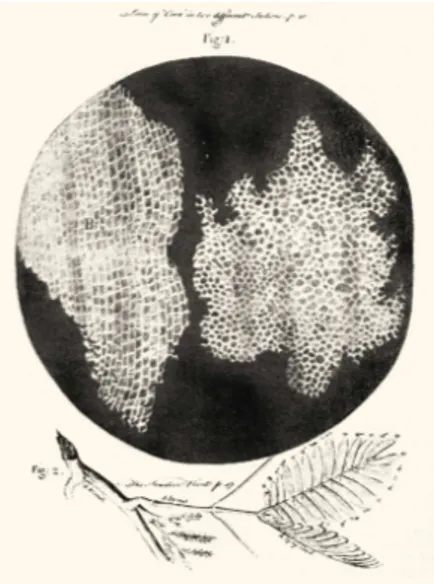
1.1 Hooke was the first person who described the cell. He applied the term cell, because plant cells, which are walled, reminded him of the cells in a honeycomb. R.H., Micrographia, 1665. . . . 2 1.2 (a) Linear excitation of fluorescein dye by focused 488nm light. (b)
Nonlinear excitation by focused fs pulses of 960nm light. [1] . . . 4 1.3 An artist’s impression of a nanorobot in the vicinity of a neuron [2] 6 1.4 (Upper panel) Schematic of the nanofabrication process when while
the laser beam is scanned over the surface. (Lower panel) SEM image of mesh nanostructures -left- and nanocircles -right-. [7] . . 8
2.1 SEM images of cells cultured on nanostructured substrates. [30] . 15 2.2 A representative picture of the implant/tissue interface. The
sur-face of titanium is in contact with the living bone [39] . . . 17 2.3 Examples of commercially-modified metal implant surfaces. (a)
Acid etched, (b) SLA, (c) Sand-blasted (d) Hydroxy-apatite-coated (M.E. archive). . . 24 2.4 Titanium surface modified with the laser parameters of 3W output
LIST OF FIGURES xiii
2.5 Depiction of the ultrashort pulsed all-fiber-integrated Yb amplifier and the biomaterial modification setup, BS: beam splitter, AOM: acousto-optic modulator, LMA: large mode area, DC: double-clad. 26 2.6 Various examples of surface modifications created by femto- and
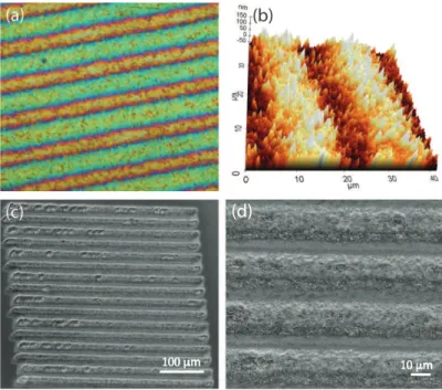
picosecond fiber lasers. Optical (a) and atomic force (b) micro-scope images of the nanoscale surface topographies. (c, d) SEM images of the microscale surface topographies. . . 28 2.7 Surface topographies created using femtosecond pulses. Optical
microscope (a) and AFM (b) images of the nanoscale surface to-pographies generated at low fluence (0.04 J/cm2
). (c, d) Scanning Electron Microscope images of the micrometer scale surface to-pographies generated at high fluences (0.89 J/cm2
). . . 29 2.8 SEM images of microscale surface topographies generated with
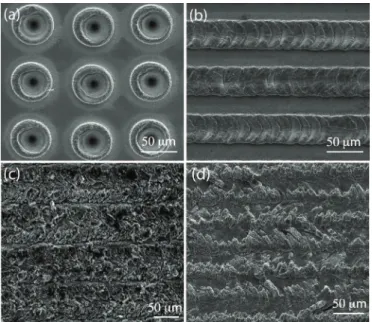
pi-cosecond pulses. Dotted (a) and line-scan (b, c, d) structures. . . 30 2.9 SEM images of micrometer-sized surface topographies created with
nanosecond pulses. Dotted (a) and line-scan (b) structures. . . 32 2.10 EDX analysis of the titanium samples: The irradiated fields are
(a) femtosecond, (b) picosecond, (c) nanosecond, and (d) non-irradiated region. The data lines are displaced vertically for clarity. 33 2.11 Raman spectra of titanium surfaces: The laser-exposed fields by
(a) femtosecond, (b) picosecond, (c) nanosecond pulses from the fiber lasers, and (d) non-irradiated field. . . 33 2.12 Cell counts for analyzing adhesion and proliferation after 36 hours
(left) and 7 days (right). p values show the significance of ex-perimental values obtained from commercial surfaces and picosec-ond laser-modified surfaces according to two tailed t-test. AE: surfaces prepared using acid-etching; SB: surfaces prepared using sandblasting; SLA: surfaces prepared using the SLA technique; Pico: surfaces prepared using the picosecond pulses. . . 34
LIST OF FIGURES xiv
2.13 (a) SEM image of SaOS-2 cells adhered to a fiber laser-modified titanium sample. The two red arrows indicate the cells aligned with linear structures. (b) Fluorescence microscope image of the same sample. The laser-modified field between the dashed (yellow) lines demonstrates a tendency of the cell population to align along the direction of the arrowheads. (c) SEM image of the cells attached on surfaces with nano-scale topography, which shows no discernible cell alignment. . . 35 2.14 (a) SEM image of laser-modified titanium surface with the dotted
pattern. (b) Fluorescent image of SaOS-2 cells stained with DAPI and cultured on the dotted pattern. SEM image of same cells on the same pattern; the red arrow indicates a cell at the edge of a hole. 36
3.1 Optical interaction modes of a tissue layer with the incident irra-diation light. [66] . . . 39 3.2 Absorption spectra of the major chromophores in tissue and the
so-called “near-infrared window in biological tissue. [70] . . . 41 3.3 Photoionization, inverse Bremsstrahlung absorption, and impact
ionization during the process of plasma formation. Repeated se-quences of inverse Bremsstrahlung events and impact ionization trigger an avalanche growth in the amount of free electrons. [74] . 45
LIST OF FIGURES xv
3.4 Laser confocal microscopic images of a fixated 3T3 fibroblast stained for F-actin with a green fluorescent dye. (A) Top view of a mid-plane horizontal section through the cell showing chan-nels and cavities produced by femtosecond laser ablation. The top and bottom channel were obtained at a pulse energy of 3 nJ; those in between at pulse energies ranging from 1.5 to 2.3 nJ. (B) Re-constructed orthogonal image of the same cell. (C) A cell that was prestained for F-actin with a green fluorescent dye immediately after ablation with 2-nJ (widechannels) and 1.5-nJ (narrow chan-nels) laser pulses. (D) The same irradiated cell after restaining for F-actin with a red fluorescent dye. [91] . . . 49 3.5 Topographical image and depth profile of two laser cuts on
chro-mosome 1. Material was partially removed, as indicated in the depth profile. A sub-100-nm FWHM cut size was determined. [93] 50 3.6 Femtosecond laser-induced fusion of two-cell porcine embryo. (a)
Irradiation of the cell-cell junction (indicated by black cross) trig-gered cell fusion. (b) Cytoplasmic streaming between both cells occurred about half an hour after laser treatment (indicated by dashed ellipse). (c) and (e) Cell fusion proceeds. Scale bar: 20 µm. [97] . . . 51 3.7 Responses of neurons to the pulsed-laser stimulation. DIC image
and fluorescence images of a Fluo-4-labeled neural circuit before and after stimulation. The lightning symbol indicates that N1 was irradiated with the femtosecond laser. [101] . . . 52 3.8 (a) Schematic of the experimental setup. FPGA: field
pro-grammable gate array; AOM: acousto-optic modulator. (b) Schematic of the laser-fluorescence microscope optics. (c) Schematic of the FPGA and analog electronic circuitry. . . 57
LIST OF FIGURES xvi
3.9 ((a) Optical spectrum of the oscillator and amplifier outputs. (b) Autocorrelation of the amplified pulses after dechirping. Inset: Close-in RF spectrum around the repetition frequency. (c) Mea-sured pulse train, exhibiting a complex pulse sequence as an ex-ample. Apparent variations in the pulse heights due to digital sampling are not real. . . 58 3.10 Mouse gastrocnemius muscle tissue slice (a) before and (b) after
laser surgery (5 parallel cuts are clearly visible); 4.08 MHz, 240-fs, 7-nJ. . . 60 3.11 Fixated SaOS-2 cells (a) and (c) before; (b) and (d) after
femtosec-ond nanosurgery; 4.08 MHz, 240-fs, 7 nJ. . . 60 3.12 (a) and (c) before; (b) and (d) after femtosecond ablation of
indi-vidual mitochondria stained with Mitotracker Red 580; 4.08 MHz, 240-fs, 2 nJ. Red arrows indicate the cell bodies. . . 61 3.13 (a) Before and (b) at the moment of laser axotomy (white arrow
indicates the incident laser beam on the axon). (c) After axotomy (white dashed arrow indicates the micro-damage); 32.7 MHz, 240-fs, 8 nJ. . . 62
4.1 The conventional form of in vivo optogenetics mounting fiber-optic cables in the rodent head. [107] . . . 69
Chapter 1
Introduction
The term biophotonics refers to the combination of biology and photonics, with photonics being the science and technology of generation and detection of pho-tons in order to probe and manipulate biological phenomena. Biophotonics can also be explained as the “development and application of optical methodologies, especially imaging, to study biological processes in cells and tissues”. One of the key advantages of using optical techniques for biology is that they are much less invasive compared to mechanical techniques, so that they highly preserve the biological integrity during examination. Thus, biophotonics has become the common term for all methodologies that contend with the interaction between biological substances and light.
1.1
History of Biophotonics
One of the earliest application fields of biophotonics is optical microscopy. More-over, given that a greater number of organisms are not visible to the naked human eye, microscopy is probably the most important among all the techniques used in biology.
Today, microscope is undoubtedly the most fundamental element of biopho-tonics research. Like many other inventions there is a debate in origins of the inventors of the microscope. The same dispute applies to who invented the micro-scope. During the late 16th century, Dutch spectacle makers were experimenting with lenses and by putting numerous lenses in a tube, they made an important discovery: The size of the view of the object near the end of the tube appeared to be increased. Although this invention of the compound lens system paves the way for the observation of small organisms and cells per se, it was Anton van Leeuwenhoek (1632-1723), again a Dutch scientist, who in the late 17th century became the first man to make and use a real microscope. Van Leeuwenhoek was the first to discover and define microorganisms, which he originally reported as animalcules. Van Leewenhoek’s work was verified and further developed by En-glish scientist Robert Hooke, who published the first work of microscopic studies by collating them in the historical book Micrographia, in 1665. Robert Hooke’s detailed studies opened up the scientific field of microbiology, and advanced bio-logical science as a whole. He is also notable for coining the biobio-logical term cell ; Figure 1.1.
Figure 1.1: Hooke was the first person who described the cell. He applied the term cell, because plant cells, which are walled, reminded him of the cells in a honeycomb. R.H., Micrographia, 1665.
In the late 19th century, Koehler illumination, which is a method of specimen illumination that permits to generate an even illumination of the sample and that eliminates the formation of the image of the illumination source in the resulting image, was invented. Further inventions such as Phase Contrast by Frits Zernike and Nomarski Interference Contrast (DIC) by Georges Nomarski in mid-1900s, allow imaging of unstained, transparent samples. With these improvements in illumination and contrast-enhancing optical techniques, the microscope as we know it today has come into being.
In parallel to these later advancements in light microscopy, ideas on LASER concept began to emerge in early 20th century. Albert Einstein is one of the pioneers who studied the fundamentals of the nature of the light-matter interac-tion and established the theoretical foundainterac-tions for the laser via a re-derivainterac-tion of Max Planck’s law of radiation, basically constructed on probability coefficients for absorption, spontaneous emission, and stimulated emission of electromagnetic radiation. Later in 1950s, Alfred Kastler proposed a technique for optical pump-ing, which was experimentally approved two years later, by Kastler, Brossel and Winter. Shortly after the first experimental demonstration of an optical or light laser in 1960s, biophotonics methodologies have been employed from ophthalmol-ogy to cosmetic surgery.
Ancient Greeks and Egyptians used sunlight for treatment, and even there is a mythological connection of such an idea that Apollo, the Greek god that is responsible for both healing and light. Although the idea of using light for curing illness has been recognized for thousands of years, it has only been since the invention of the laser in 20th century, which has revealed the potential of light for medicinal and biological purposes. The exceptional features of lasers make them much suitable than sunlight or other light sources for biological appli-cations. Lasers operate within a specific narrow wavelength range and the light emitted is coherent, together with their capacity of reaching high optical powers. Their beams can be focused to very small points, which also enables high optical power densities. Such features have led lasers being utilized in a wide range of biomedical and biological research, e.g. Laser Scanning Confocal Microscopy and Laser Surgery are two of the most recognized examples of lasers contribution into
such research fields.
Discovery of the lasers has revolutionized biology and medicine. Especially with the demonstration of femtosecond lasers in the 1980s, investigators from a wide variety of research fields have acquired a versatile tool for probing and manipulating diverse range of natural phenomena. What makes ultrafast lasers particularly useful for biological and medical research is that they mostly operate in near infrared wavelength, which has a high penetration depth in biological tissue. Additionally, their ultrashort pulse duration makes it possible to reach high peak powers at very low pulse energies and average optical powers. For instance, an ideal laser system can generate 1 Joule of energy with 20 femtosecond pulses and the peak fluence value at the focal point can exceed 1020
W cm−3, this
is orders of magnitude higher when compared to the total solar flux at the Earth, which is approximately 1017
W.
An important advantage of ultrashort pulse lasers is that they enable time-resolved experiments with which ultrafast phenomena can be probed. Another biophotonic application in which ultrashort pulse lasers are utilized is multiphoton imaging of biological materials. In multiphoton microscopy, a fluorescent dye is excited through nonlinear absorption in a volume that is strictly limited to the focus of the objective lens, which permits for optical 3D sectioning as shown in Figure 1.2.
Figure 1.2: (a) Linear excitation of fluorescein dye by focused 488nm light. (b) Nonlinear excitation by focused fs pulses of 960nm light. [1]
An alternative and unique application of ultrashort pulse lasers takes ad-vantage of the potential of these lasers on reaching enormous peak powers with moderate energies and optical powers. Through nonlinear interactions, ultrashort pulses are able to produce highly localized effects in materials. Hence, this makes ultrashort pulses especially suitable for ablation of biological materials, fragmen-tation (e.g. DNA into fragments that may be analyzed via Mass Spectroscopy), etc. In addition to these, another recent application field is ultrashort pulse laser micromachining of materials. Such ultrashort pulses are expected to result in cleaner cuts, because ultrashort pulse durations do not let the temperature to increase up at the irradiated area.
In parallel to all these advancements in optics and photonics, fiber laser sys-tems have increasingly been utilized in a wide range of scientific and biomedical applications. Compatibility of a system for being used outside the research lab-oratory is a fundamental requirement for industrial and biomedical applications. Fiber lasers are clearly advantageous in this respect. In addition to their long-term stability in operation, the optical fibers provide isolated paths for light propagation, which minimizes the effects from the environment that may cause optomechanical misalignments throughout the system.
1.2
Future of Biophotonics: Nanobiophotonics
Nanobiophotonics is the next step of photonics in biological and biomedical ap-plications. This step will literally be a small (nano) one, but will be a giant leap for mankind. The fantastic, though inevitable, combination of nanotechnology and biophotonics offers a great potential especially for the delivery of light in an extremely specific and controlled manner into living systems, together with the capability collection of data at the same precision.
A fictional, yet probable, world in which light-emitting and image-capturing nanorobots capable of navigating through tissues of organisms would be the cul-minant therapeutic technology for most of the diseases that the humankind has
encountered so far. In a state-of-the-art case, these nanorobots would circulate throughout the body with specific purposes, e.g. for detecting and counteract-ing cancer cells or for ultrahigh-resolution mappcounteract-ing of the neuronal networks in the brain as illustrated in Figure 1.3. Although such practices, at least for now, appear to be the subjects of far-future technologies, the integration of nanotech-nology and nanophotonics into biological applications has already begun, so that novel terms such as nanobiotechnology and nanobiophotonics has emerged.
Figure 1.3: An artist’s impression of a nanorobot in the vicinity of a neuron [2]
The ideal integration of photonics into nanobiotechnology and thereby the technological achievements imagined are fundamentally dependent on the ad-vancements in nanophotonics. Nanophotonics is a field that investigates the be-haviour of light on the nanometer scale, conceptually on a scale much smaller compared to the wavelength of light used, and of the interaction of nanometer-scale objects with light [3]. In order for this integration to occur as envisioned, nanoscale confinement in different methodologies of photonics must be achieved, and these can basically be categorized as nanoscale confinement of radiation,
nanoscale confinement of matter and nanoscale photoprocesses [4].
Today, nanoscale confinement of radiation is conceptually the subject of near-field optics, a branch of optics that deals with exceeding the optical resolution limit (mainly diffraction limit) by engineering the properties of evanescent waves. Contemporarily, near-field optics and microscopy has emerged as a powerful bio-logical research methodology to observe submicron-sized biobio-logical structures and to probe functions of nanoscale phenomena.
The optical resolution limit of conventional microscopes, that is diffraction limit, is approx. half the wavelength of the illumination light. Thereby, while imaging at visible spectrum, the minimum resolvable structures are in the order of hundred nanometers. In contrast, by exploiting near-field optical methods, it has been achieved to optically resolve structures as small as a couple of tens of nanometers in size. At this point, it is important to note that the most widely employed near-field apparatuses as near-field probes are tapered optical fibers. The light, either delivered or collected, propagating through the fiber leads to a resolution determined by the dimensions of the tip of the fiber and its distance from the specimen. The size of the fiber tip is usually around a few tens of nanometers and the resolution of a near-field system is mainly dependent on the probe size, thus, such factors necessitates further developments in nanoscale confinement of matter in terms of producing nanometer-sized probes and light sources.
Nanoscale confinement of matter considering nanophotonic applications ex-ploits the nanoscale manipulation of molecular architecture and anatomy, which allows to precise control over the optical and electronic properties of a mate-rial. For instance, if light can be transmitted in a confined manner, it can also be detected with a confined detector. In parallel to these, researches that deal with reducing the sizes of light sources such as lasers and LEDs into nanometer scale will result is an the development of ultra-compact light emitters exhibiting both optical and electrical functionalities. Such technological innovations require a priori knowledge and a better understanding of fundamentals of nanophoton-ics; thus, contemporary sciences of optics and photonics are now at this phase. Two interesting examples are generation of light with nanolasers [5] and non-linear plasmonics [6]. The initial applications of the technological achievements in nanophotonics would likely be into the data transmission and communication systems, but as happened previously in the invention of the lasers and their em-ployement in biological applications, it will not take much time to convert the methodologies of nanophotonics into tools of nanobiophotonics.
Nanoscale photoprocesses such as optical lithography offer facilities for nanofabrication. For instance, near-field photolithography can be employed for
creating nanometer scale chip-arrays for nucleic acid detection. Such nanometer-scale photoprocesses enables the production of ultra-high density chip-arrays, thereby allowing the use of lower quantities of specimens. This holds an excel-lent potential for analysis of biochemicals with minuscule amounts, e.g. even at the nucleic acid levels for which there is no possibility of PCR amplification that enables detection. Such a methodology will allow to earlier diagnosis of diseases when compared to current molecular and biochemical diagnosis technologies, even at the moment of infection.
Figure 1.4: (Upper panel) Schematic of the nanofabrication process when while the laser beam is scanned over the surface. (Lower panel) SEM image of mesh nanostructures -left- and nanocircles -right-. [7]
Thus, micro- and nanofabrication on surfaces is increasingly becoming impor-tant and widespread in nanotechnology researches. There are already established methods such as e-beam lithography, photolithography and interference lithogra-phy. Recent developments such as the demonstration of a technique that utilizes nonlocal feedback mechanisms for ultrashort pulse laser-induced nanostructuring
with extreme uniformity and speed, even on non-planar surfaces, is especially ex-citing and promising [7]. The capability of the methodology allowing structuring of indefinitely large areas even at sub-nanometer precision, as shown in Figure 1.4, is particularly promising for nanophotonic, plasmonic, photon detection, nano-electronic applications and especially for engineering superior quality biomaterial surfaces for the manipulation and control of cell behaviours in nanometer preci-sion.
1.3
Overview of the Thesis
Considering all these, as a part of this thesis study, we develop ultrashort pulse fiber lasers and demonstrate their use in biomaterial processing. We demonstrate that different surface topographies with micro- and nano-scale features can be generated at high speed, precision and repeatability. The outcomes of biomaterial surface modification with alternative laser parameters are compared in terms of topographical uniformity and repeatability. Additionally, the results of various different surface modifications are evaluated with respect to the efficiency of cell attachment and proliferation on the medical implants.
As the second part of this thesis, we develop a custom-built ultrafast fiber laser-based microscope system for nanosurgery and tissue ablation experiments. Furthermore, we applied this system for doing high-precision cuts, to various biological specimens ranging from the tissue level to subcellular level, such as a part of an axon or a single organelle. Finally, we improve this integrated system such that, through the use of AOM’s and custom-developed FPGA’s, it can generate arbitrary pulse patterns with no limitations.
Chapter 2
Biomaterial Surface Modification
Biomaterial is a term that specifies any substance that has been engineered to take a form, and alone or as part of a composite system, to regulate the progress of any therapeutic or diagnostic procedure, by controlling the interactions be-tween the constituents of living systems. During these procedures, the main part of the biomaterial that directly contacts with the living system is the surface of the biomaterial. Hence, biomaterial surface modification have an important role in manipulating the interactions between the biomaterials and living sys-tems. Through appropriate modifications of their surfaces, biomaterials can be altered to improve biocompatibility, attachment and tissue interactions. Thus, surface modification is critical in the design and development of novel bioma-terials and medical apparatuses. Thereby, modification of biomaterial surfaces aims to enhance biological performance by optimizing the interactions within the living system.
2.1
Theoretical Background of
Biomaterial-Tissue Interaction
The subject of biomaterials embodies the combination of physical and biological sciences. As a matter of course, it is of great consequence that these aspects are combined to develop eligible biomaterials that function optimally in biolog-ical environment. The key to this issue remains in the interactions between the biological substance of interest and biomaterials . The recent scientific and tech-nological advances in fields such as molecular and cellular biology, biotechnology and tissue engineering as in other connected disciplines, gave rise to an importor-tant increase in the development and utilization of apparatuses with bio-logical/-medical purposes, such as catheters [8], heart valves [9], and scaffolds for tissue engineering [10, 11]. Nevertheless, mainly because of the fact that an implanted biomaterial naturally undergoes a functionality loss through time after implanta-tion, it is critical to establish an effective and mechanically stable integration of the biomaterial to the surrounding tissue. Inadequate in vivo functionality and permanency of this integration are essential issues, originating either from the normal homeostatic or the abnormal reactions against the implantation, or even to the lack of biocompatibility per se, between the biomaterial and the living system [12, 13, 14].
There are many factors having a role in the efficiency of the biomaterial used as an implant, and being influential on the fate of the implant in the living system. In addition to the physical and mechanical features of biomaterials, the core and determinative factor is the interaction between the biomaterial and the surrounding tissue. These interactions are predictable only to some extent due to their complex nature and comprise many factors, elements and elaborate interactions; yet, a brief but practical categorization has been made by D.F. Williams, considering the essential phases of the integration of a biomaterial to a living system [12]:
(1) “The initial events that take place at the interface, largely concerned with the physicochemical phenomena that take place in times measured in seconds or
minutes following contact between biomaterial and tissues;
(2) “the effect that the presence of a foreign body has on the tissue surrounding the implant, which may be seen at any time ranging from minutes to years;
(3) “the changes seen in the material as a result of its presence in the tissues, usually described under the headings of corrosion or degradation; and
(4) “the sequelae of the interfacial reaction that are seen systemically (that is, throughout the body) or at some specific but remote site.
These events cooperatively outline the biocompatibility phenomenon.
One of the fundamental concerns in biomaterials studies is biocompatibility. In brief, biocompatibility is general a term for the nature and the status of interac-tion between biomaterials and living systems [15, 16]. Given that the response to different materials could also differ from one application to another, biocompati-bility could not only be dependent on the properties of material itself, but also has to be related to the situation in which the biomaterial is employed. Moreover, an increasing number of studies demonstrated that the biomaterial should respond to the tissues in concordance with the planned application, rather than be ig-nored by them [17]. In relation to this, it is necessary for some applications that the biomaterial should decompose or degrade over time in the body, rather than staying indefinitely. Hence, a more accurate and appropriate definition would be:
“Biocompatibility is the ability of a material to perform with an appropriate host response in a specific situation [17].
It seems that there are numerous distinct ways in which biomaterials and living systems interact with each other so that it is usually challenging to un-ravel the underlying mechanisms. Collection of information and comprehension of these interactions is essential for designing of biomaterials that give the opti-mized performance and these are primarily evaluated in the broad context of the biocompatibility phenomenon.
as a medical implant used in biomedical applications, or by mistake as when a splinter penetrates into the tissue gives rise to the formation of interfaces between the material and the surrounding tissue. In terms of the kinetics and thermody-namics, surfaces have different characteristics compared to corresponding bulk of the material and contain reactive bonds, which in turn lead to the formation of surface reactive layers such as the surface oxide layers on metals [18, 19]. Meet-ing with the livMeet-ing system precedes surface reactions that alters the surface, and causes to the adsorption of water, ions, and biomolecules, which are in a dynamic equilibrium within the living system. Interruption of the exact nature of these dynamics influences the behaviour of cells connecting the material surface, and hence the tissue response.
The surface of a material is a termination of a three-dimensional structure, and therefore, as a rule, corresponds to an increase in energy; at the atomic level, this energy is represented as unsaturated bonds and if there is a reactive environment as in air or water meets a metal surface, these terminals readily react to create new bonds and compounds, thus lowers its surface energy [19]. In parallel to these, living systems consist of a mixture of biomolecules such as water, oxygen, cations, anions, proteins etc. The biological and non-biological substances contact and interact at the interface, which might react to lower the surface energy of the system. There are two key factors that disable such undesired reactions; one of which is that when the material and the living system are disconnected, both substances have their lowest thermodynamic state already, and the other factor is that the inherent kinetic barriers prevent all possible reactions [19, 20].
The chemical components of the biological milieu effect the interactions at the interface [19, 20]. For instance, in case the biomaterial is metal-based, corrosion may cause the release of metal ions from the metal-oxide surface into the milieu within the biological system, which has a potential for triggering adverse systemic effects [21, 22]. In addition to this, a biomolecule at the biological side of the interface and the biomaterial surface at the other side may form momentary van der Waals bonds or even covalent bonds, which are stronger and stable. In case that such interactions are so strong, macromolecules may irreversibly become denatured [23].
In the medium and long term, the larger and more complex elements like cells communicate the interface with their membrane. In view of the fact that the cell membrane as a composite bioorganic structure and the biomaterial surface are both active, they may develop a complex and dynamic interface. Depending on the properties of the surface of the material, cells respond differently to different biomaterials.
Eventually, the ultimate aim of engineering the tissue-biomaterial interface is to control cellular responses. The basic approach to this this phenomenon is to consider the factors those are used by cells when they are interaction with the surrounding substrate, and the parameters those are utilized by surface scientists when they are trying to control the interactions between different materials. Such factors or cues to which a cell will respond can principally be categorized into three categories: Chemical, topographical, and mechanical. These three different types of factors can have related effects [23]. It is recognized that the structure of a biomaterial surface rules the phenotypic response of interacting cells [24]. Accordingly, surface features such as chemical composition, hydrophobicity and roughness are all known to have influence on the response of cells that interact with a biomaterial. Moreover, there is an intertwined relationship between these diverse properties of a surface and altering one feature practically alters others as well. For instance, altering hydrophobicity of a polymer biomaterial is accom-panied with a change in the chemical composition of side groups at the surface of the biomaterial, hence its surface energy [25, 26].
On the other hand, recent maturation of biological studies in parallel to the outcomes of the Human Genome Project, along with accompanying technological progresses in molecular and cellular biology, places the strategies and method-ologies of molecular and cellular biology into a distinctive position. By being equipped with the capability of examining the gene/protein expression changes within biological systems of interest via transcriptomic and proteomic methodolo-gies, molecular biology has led to further understanding of the molecular basis of a broad range of physiological activity at the biomaterial-tissue interface [27, 28]. In addition to the physicochemical properties of the surfaces of biomaterials,
cells also substantially interact with the topographical features on these surfaces, as shown in Figure 2.1, principally through a phenomenon known as contact guidance [29].
Figure 2.1: SEM images of cells cultured on nanostructured substrates. [30]
The topography of material surfaces regulates a range of cellular processes such as cell adhesion, cell shape and cellular differentiation [31]. For instance, epithelial cells attach and align along grooves and ridges with feature dimensions smaller than 100 nm [30]. Nanoscale topographic textures induce mineralization of human mesenchymal stem cells [32]. Another example to such topographical guidance behaviour is that human SaOS-2 osteosarcoma cells cultured on metal surfaces with parallel lines of different widths and distances often orientate along these parallel lines [33]. Besides, tissue-biomaterial interactions are also directly related to the fact that different cell types respond differently to alterations in the same surface property. For example, increasing surface hydrophobicity in-duces adhesion of endothelial and epithelial cells to the surface [26], but rein-duces adhesion of osteoblasts [34].
Consequently, a complete understanding of tissue-biomaterial interactions is the key parameter that will pave the way for designing and developing functional biomaterials and medical implants. And, because of the fact that the biomaterial surface is the major portion that directly interacts with the surrounding tissue, it is critical to have methodologies that allow the modification of surface properties
such as topography. Such methodologies would lead to development and costless production of higher quality medical implants with optimum performance.
2.2
Surface Modification of Medical Implants
Hard tissues are mineralized biological tissues that contain minerals in soft matri-ces. Usually these tissues provide a structural support or protective shield [35]. In addition to the mollusc shells, radiolarians, diatoms etc.; there are several mineralized tissue types, and so called hard tissues, such as bones, tooth enamel, dentin, tendons and cartilage found in the human body [35, 36].
On the other hand, bone is one of the few tissues in the human body that is capable of undergoing spontaneous regeneration and having a high capacity of remodeling its micro- and macro- structure. This is accomplished through a precise balance between the osteogenic (bone forming) and osteoclastic (bone removing) activities [37], which enables bone tissue to adapt to different mechan-ical environments by allowing it to adjust the dynamic balance between these two biological processes.
As hard tissue replacements have become an conceivable treatment for pa-tients, it also has become even more evident that the interaction between host tissue and the implanted biomaterial surface is of critical importance. The whole biological response of host tissue to medical implants can be separated as two different, but interconnected periods. Initial period comprises the biological pro-cesses of a clinical healing, which immediately follows the implantation of the bio-material. During this recovery period, the early biological activities and molecular deposition on the biomaterial surface are followed by cell adhesion, migration, and differentiation. It is thus crucial to realise the characteristics of the biomaterial, which have an influence on the initial formation of the host tissue-implant inter-face. These early tissue activities result into cellular expression and extracellular matrix formation and eventually into the development of bony interfaces with the implant [38]. With Branemarks and his colleagues huge amount of contribution
to this research field, the characteristics of the implant substrate which permit biomaterial-tissue integration has been realized. When the early healing period is completed, within months, the maturing interface transforms as biomechanical stresses are placed on the implant, and this also is closely interrelated to the initial degree of tissue-implant surface interaction [39].
At this point, osseointegration phenomenon becomes critical and determina-tive. The concept has been portrayed by Branemark as the direct contact between bone and implant, principally on the light microscopic level [40], as illustrated in Figure 2.2.
Figure 2.2: A representative picture of the implant/tissue interface. The surface of titanium is in contact with the living bone [39]
Currently, an implant is regarded as osseointegrated if there is no progressive relative movement between the implant and the bone with which it is directly con-tacted. During the osseointegration process, mesenchymal cells and osteoblasts migrate and attach to the surface of the biomaterial, beginning from the early stages of implantation. With the accumulation of bone-associated proteins and
establishment of a extracellular layer on the implant surface that controls cell attachment and mineralization, integration of the bone tissue and the biomate-rial begins [41]. Subsequent to this, primary bone formation occurs on implants to reestablish continuity. Primary bone structure fills the initial space at the implant-bone boundary. The physical architecture of a three-dimensional regular network provides a biological scaffold for cell attachment and bone sedimentation, [42]. Eventually, bone in contact with the implant surface goes through morpho-logical remodeling as adaptation to biomechanical stress and loading. During the remodeling of this peri-implant bone, new osteons circle around the implant perpendicular to the long axis of the implants. Osteoid tissue is generated by osteoblasts, which indicates that osteogenesis is in progress. A remodeled bone at its later stages can expand up to 1 mm from the implant surface [42].
Different materials and implant surface treatment together with surface coat-ings have been proposed to enhance the quality of osseointegration. The biocom-patibility of the biomaterial is of great importance and a necessity for osseointe-gration. In this regard, titanium metal is widely used as a hard tissue replacement implant material. Titanium has significant advantages with its great biocompat-ibility, resistance to corrosion and lack of toxicity on living systems with its little, if any, inflammatory response in peri-implant tissues [43]. Titanium’s low density [4.5 g/cm3] and good mechanochemical characteristics are main parameters for implant application. There are various types of commercially available titanium and titanium alloys for surgical implant applications. Ti6Al4V titanium alloy is extensively utilized to construct implants. The core alloying elements of the Ti6Al4V are aluminum (5.5 6.5%) and vanadium (3.5 4.5%). The addition of al-loying elements to titanium allows it to gain a numerous features e.g. Aluminum and Vanadium serves for the resistance to the external forces and strengthens against mechanical loads [43].
Metal surface modification is frequently utilized for enhancing osseointegra-tion by means of the manipulaosseointegra-tion of the dynamics at the tissue- biomaterial interface. Cells at the interface and their secreted biochemicals involved in the course of osseointegration change the organization and physiochemical features of the biomaterial surface. Interconnectedness in the case of macro-featured surfaces
and surface roughness in the case of micro-featured surfaces are generally consid-ered as suitable surface structures for osseointegration [44]. Besides, surfaces of biomaterials are rarely smooth at the molecular level mostly due to the processes the fabrication of the biomaterial and these surface features, such as roughness may or may not be formed intentionally. On the other hand, micron-sized topog-raphy has been demonstrated to have a central role in regulating cell adhesion and tissue-biomaterial integration [45]. Biomaterial modifications are recently of particular interest, with the aim of controlling cell adhesion and spreading. In this regard, modification of biomaterial surfaces, particularly by considering the topographical surface properties, is at utmost importance [46]. There are vari-ous kinds of metal implant surface modification targeting topographical features such as titanium plasma spraying, hydroxyapatite coating, machine processing, polishing, sandblasting and acid etching. These techniques are among the most commonly used commercial surface modification methods [47].
On the other hand, these techniques commonly used to create surface textures in commercial applications are all incapable of creating selective and tailored-topography, which is recently of particular interest for the control of osseoin-tegration in a precisely regulated manner. Recently, laser surface texturing has emerged as a novel technology for creating a large variety of micro- and even nano-structured biomaterials including medical implants. Laser texturing of material surfaces has exceptional advantages over other surface structuring techniques, e.g. photolithography, such as:
(1) Capability of modification of virtually all types of materials, including metals and polymers;
(2) Competence for texturing of non-planar surfaces;
(3) Capacity to create or nano-structures on surface areas from micro-scale to macro-micro-scale;
(4) Maskless single-step processing even at high speeds under normal environ-mental conditions, without the requirement of a clean room facility.
Although surface patterning can be performed using continuous lasers, nanosecond or longer-pulsed lasers, they lead to unwanted thermal effects, which is restrictive for the degree of precision. Thermophysical response of the mate-rial is dependent on the pulse length of the laser and shows substantial differ-ences below and above the picosecond regime. Ultrashort pulses in the pico- and femtosecond regimes have significantly reduced thermal effects, enabling high-precision and confined processing with little or no effect to the incident region on the textured material surface [48].
In this regard, high precision can be achieved by employing ultrashort-pulsed solid-state lasers [49]. Nevertheless, they are complicated and costly devices; hence, application of them on surface modification of biomaterials has been re-stricted mostly to laboratory experiments. Ultrafast fiber lasers offer excellent potential for biomaterial surface modification at micro- and nanoscale with an exceptional spatial control. Operating at high speed and together with their de-sirable features such as robust operation, low cost, compact size, low intensity noise and diffraction-limited beam quality; modification of biomaterial surfaces using ultrafast fiber lasers is a promising methodology.
2.3
Experimental Methods
2.3.1
Sample Characterization
2.3.1.1 Scanning Electron Microscopy (SEM)
Micro- and nanostructural observations of the titanium surfaces are carried out using the scanning electron microscope in UNAM, Bilkent University; E-SEM, Quanta 200F, FEI on ultra-high resolution mode with ETD detector under ul-tralow vacuum conditions are used with approx. 10 kV. Imaging of cell attach-ment on titanium surfaces are performed with the same microscope by using GSED detector under low vacuum conditions without application of any conduc-tive coatings in order to directly observe the morphologies, operated at approx.
5 kV.
2.3.1.2 Atomic Force Microscopy (AFM)
The surface topography of the titanium implants are analyzed using the atomic force microscope in UNAM, Bilkent University; AFM, XE-100E, PSIA in non-contact mode.
2.3.1.3 Energy-dispersive X-ray Spectroscopy (EDX)
The chemical structure of the modified areas are identified through EDX analyses. EDX analyses are performed using Bruker AXS detector with ultra-thin window attached to the Carl Zeiss Evo40 SEM in Dept. of Chemistry, Bilkent University.
2.3.1.4 Raman Spectroscopy
Further chemical structure analysis of the modified areas are carried out through the Raman spectroscopy in Dept. of Chemistry, Bilkent University. Horiba Jobin Yvon micro-Raman equipment is used for Raman analyses, where the source of radiation was a laser operating at a wavelength of 632.8 nm and a power of 25 mW.
2.3.1.5 Bright-field Optical Microscopy
Optical imaging of nanostructural textures is performed with the light microscope in UNAM, Bilkent University; Axio Scope.A1, Carl Zeiss at room temperature conditions.
2.3.2
Cell Culture
2.3.2.1 Cell lines and Growth conditions
Human osteosarcoma cell line SaOS-2 is used for cell attachment and prolifera-tion. The cells are cultured in McCoys 5A medium supplemented with 15% fetal bovine serum, 2 mM L-glutamine, streptomycin/penicillin 100 U/mL, and in a humidified atmosphere of 95% air and 5% CO2 at 37◦C.
2.3.2.2 Attachment and Proliferation Assays
Cells are seeded onto 8-mm diameter Ti6Al4V disks with three different commercially-modified surfaces and different fiber laser-modified surfaces. Before use, all samples are sterilized for 30 minutes in 10% NaClO with an ultrasonic cleaner and placed in 24-well cell culture plates with a density of 100,000 cells/ml. Separate attachment tests are done for incubation periods of 36 hours and 7 days.
2.3.2.3 Fixation and Fluorescent Imaging of Cells Attached on Im-plants
At the end of the incubation periods, disks are washed with PBS, treated with trypsin/EDTA solution for 20 seconds to eliminate the poorly attached cells, and then fixed in 0.1 M sodium cacodylate buffer with 3% glutaraldehyde, pH 7.2 at 4◦C overnight. For each surface type, 3 Ti disks are used (samples are triplicated),
hence 12 Ti disks are used for each incubation period in total. Following up the fixation stage, cells are stained with 300 nM 4, 6-diamidino-2-phenylindole, dilactate (DAPI, dilactate) in PBS and with Mitotracker Red 580 (Invitrogen) in McCoys 5a and subsequently counted under a fluorescent microscope (Nikon Eclipse Ti/U).
2.3.2.4 Cryopreservation of Stock Cells
Exponentially growing cells in 100 mm cell culture plate were harvested by trypsinization and collected in 5 ml growth medium. Then, cells were precip-itated at 1500 rpm for 3 min. The pellet was suspended in a freezing medium (8% DMSO, 92% FBS). Pellets were resuspended in 1 ml freezing medium in cryotubes and they were left at -20◦C overnight. The next day cells were stored
at -80◦C for 1 day to 1 month. Finally, the tubes were transferred into the liquid
nitrogen storage tank for future experiments.
2.3.2.5 Thawing of Frozen Cells
The frozen cell line was taken from the liquid nitrogen tank and immediately put on ice and then placed into a 37◦C water bath for 1-2 minutes. The cells were
transferred into 15 ml tubes and resuspended in 9 ml growth medium. The cells were centrifuged at 1500 rpm for 3 minutes. Supernatant was discarded and the pellet was resuspended in 10 ml culture medium to be plated into 100 mm dish. After overnight incubation culture mediums were replenished.
2.4
Results
2.4.1
Surface Modification with Pulsed Fiber Lasers
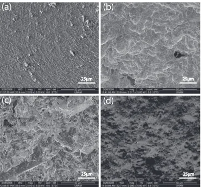
Biomaterial surface modification is currently of special interest, with the aim of directing cell attachment and spreading. In this context, modification of bio-material surfaces, especially in consideration of the surface topographies, is at utmost importance [46]. There are different, commercial, metal implant surface modification types, focusing on the generation of discernible topographical fea-tures e.g. titanium plasma spraying, hydroxyapatite coating, machine processing, polishing, sandblasting and acid etching (For some examples, Figure 2.3).
(a)
(b)
(c)
(d)
25µm 25µm
25µm 25µm
Figure 2.3: Examples of commercially-modified metal implant surfaces. (a) Acid etched, (b) SLA, (c) Sand-blasted (d) Hydroxy-apatite-coated (M.E. archive).
Although these techniques are among the most commonly-used commercial surface modification methods [47], they are all incapable of creating selective and tailored-topography, which is recently of particular interest for the control of osseointegration in a precisely controlled manner. On the other hand, laser-based surface modification has emerged as a novel methodology for generating a great diversity of micro- and even nano-structured biomaterial surfaces including medical implants.
Laser surface modification has been increasingly used as a novel methodol-ogy for creating precisely-controlled surface topopgrapies on biomaterials such as metal medical implants. Laser modification of biomaterial surfaces has significant advantages over other surface modification techniques such as being capable of modification of almost all types of materials, and of creating fine structures on surface areas from micro- to macro-scale (e.g. Figure 2.4).
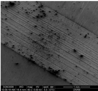
Figure 2.4: Titanium surface modified with the laser parameters of 3W output power, 80 ps pulse duration (M.E. archive).
For the purpose of surface modification, we develop and make use of short-and ultrashort pulse fiber lasers with MHz-repetition-rate, microjoule- short-and sub-microjoule-energy pulses [50]. Modification of Ti6Al4V alloy-based biomaterial surfaces is performed using home-built femtosecond and picosecond pulsed fiber lasers. Additionally, we make a comparison of the effects of these regimes with nanosecond regime by using a commercial nanosecond fiber laser.
We utilized home-built picosecond and femtosecond pulse fiber laser systems in order to assess the effects of MHz-level repetition rates and of relatively low pulse energies on biomaterial surface modification. We demonstrate that a range of surface topographies with micro- and nano-scale features can be created at high speed with high precision and repeatability. We compared the results of picosecond and femtosecond pulses with those of commercial nanosecond fiber laser and as a result, we showed that there are significant thermal effects with reduced precision in nanosecond regime. In the end, effects of various surface topographies on cell adhesion and proliferation is investigated.
high-power laser diodes
isolator
signal-pump combiner LMA-DC Yb-doped fiber collimator
with isolator
objective
neutral density filter grating compressor Yb-doped fiber AOM stretching fiber pump laser pump protection filter WDM isolator polarization controller collimator coupler polarization controller collimator grating compressor coupler Yb-doped fiber pump protection filter pump laser WDM BS 3D motorized stage OSCILLATOR AMPLIFIER POWER AMPLIFIER fs pulses ps pulses polarization port
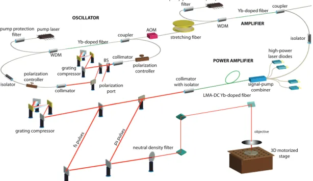
Figure 2.5: Depiction of the ultrashort pulsed all-fiber-integrated Yb amplifier and the biomaterial modification setup, BS: beam splitter, AOM: acousto-optic modulator, LMA: large mode area, DC: double-clad.
pico- and femtosecond fiber laser systems and a commercial nanosecond fiber laser (FL-NS-8W, FiberLAST) are utilized in this research. The nanosecond laser generates 70 ns-long pulses at 20-200 kHz repetition rate and maximum average optical power of 8 Watts.
One of the ultrashort pulse laser systems is seeded by an all-normal-dispersion (ANDi) mode-locked Yb oscillator [51] with a central wavelength of 1060 nm and the other one by a self-similar mode- locked Yb oscillator [52] with a central wavelength of 1035 nm. The oscillator repetition rates are 43 MHz and 28 MHz, respectively. Both oscillators produce few picosecond-long, chirped pulses, which are fiber-coupled to all-fiber-integrated and misalignment-free amplifiers. First, the pulses are temporally stretched in fiber stretchers to reduce nonlinear effects, then traverse preamplifiers, which boost the power to 100-150 mW, which is sufficient to seed the power amplifiers. Both systems incorporate 10 ns-risetime fiber-coupled acousto-optic modulators (AOM) to optionally reduce the repetition rate to 1 MHz prior to amplification. This first system generates an average power of up to 16 W, corresponding to an estimated peak power of 20 kW for 20 picosecond pulses at 43 MHz of repetition rate. Following pulse compression with the gratings, pulse duration is measured to be 200 femtosecond full-width at half-maximum (FWHM) [53].
The second system is utilized only at the lower repetition rate of 1 MHz in order to increase the pulse energy. The pulses are stretched to 150 picosecond in a 100-m long fiber stretcher and amplified to 104 nanojules in the preamplifier. The nonlinear chirped-pulse power amplifier generates pulses with up to 4 µJ energy at 1 MHz repetition rate. The pulse duration is reduced to 80 picosecond during amplification as a result of gain filtering clipping the edges of the pulse in the time domain. The maximum peak power of amplified pulses is 57 kW and the compressed pulse duration is 150-200 femtoseconds [54].
Both laser systems can operate in picosecond mode by simply bypassing the pulse compressor. When the compressed pulses are utilized, they are always linearly polarized due to the grating compressor. Uncompressed pulses can either be unpolarized or optionally linearly polarized with a polarizer.
For modification with the home-built ultrashort pulse fiber laser systems, the beam is focused to a focal diameter, changeable in the range of 10-15 µm using a high-power and 1 µm-wavelength-compatible microscope objective. The pro-cessing setup includes a collimator telescope, an objective and a 3-axis motorized and computer-controlled translation stage. Numerous surface textures can be generated by moving the translation stage on which the samples are located. For texturing with the commercial ns-pulsed fiber laser, the beam is scanned over the sample with a computer-controlled galvanoscanner, using a special objective designed to preserve a uniform spotsize irrespective of the deflection angle. The focal diameter in the focal plane is approximately ∼40 µm. All three laser sys-tems have nearly diffraction-limited beam quality (M2
< 1.2) and they can be switched off within microseconds for jumping from one point to another. Scan rates used in the experiments range between 3 µm/s and 50 µm/s.
(a)
(a)
(b)
(c)
(d)
75 µm 75 µm
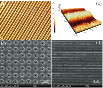
Figure 2.6: Various examples of surface modifications created by femto- and picosecond fiber lasers. Optical (a) and atomic force (b) microscope images of the nanoscale surface topographies. (c, d) SEM images of the microscale surface topographies.
By employing the systems mentioned above, we demonstrate that a great di-versity of surface patterns can be generated using both the femto- and picosecond pulses by adjusting the pulse energy, duration, exposure time and the scanning texture. Samples of selected topographies are shown in Figure 2.6.
For the generation of micron-sized surface topographies, picosecond and fem-tosecond pulses yield similar results, as predicted. By employing fs pulses at sufficiently low fluences (approx. 0.04 J/cm2
), it is possible to generate nanoscale surface roughening. This seems not to be possible with ps pulses, which sim-ply result in generation of micron-sized structures dependent on the laser spot size on the sample. We refer this difference to thermal effects resulting from the picosecond-long pulse, which clears away the small nanometer-sized details, even though the thermal effects are eminently decreased compared to the nanosecond regime.
(d)
100 μm 10 μm
Figure 2.7: Surface topographies created using femtosecond pulses. Optical mi-croscope (a) and AFM (b) images of the nanoscale surface topographies generated at low fluence (0.04 J/cm2
). (c, d) Scanning Electron Microscope images of the micrometer scale surface topographies generated at high fluences (0.89 J/cm2
By utilizing femtosecond pulses, we generate nanometer scale surface topogra-phies at low fluences and microscale surface topogratopogra-phies at high fluences. Line textures are generated on the titanium surface with incident power of 1.4 W, repetition rate of 43 MHz, pulse length of 300 femtosecond, scan rate of 4 µm/s, and focal diameter of 10 µm (Figure 2.7(a) and (b)). The corresponding fluence is 0.04 J/cm2
. These parameters result in nanoscale surface modification with a mean roughness of 100 nm.
On the other hand, a similar line texture, however of microscale height, is generated by employing incident power of 0.7 W, repetition rate of 1 MHz (corre-sponding to a pulse energy of 700 nJ), pulse length of 400 femtosecond, scan rate of 3 µm/s, and focal diameter of 10 µm (Figure 2.7(c) and (d)). The correspond-ing fluence here is 0.89 J/cm2
. When using fs pulses, there is no heat-affected zone (HAZ) that can be distinguished through scanning electron microscope (SEM) and atomic force microscope (AFM) images. Decreasing heat affected zones is critical since these HAZ are more susceptible to the generation of cracks that decreases the life-time of the biomaterial [55, 56].
Figure 2.8: SEM images of microscale surface topographies generated with pi-cosecond pulses. Dotted (a) and line-scan (b, c, d) structures.
By using picosecond pulses, microscale surface topographies with 10-20 µm feature sizes and height differences of approx. 5-10 µm are efficiently generated. Figure 2.8(a) shows SEM image of dot-pattern generated on the titanium surface employing incident optical power of 2 W, repetition rate of 43 MHz, pulse duration of 20 picosecond, and approx. 40 µm of focal diameter. The corresponding fluence here is 0.0037 J/cm2
.
A line texture is shown in Figure 2.8(b), generated using incident laser power of 1 W, repetition rate of 1 MHz, pulse length of 80 picosecond, scan rate of 3 µm/sec, and 10 µm of focal diameter. The corresponding fluence is 1.27 J/cm2
. Another texture is shown in Figure 2.8(c), generated using incident laser power of 2 W, 43 MHz repetition rate, pulse length of 25 picosecond, focal diameter of 10 µm, scan rate of 50 µm/sec and fluence of 0.0059 J/cm2
. The texture is gener-ated by line scans, with the parallel scan lines scarcely touching each other at the edges. The line texture in 2.8(d) is generated using the same parameters as in Figure 2.8(c), but the parallel lines are overlapped by more than few micrometers at the edges. In the picosecond regime, a little Heat Affected Zone is distinguish-able around the microscale structures, appearing as contrast changes in the SEM images and with an extent of 10-15 µm. The surface textures are mechanically stable and strong. The processed titanium surfaces were repetitively subjected to 10% NaClO in an ultrasonic cleaner and to solutions such as proteolytic enzymes during cell adhesion experiments. Moreover, after almost a year, during which a vast of experiments have been performed, the laser-modified surfaces seem to be unaltered with regard to more recent scanning electron microscope images.
In order to compare the differences between the pulse regimes, we also em-ploy the nanosecond fiber laser to generate surface modifications. A dot-pattern, which is shown in Figure 2.9(a) is created using incident laser power of 1 W, pulse length of 71 nanosecond, repetition of 25 kHz, scan rate of 5 mm/sec, and focal diameter of approx. 40 µm. The corresponding fluence is 3.2 J/cm2
. SEM image of a line texture generated with same laser parameters are demonstrated in Figure 2.9(b). The ns-pulsed laser significantly results in more noticeable Heat Affected Zone, extending up to 50 µm, as well as a relatively decreased repeata-bility and precision. It seems that for most surface modification applications, the
(a) (b)
100 μm 50 μm
Figure 2.9: SEM images of micrometer-sized surface topographies created with nanosecond pulses. Dotted (a) and line-scan (b) structures.
micrometer scale control afforded by the picosecond fiber laser to be enough. [57]. For a better understanding of the processes behind the formation of the struc-tures, we utilize energy-dispersive X-ray spectroscopy (EDX) analysis and Raman spectroscopy of the modified surfaces. It is possible to generate microstructures, which are pronounced as either protrusions or depressions on the surface. The height differences are around few micrometers and dependent on the laser pa-rameters. The EDX results show that oxygen is present in the modified regions, while it is absent in the non-irradiated fields as shown in Figure 2.10. The con-centration of oxygen in surface structures seems to vary in the range of 25% to 35% for any pulse duration. This is consistent with the generation of TiO2 as a
result of the laser modification.
Figure 2.11 depicts the Raman spectra covering 150-750 cm−1 of the modified
titanium surfaces. As control, the unexposed field of the titanium samples does not show Raman activity. The exposed fields result in three significant peaks located at 241, 439 and 613 cm−1, which can be referred to the multi-photon
process, Eg, and A1g active Raman modes for the tetragonal rutile structure of
TiO2, respectively [58, 59]. Thereby, we conclude that the protruding structures
generated by laser exposure are mostly composed of TiO2 in the rutile phase,
![Figure 1.3: An artist’s impression of a nanorobot in the vicinity of a neuron [2]](https://thumb-eu.123doks.com/thumbv2/9libnet/5615481.111063/23.892.362.600.393.574/figure-artist-s-impression-nanorobot-vicinity-neuron.webp)
![Figure 2.1: SEM images of cells cultured on nanostructured substrates. [30]](https://thumb-eu.123doks.com/thumbv2/9libnet/5615481.111063/32.892.287.675.289.510/figure-sem-images-cells-cultured-nanostructured-substrates.webp)
![Figure 2.2: A representative picture of the implant/tissue interface. The surface of titanium is in contact with the living bone [39]](https://thumb-eu.123doks.com/thumbv2/9libnet/5615481.111063/34.892.292.676.476.840/figure-representative-picture-implant-interface-surface-titanium-contact.webp)