DESIGN AND SYNTHESIS OF MONOSACCHARIDE
FUNCTIONALIZED CONJUGATED POLYMERS, POLYROTAXANES
AND OLIGOMERS FOR BIOLOGICAL APPLICATIONS
A THESIS SUBMITTED TO
THE GRADUATE SCHOOL OF ENGINEERING AND SCIENCE
OF BILKENT UNIVERSITY
IN PARTIAL FULFILLMENT OF THE REQUIREMENTS FOR
THE DEGREE OF
MASTER OF SCIENCE
IN
CHEMISTRY
By
Esra Deniz Soner
September, 2015
ii
DESIGN AND SYNTHESIS OF MONOSACCHARIDE FUNCTIONALIZED CONJUGATED POLYMERS, POLYROTAXANES AND OLIGOMERS FOR BIOLOGICAL APPLICATIONS
By
Esra Deniz Soner September 2015
We certify that we have read this thesis and that in our opinion it is fully adequate, in scope and in quality, as a thesis for the degree of Master of Science.
_______________________
Assoc. Prof. Dönüş Tuncel
_______________________ Prof. Dr. Engin Umut Akkaya
_______________________ Assist. Prof. İrem Erel-Göktepe
Approved for the Graduate School of Engineering and Science:
_______________________ Prof. Dr. Levent Onural Director of the Graduate School
iii
ABSTRACT
DESIGN AND SYNTHESIS OF MONOSACCHARIDE FUNCTIONALIZED CONJUGATED POLYMERS, POLYROTAXANES AND OLIGOMERS FOR
BIOLOGICAL APPLICATIONS Esra Deniz Soner
M.Sc. in Chemistry
Advisor: Assoc. Prof. Dr. Dönüş Tuncel September, 2015
In this work, the design, synthesis and characterization of fluorescent, water-soluble, multivalent glycoconjugates for their potential applications in active-targetted cellular theranostics through receptor-mediated endocytosis are presented. Gluco-functionalized thiophene monomers are utilized for the pre-functionalized Suzuki coupling polymerization of glycopolythiophenes and glycopolythiophenerotaxanes. The pre-functionalized glycopolythiophenerotaxane synthesis route was designed to provide in situ complexation between boronic ester thiophene monomer and water-soluble macrocycle cucurbit[7]uril, for the Suzuki coupling with the glycothiophene monomer in water.
Red emitting oligomers carrying azide groups were utilized for the synthesis of post-functionalized glycoconjugate oligomers.
These functionalizations were carried through 1,3-dipolar cycloaddition (click reaction) between azide groups and alkyne-functionalized monosaccharides (mannose or glucose). Structural and photophysical properties of glycopolythiophenes were investigated through ¹H-NMR, UV-VIS, and Fluorescence Spectroscopy. Monomers in synthetic steps were analysed through ¹H-NMR, IR, and ¹³C-NMR. Structural, photophysical and morphological properties of red oligomers were investigated through ¹H-NMR, HRMS-TOF, DLS, SEM.
iv
ÖZET
BİYOLOJİK UYGULAMALARA YÖNELİK MONOSAKKARİT FONKSİYONLU KONJUGE POLİMER, POLİROTAKSAN VE OLİGOMERLERİN TASARIM VE
SENTEZİ Esra Deniz Soner
Kimya Bölümü Yüksek Lisans Tezi Tez Danışmanı: Doç. Dr. Dönüş Tuncel
Eylül, 2015
Bu çalışmada reseptör aracılığıyla endositoz yoluyla aktif-hedeflenmiş hücre teranöstiği uygulamalarına olanaklı floresan, suda çözünebilen, çoklu- glikokonjugelerin tasarım, sentez ve karakterizasyonu sunulmaktadır. Gluko-fonksiyonel tiyofen monomerleri, Suzuki eşlenme polimerizasyonu öncesinde fonksiyonelleştirilmiş glikopolitiyofen ve glikopolitiyofen rotaksanları yapımında kullanılmıştır. Önceden fonksiyonelleştirilmiş glikopolitiyofen rotaksanının sentez yöntemi, boronik ester ve suda-çözünen bir makroçember olan kükürbituril-7 arasında, glikotiyofen monomeriyle suda Suzuki eşlenmesi yerinde kompleksleşme sağlamak amacıyla tasarlandı.
Azit grubu taşıyan kırmızı ışıyan oligomerler, sonradan fonksiyonelleştirme sentezi ile glikokonjuge oligomer yapımında kullanıldı.
Bu fonksiyonelleştirmeler azit grupları ve alkin-fonksiyonel monosakkaritler (mannoz veya glikoz) arasında 1,3-dipolar sikloekleme (çıtçıt reaksiyonu) ile yürütüldü.
Glikotiyofenlerin yapısal ve fotofiziksel özellikleri ¹H-NMR, UV-vis ve Floresans Spektroskopisi kullanılarak incelendi. Sentez adımlarında kullanılan monomerler ¹H-NMR, IR, ve ¹³C-NMR kullanılarak incelendi. Kırmızı oligomerlerin yapısal, fotofiziksel ve morfolojik özellikleri ¹H-NMR, HRMS-TOF, DLS ve SEM kullanılarak incelendi.
Anahtar sözcükler: Konjuge polimerler, Glikokonjuge, Kükürbituril, Polirotaksan, Çıtçıt reaksiyonu.
v
Acknowledgement
I would like to express my appreciation to Assoc. Prof. Dr. Dönüş Tuncel, for first ingraining the love of organic chemistry in me through her lectures, then for her dedication to the lab, her
enthusiasm for trying new things, her valuable feedback and her supervision in my research. I am sincerely thankful to the examining committee members, Prof. Dr. Engin Umut Akkaya and Assist. Prof. İrem Erel-Göktepe for their valuable time and feedback. I thank TUBITAK
(project no: 112T058) for the financial support.
I am grateful to former or present -all my labmates Muazzam İdris, Özlem Ünal, Alp Özgün, Dr. Rehan Khan, Dr. Josheed PK, Hamidou Keita, Obadah Albahra, Emre Köken, Sinem
Gürbüz, Ahmet Koç and Dr. Maasoomeh Bazzar (aka. Masi) for their constant help and support. I thank Masi again for synthesizing the beautiful oligomer. I would also like to express my gratitude to former UNAM member Dr.Ashif Shaikh for his practical advice in
carbohydrate chemistry. I am especially grateful to Hamidou for his inspirational peaceful mindset and subtle awesomeness, and Obadah for his innate kindness, exquisite
companionship and matchless generosity.
I would like to express my heartfelt gratitude to friends with whom I was able to share a mutual understanding, especially for having my chemistry BFFs Cansu Kaya and Aykut Aydın with me through this journey. I am deeply grateful to have Jan Sayılgan’s support along the way; sharing the meta perception of things. I have been very lucky to have this campus as my neighborhood, I’m grateful for the peace it has given me throughout my
studies, with its beautiful flora and wildlife.
I owe the deepest gratitude to my everloving and endlessly supporting family; my grandma who unites the big family, my aunt who always knows how to sort things out, my sister who made every step in life feel easier by leading the way, my father and my mother who filled me
with love of nature, arts and ideas.
I dedicate this thesis to my beloved nephew Halil, the newest member of our family, who fills our hearts with joy.
vi
TABLE OF CONTENTS
Chapter 1 Introduction
...1 1.1 Conjugated Polymers...1 1.1.1. Polythiophenes...5 1.1.2 Oligomers...91.2 Polyrotaxanes and Cucurbituril...10
1.3 Nanoparticles of Conjugated Materials...15
1.4 Biomedical Applications of Conjugated Materials...16
1.4.1. Glycoconjugates for Active Targetting...20
1.4.1.2. Monosaccharide Pendant Groups...21
1.5. Aim of the Thesis...29
Chapter 2 Experimental...30
2.1 Materials...30 2.2 Instrumentation...30 2.2.1. 1H-NMR and 13C-NMR Spectroscopy...30 2.2.2. UV-VIS Spectroscopy...30 2.2.3. Photoluminescence Spectroscopy...30 2.2.4. FT-IR Spectroscopy...30 2.2.5. Flow-Syringe Pump...31 2.2.6. Mass Spectroscopy ...312.2.7. Dynamic Light Scattering...31
2.2.8. Scanning Electron Microscopy...31
2.3 Synthesis...32
2.3.1. Synthesis of 2-(2,5-dibromothiophen-3-yl)ethanol (M1) ...32
2.3.2. Synthesis of 2,5-dibromo-3-(2-bromoethyl)thiophene (M2) ...32
2.3.3. Synthesis of 3-(2-azidoethyl)-2,5-dibromothiophene (M3) ...33
2.3.4. Synthesis of 1,2,3,4,6-Penta-O-acetyl-D-glucopyranose (S1) ...33
2.3.5. Synthesis of 2,3,4,6-Tetra-O-acetyl-O-prop-1-yl-β- D-glucopyranoside (S2)...34
2.3.6. Synthesis of 1,2,3,4,6-Penta-O-acetyl-D-mannopyranose (S3)...35
2.3.7. Synthesis of 2,3,4,6-Tetra-O-acetyl-O-prop-1-yl- α - D-mannopyranoside (S4)...36
vii
2.3.8. Synthesis of (2,5-dibromothiophen-3-yl)ethyl)-prop-1,2,3-triazol-4-yl-tetra-O-acetyl β-D-glucopyranoside (M4)...36 2.3.9. Synthesis of 2,5-thiophenediboronic ester (M5)...37 2.3.10. Synthesis of Cucurbit[7]uril (M6)...38 2.3.11. Synthesis of poly[(3-
triazol-β-D-glucopyranosyl-thiophene)-co-(2,5-thiophene)] (P1)...39 2.3.12. Synthesis of poly[(3-
triazol-β-D-glucopyranosyl-thiophene)-co-(2,5-thiophene)] with Cucurbit[7]uril (P2)...40 2.3.13. Synthesis of 4,7-bis((E)-2-(9,9-bis(3- triazol- tetra-O-acetyl- α – D-
mannopyranosyl)-9H-fluoren-2-yl)vinyl)benzo[c][1,2,5]thiadiazole (O1)...41 2.3.14. Synthesis of 4,7-bis((E)-2-(9,9-bis(3- triazol- α – D- mannospyranosyl)-9H-fluoren-2-yl)vinyl)benzo[c][1,2,5]thiadiazole (O2)...42 2.3.15. Synthesis of 4,7-bis((E)-2-(9,9-bis(di-(3- triazol- tetra-O-acetyl- α – D-
mannopyranosyl))-
(di-(3-azidopropyl))-9H-fluoren-2-yl)vinyl)benzo[c][1,2,5]thiadiazole (O3) ...42
Chapter 3 Results and Discussions...44
3.1 Section 1: Synthesis of Thiophene-based monomers, polymer andpolyrotaxane...44 3.1.1. Synthesis and Characterization of 2-(2,5-dibromothiophen-3-yl)ethanol (M1)...48 3.1.2. Synthesis and Characterization of 2,5-dibromo-3-(2-bromoethyl)thiophene (M2)...49 3.1.3. Synthesis and Characterization of 3-(2-azidoethyl)-2,5-dibromothiophene (M3)...51 3.1.4. Synthesis and Characterization of 1,2,3,4,6-Penta-O-acetyl-D-glucopyranose (S1)...52 3.1.5. Synthesis and Characterization of 2,3,4,6-Tetra-O-acetyl-O-prop-1-yl-β- D-glucopyranoside (S2)...54 3.1.6. Synthesis and Characterization of (2,5-dibromothiophen-3-yl)ethyl)-prop-1,2,3-triazol-4-yl-tetra-O-acetyl β-D-glucopyranoside (M4)...55 3.1.7. Synthesis and Characterization of 2,5-thiophenediboronic ester (M5)...58 3.1.8. Synthesis and Characterization of Cucurbit[7]uril (M6)...59
viii
3.1.9. Synthesis and Characterization of poly[(3- thiophene)-co-(2,5-thiophene)] (P1) and poly[(3-
triazol-β-D-glucopyranosyl-thiophene)-co-(2,5-thiophene)] with Cucurbit[7]uril (P2)...60
3.1.10 Photophysical properties of P1 and P2...62
3.2 Section 2: Functionalization and Nanoparticle formation of Red-emitting oligomers...64
3.2.1. Synthesis and Characterization of 1,2,3,4,6-Penta-O-acetyl-D-mannopyranose (S3)...65
3.2.2. Synthesis and Characterization of 2,3,4,6-Tetra-O-acetyl-O-prop-1-yl- α - D-mannopyranoside (S4)...66
3.2.3. Synthesis of 4,7-bis((E)-2-(9,9-bis(3- triazol- tetra-O-acetyl- α – D- mannopyranosyl)-9H-fluoren-2-yl)vinyl)benzo[c][1,2,5]thiadiazole (O1)...67
3.2.4. Synthesis of 4,7-bis((E)-2-(9,9-bis(3- triazol- α – D- mannospyranosyl)-9H-fluoren-2-yl)vinyl)benzo[c][1,2,5]thiadiazole (O2)...68
3.2.5. Synthesis of 4,7-bis((E)-2-(9,9-bis(di-(3- triazol- tetra-O-acetyl- α – D- mannopyranosyl))- (di-(3-azidopropyl))-9H-fluoren-2-yl)vinyl)benzo[c][1,2,5]thiadiazole (O3)...69
3.2.6. Photophysical properties of oligomers...70
3.2.7. Morphological properties of oligomer nanoparticles...71
Conclusion...75
ix
Abbreviations
1H-NMR Proton-Nuclear Magnetic Resonance spectroscopy 13C-NMR Carbon-Nuclear Magnetic Resonance spectroscopy FTIR Fourier Transform Infrared spectroscopy
HRMS-TOF High Resolution Mass Spectrum Time of Flight UV-Vis Ultraviolet-Visible spectroscopy
PL Fluorescence spectroscopy DLS Dynamic Light Scattering SEM Scanning Electron Microscope PA trans-Polyacetylene PPP Poly(p-phenylene) PF Polyfluorene PPV Poly(p-phenylenevinylene) PP Polypyrrole PT Polythiophene PDV Polydiphenylenevinylene CD Cyclodextrin CDCl3 Deuterated Chloroform D2O Deuterated water
d-DMSO Deuterated dimethylsulfoxide DMSO Dimethylsulfoxide
DMF Dimethylformamide THF Tetrahydrofuran CP Conjugated Polymers NPs Nanoparticles
CPNs Conjugated Polymer Nanoparticles BT Benzothiadiazole
PEG Polyethylene glycol CB Cucurbitur[n]uril
M1 2-(2,5-dibromothiophen-3-yl)ethanol M2 2,5-dibromo-3-(2-bromoethyl)thiophene M3 3-(2-azidoethyl)-2,5-dibromothiophene S1 1,2,3,4,6-Penta-O-acetyl-D-glucopyranose
x S2 2,3,4,6-Tetra-O-acetyl-O-prop-1-yl-ß- D-glucopyranoside S3 1,2,3,4,6-Penta-O-acetyl-D-mannopyranose S4 2,3,4,6-Tetra-O-acetyl-O-prop-1-yl- ? - D-mannopyranoside M4 (2,5-dibromothiophen-3-yl)ethyl)-prop-1,2,3-triazol-4-yl-tetra-O-acetyl ß-D-glucopyranoside M5 2,5-thiophenediboronic ester M6 Cucurbit[7]uril P1 Poly[(3- triazol-ß-D-glucopyranosyl-thiophene)-co-(2,5-thiophene)] P2 Poly[(3- triazol-ß-D-glucopyranosyl-thiophene)-co-(2,5-thiophene)] with Cucurbit[7]uril OA 4,7-bis((E)-2-(9,9-bis(3-azidopropyl)-9H-fluoren-2-yl)vinyl)benzo[c][1,2,5]thiadiazole
O1 4,7-bis((E)-2-(9,9-bis(3- triazol- tetra-O-acetyl- α– D- mannopyranosyl)-9H-fluoren-2-yl)vinyl)benzo[c][1,2,5]thiadiazole
O2 4,7-bis((E)-2-(9,9-bis(3- triazol- α – D- mannospyranosyl)-9H-fluoren-2-yl)vinyl)benzo[c][1,2,5]thiadiazole
O3 4,7-bis((E)-2-(9,9-bis(di-(3- triazol- tetra-O-acetyl- α – D-
xi
List of Figures
Figure 1.1. Common conjugated polymer structures...1
Figure 1.2. A simplified Jablonski Diagram for photoluminescent phenomena...2
Figure 1.3a. Similar fluorene-based structures and color of emissions in water...3
Figure 1.3b Bandgap engineered fluorene-based LEDs of colors blue, green, yellow, red...3
Figure 1.4. A schematic summary including a) General catalytic cycle for Pd-catalyzed cross-coupling reactions. b) Simplified reaction schemes for Negishi, Heck, and Suzuki reactions, respectively. ...4
Figure 1.5a. PT synthesis using chemical oxidation polymerization. ...5
Figure 1.5b. Thiophene carbon sites and heterocyclic resonance towards an electrophilic attack...5
Figure 1.6. Polythiophenes with various ionic functionalities...6
Figure 1.7. Copper-catalysed 1,4-cycloaddition...7
Figure 1.8. PT functionalization using click chemistry...7
Figure 1.9. Conformations and corresponding absorption of a polythiophene...8
Figure 1.10. Hybridized DNA sensing cationic PT...8
Figure 1.11. Fluorescent amphiphilic oligomers...9
Figure 1.12. Molecular structures of CB6 and CB7, respectively...10
Figure 1.13. Common guests for CB homologues...11
Figure 1.14. PPP Conjugated polyrotaxane...13
Figure 1.15. Post-polymerization pseudopolyrotaxane having PT backbone...13
xii
Figure 1.17. Side chain- CB7 pseudorotaxane of red conjugated oligomer...14
Figure 1.18. Amphiphilic nanoparticle formation...15
Figure 1.19. Nano-precipitation method using a) an additional amphiphilic matrix b) intrinsically amphiphilic CP...15
Figure 1.20. Schematic representation of CPNs in biological applications...16
Figure 1.21. Pinocytic cellular internatization pathways of linear or nanoparticular polymers...17
Figure 1.22. Transportation mechanism of nanoparticles to tumor microenvironment...18
Figure 1.23. Polythiophene – porphyrin dyad for photodynamic therapy...19
Figure 1.24. An oligonucleotide carrier π- conjugated oligomer...19
Figure 1.25. Receptor-mediated endocytosis of a glycoconjugate through “cluster glycoside effect”...21
Figure 1.26. Equilibrium and anomeric forms of glucose...22
Figure 1.27. Cyclic and acyclic forms of glucose and its stereoisomers mannose and galactose...22
Figure 1.28. Anomer-selective activated glucoside and mannoside formation with an alcohol substituent...23
Figure 1.29. Direct detection of lectins with “turn on” luminescent probes...25
Figure 1.30. Benzothiadiazole-fluorene- based conjugated glycopolymer for Con A sensing...27
Figure 1.31. Mannose-functionalized oligomer for ConA and E. coli detection...27
Figure 1.32. Mannose-functionalized homo polythiophene for Con A sensing...28
Figure 1.33. Pre-functionalized galactoside-functionalized monomer for Suzuki Coupling...28
xiii
Figure 3.1a. Initial design for water soluble, CB-threaded thiophene-based conjugated
polymers...44
Figure 3.1b. Elimination product in basic and heated Suzuki conditions...44
Figure 3.1c. Initial design for the gluco-substituted alternative...45
Figure 3.1d. Acetylated product in SN2 conditions...45
Figure 3.1e. New design for the soluble CB-threaded polythiophenes using click chemistry...45
Figure 3.2a. ¹H-NMR (400Hz, CDCl3, 25 C) spectrum of M1...48
Figure 3.2b. FT-IR spectrum of M1...49
Figure 3.3a. ¹H-NMR spectrum of M2...49
Figure 3.3b. FT-IR spectrum of M2...51
Figure 3.4a. ¹H-NMR spectrum of M3...51
Figure 3.4b. FT-IR spectrum of M3...52
Figure 3.5a. ¹H-NMR spectrum of S1...53
Figure 3.5b. FT-IR spectrum of S1...54
Figure 3.6a. ¹H-NMR spectrum of S2...54
Figure 3.6b. FT-IR spectrum of S2...55
Figure 3.7a. ¹H-NMR spectrum of M4...57
Figure 3.7b. ¹³C-NMR (100Hz, CDCl3, 25 C) spectrum of M4. ...57
Figure 3.8a. ¹H-NMR spectrum of M5. ...58
Figure 3.8b. ¹³C-NMR spectrum of M5. ...58
Figure 3.9. ¹H-NMR (400Hz, D2O, 25 C) of M6...59
xiv
Figure 3.11 a. UV-Vis and Fluorescence spectra of P1 b. UV-Vis and Fluorescence spectra of P2 c. Fluorescent appearance of synthesized conjugated materials under longwave UV
light...63
Figure 3.12. ¹H-NMR spectrum of S2...65
Figure 3.13. ¹H-NMR spectrum of S4...66
Figure 3.14. ¹H-NMR spectrum of O1...67
Figure 3.15. ¹H-NMR (400Hz, d-DMSO, 25 C) spectrum of O2...68
Figure 3.16. ¹H-NMR spectrum of O3...69
Figure 3.17. HRMS-TOF spectrum of O3...70
Figure 3.18. UV-Vis and Fluorescence spectra of O2...70
Figure 3.19. UV-Vis and Fluorescence spectra of O3...71
Figure 3.20. DLS measurement of O2 nanoparticles, size distribution of by number...72
Figure 3.21. SEM image of O2 nanoparticles...72
Figure 3.22. DLS measurement of O2 nanoparticles after 3days, size distribution of by number...73
Figure 3.23. DLS measurement of O3 nanoparticles, size distribution of by number...74
List of Schemes
Scheme 3.1. Synthetic pathway of monomers...46Scheme 3.2. Synthetic pathway of glucothiophene polymer...47
Scheme 3.3. Synthetic pathway of glucothiophene polyrotaxane...47
Scheme 3.4. Synthesis mechanism of M1...48
Scheme 3.5. Synthesis mechanism of M2...50
xv
Scheme 3.7. Synthesis mechanism of S1...53
Scheme 3.8. Synthesis mechanism of M4...56
Scheme 3.9. Magnetic anisotropy shielding/deshielding cones of a carbonyl group...59
Scheme 3.10. Suzuki coupling reaction between M4 and M5 to yield P1...61
Scheme 3.11. Suzuki coupling reaction between M4 and M5 with M6, to yield P2...61
Scheme 3.12. Red-oligomer functionalization pathway to respectively yield O1 and O2...64
Scheme 3.13. Partial functionalization pathway for red-oligomer to yield O3...65
List of Tables
Table 1.1. Size parameters for the major CBn species...10Table 1.2. Monomers for Suzuki coupled conjugated polyrotaxanes based on macrocycle ß-Cyclodextrin...12
1
Chapter 1 INTRODUCTION
1.1 Conjugated Polymers
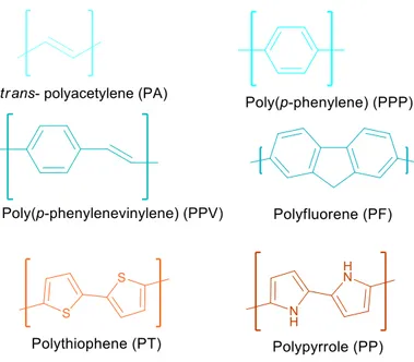
Conjugated polymers are synthetic organic materials with electroluminescent and photoluminescent properties. Conjugation refers to the backbone of the molecular structure where there is a system of alternating single and multiple bonds. This system results in the delocalization of π-electrons along the chain to establish energy stabilization throughout the overlapping p-orbitals. The properties of such systems were first being discovered in late 70’s. In 1977 Alan G. MacDiarmid, Alan J. Heeger and Hideki Shirakawa were the ones to discover the drastic change in the conductive properties of polyacetylene (PA) upon doping.[1] Soon later, the Nobel Prize in Chemistry 2000 was awarded jointly for these three scientists "for the discovery and development of conductive polymers".[2] Besides the simple alternating double bond structure of PA, structures with aromatic cycles on the backbone (i.e. PPP, PF), with both double bonds and aromatic cycles (i.e. PPV), and with heteroatom containing aromatic cycles (i.e. PP, PT) are some common conjugated polymer structures then reported.[3] Figure 1.1 shows some common conjugated polymer structures.
Figure 1.1. Common conjugated polymer structures.
These polymers were shown to be applied in many interesting fields of optoelectronics, such as in organic light-emitting diodes (screen displays) [4], solar cells (photovoltaics) [5], and electronic circuits.[6] Performance enhancements rapidly progress for optoelectronics, in which the use of conjugated polymers are well-established by now. Additionally, conjugated
2
systems have found applications through their photoluminescent properties in other fields, like chemosensing [7], biosensing [8], and biomedicine.[9,10]
How these applications are all possible for conjugated polymers lies in their unique electronic properties. Molecular orbital diagrams for such well defined molecules indicate whether the material can conduct electricity, as well as the types of relaxations it can undergo when its electrons are excited by light. The energy difference between the highest occupied molecular orbital (HOMO) and lowest unoccupied molecular orbital (LUMO) is called the band gap. These orbitals are also referred to as π-level (or So), and π*-level (or S1), respectively. The electrical conductivity of such materials increase as the band gap decreases; the typical band gap for these materials being in the range of 1.5 eV to 4 eV.[11] Also, electron excitations can occur upon photon absorption at these molecular energy levels, and these excitations can relax back to the groundstate through several paths. If the energy is released as photon emission from the lowest singlet excited state, it is called fluorescence. If the electrons first cross to a triplet state, then photon emission occurs as electrons return to singlet ground state from this triplet state which is lower energy than the lowest singlet excited state, it is called phosphorescence. Conjugated polymers exhibit fluorescence.
Figure 1.2. A simplified Jablonski Diagram for photoluminescent phenomena.
The fluorescence of conjugated polymers can be altered through the monomers involved for color tuning. The energy of the emitted photon is directly determined by the band gap energy (Figure 1.2). The band gap is primarily governed by the extend of conjugation; as the number
3
of repeating units increase, more electrons are delocalized, lower the energy gap.[12] Also, copolymer designs of alternating electron donating and withdrawing groups affect the band gap of the molecule. For example, benzothiadiazole (BT) is a commonly used electron acceptor monomer with a low LUMO, which can be conjugated with an electron donating monomer with a high HOMO energy level such as a fluorene unit to produce oligomers or polymers with small band gap energies and a broad absorption. Similar band gap engineering is shown in Figure 1.3a, comparing the colors of two fluorene co-polymers, and Figure 1.3b gives common monomers and corresponding colors of resulting copolymers.
Figure 1.3a. Similar fluorene-based structures and color of emissions in water (ref [13] and
[14] respectively).
Figure 1.3b Bandgap engineered fluorene-based LEDs of colors blue, green, yellow, red. ref
[15]
Besides the factors determined by the band gap through the backbone, pendant groups (side chain functionalities) can also affect the photoluminescence. Large bulky groups can indirectly improve fluorescence by reducing aggregation induced quenching, and with polar
4
groups, conjugation may be enhanced or disrupted through changing the planarity of the backbone by forcing the molecule into a specific conformation depending on its interactions with the solution.[16] Effects of electron donating or accepting side chains are also investigated. The end-groups of the monomers can be designed for the synthesis of these polymers using facile palladium catalyzed C-C bond forming cross-coupling reactions in solution. Palladium catalyzed cross-coupling is a commonly used technique of polymerization in the field, due to high efficiency, and the flexibility they provide in terms of monomer combination, reaction media, and due to the fact that they do not require a special experimental set-up as in the electrochemical techniques. The Nobel Prize in Chemistry in 2010 was awarded to Richard F. Heck, Ei-ichi Negishi and Akira Suzuki who developed unique and advantageous variations of such Pd-catalysis. [17]
Figure 1.4. A schematic summary including a) General catalytic cycle for Pd-catalyzed
cross-coupling reactions. b) Simplified reaction schemes for Negishi, Heck, and Suzuki reactions, respectively.
In fact, the main advantage of these three reactions (Figure 1.4) over other Pd-catalyzed techniques is that no other transition metal or heavy metallic moiety is needed [18], Suzuki coupling being the most prominent since it can be carried out in aqueous solutions, as well as being tolerant to different organoborane functionalities and environmentally friendly.[19] In Suzuki, the Pd-catalyzed cross coupling takes place between the organoboronic species and the aryl halide, and this C-C bond forms in the presence of a base.[20] For conjugated polymers, Suzuki reaction is exceptionally important for in situ or one-pot synthesis of water-soluble products.
5 1.1.1. Polythiophenes
Polythiophenes (PTs) are one of the conjugated polymers (Figure 1.1) to find a wide range of synthetic approaches [21a,b] and applications.[22] Polythiophenes have highest charge carrier mobilities reported for conjugated polymers.[3] They are also good candidates for biological applications since they are biocompatible [23], and their absorbance range is beyond the harmful UVB region. Various water-soluble thiophene species have been synthesized and reported in literature, to find various biological applications.[9] Biocompatibility of these conjugated polymers can easily be improved through functionalizing the side chains. Although the most reported, and possibly the simplest method for PT synthesis is chemical oxidation polymerization, it is useful only when regioregularity and/or solubility is of no importance, and one type of monomer is used for the polymerization (Figure 1.5a).
Figure 1.5a PT synthesis using chemical oxidation polymerization.
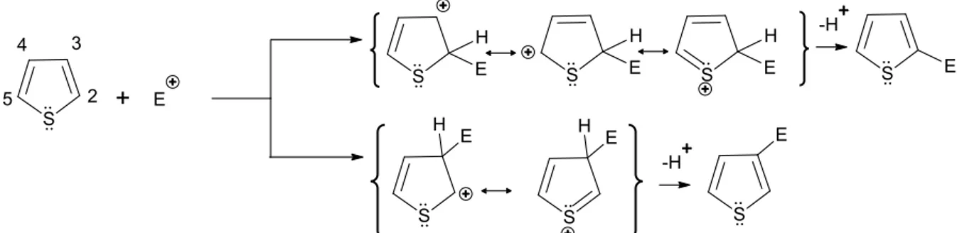
Figure 1.5b Thiophene carbon sites and heterocyclic resonance towards an electrophilic
attack
The heterocyclic chemistry of thiophene favors site-selective functionalization. C2 and C5 are highly susceptible to an electrophilic attack, and this α-selectivity is due to the higher charge delocalization stabilizing the intermediate of C2 attack compared to C3 attack (Figure 1.5b). Commercial thiophene reagents for polymer applications have C2 and C5 sites available for desired substitutions for head–tail (2,5'), head–head (2,2'), or tail–tail (5,5') coupling. 3-Alkylthiophene is the most used thiophene monomer. Since it is not a symmetrical molecule, there are three relative orientations available. Head-head
6
or tail-tail couplings lead to regio-irregular PTs due to steric effects of the 3-substituent that leads to a backbone twist, and loss of conjugation.[21a] Head-tail coupling PTs are regioregular. This can be controlled through the choice of end-groups and polymerization.[21b] For Pd-catalyzed cross-coupling reactions, the end-groups can be designed as shown in Figure 1.4. Co-polymers can also be achieved through end-group design.
Figure 1.6. Polythiophenes with various ionic functionalities, ref [7]
Polythiophenes can thus be side-chain functionalized either pre-polymerization or post-polymerization. In the literature, the monomer designs usually consist of pre-polymerization attachment of a spacer followed by a post-pre-polymerization functional group addition. For PEG-based water soluble designs the spacer is the functionality.[24] Some ionic functional groups to achieve water solubility reported include carboxylic acids [26], amino acids [27], sulfonates [28], amines [25], imidazolium [29] moieties.(Figure 1.6) Spacer length is important for side chain stability, since conjugation of the backbone can end up extended through elimination if possible during polymerization for pre- functionalization.
Side chains can be functionalized using different approaches, mainly through substitution-type reactions with halide or better leaving groups, however this may not be preferred in the possibility of an adverse side reaction, or problems of stability in harsh reaction conditions. One of the most reported alternative is a 1,4-cycloaddition reaction, often referred as the “click reaction”. 1,4-substituted click reaction occurs easily in mild conditions between an azide and an terminal alkyne to form 1,2,3-triazoles in the presence of Cu(I) catalyst (Figure1.7). This reaction was used to functionalize [30] or cross link [31] various conjugated polymers. One recent example is shown in Figure 1.8
7 Figure 1.7. Copper-catalysed 1,4-cycloaddition.
Figure 1.8. PT functionalization using click chemistry. [32]
Along with the advantages of synthetic feasibility and low toxicity, various applications have been monopolized specifically by PTs due to the unique regio-photochemistry they can provide. When the backbone can be forced into a more planar conformation from a non-planar one, it results in a significant red-shift in absorbance (Figure 1.9). Using these properties, water soluble polythiophenes have been designed and applied as sensing probes for detection of DNA (Figure 1.10) and fibrillar proteins like ß-amyloid [33b] through conformational changes. Although long polymeric chains of thiophene are usually preferred in applications of conformation monitoring, it is also a choice for biomedical applications. Fluorescent oligomers have been designed with similar monomers too.
8
Figure 1.9. Conformations and corresponding absorption of a polythiophene.
Reproduced with permission from ref. [33c], Copyright © 2008 American Chemical Society
Figure 1.10 Hybridized DNA sensing cationic PT. Reproduced with permission
from ref [33a], Copyright © 2005 Wiley-VCH Verlag GmbH & Co. KGaA, Weinheim
9 1.1.2 Oligomers
Oligomers that have convergent molecular properties with that of polymers have been of great interest especially in terms of synthesis; due to synthetic reproducibility, and the ease of establishing uniformity, having higher control over end-groups and molecular weight. The structure-property relationships of oligomers have been long investigated also to better understand longer conjugated materials.[34] Oligomers may have advantage over polymers depending on the intended application; oligomers may be preferred over their analogous polymers which are poorly soluble or intractable, which is especially important in photovoltaic device fabrication.[35] Especially symmetrical donor-acceptor-donor (D-A-D) type conjugated oligomers have been studied for solar cell and field effect transistors.[36]
Monodisperse amphiphilic π-conjugated oligomers have been designed for imaging too. Oligomers with fluorescence colours spanning the entire visible spectrum have been made amphiphilic to form water solubile, extremely stable self-assembled particles in water (Figure 1.11) With the use of oligomeric moieties that self-assemble, it gives the possibility co-assemble multi-targeting agents for biological imaging.[37]
Figure 1.11 Fluorescent amphiphilic oligomers. [37]
Oligomers were made far-red near-infrared (NIR) emitting for light emitting diodes [38] through bandgap engineering. Establishing efficient photoluminescence in
NIR-10
region is also very important for biological applications. NIR fluorescent probes have been widely used because NIR light has the ability to deep-penetrate in tissue, and also preferable in cellular level because it causes minimal cellular autofluorescence in imaging.[39] The ultimate goal is to achieve NIR-emitting bio-active or –compatible oligomers to form nanoparticles.
1.2. Polyrotaxanes and Cucurbituril
Cucurbiturils (CB) are bulky and symmetrical pumpkin shaped macrocyclic oligomers formed from glycoluril units (n= 5,6,7,8 or 10) linked through methylene bridges.[40] These CB homologues all consist of portals decorated with polar carbonyl groups that enable strong dipole-dipole or ion-dipole interactions with the environment, and a hydrophobic cavity, but they all have different physical properties than one another. CBs are prepared from the acid-catalyzed condensation polymerization of glycoluril with formaldehyde.[42]
Figure 1.12 Molecular structures of CB6 and CB7, ref [41a] modified and [41b]
reproduced, respectively.
11
Most important difference between the physical properties is the solubility. The homologues are thus seperated from the reaction mixture using solubility differences, using acid, methanol and water.[43] Efficient solubility in water is crucial for any application that aims to benefit from these amphiphilic structures, which only the unique structural parameters of CB5 and CB7 (Table 1.1) was discovered to provide. CB7 is used since CB5 is too small in size and its abundance in a reaction mixture. CB7 also has very low toxicity and is safe even in vivo.[44]
Cucurbiturils have found interesting applications in supramolecular polymerization [45], charge-transfer complexes [46], molecular switches [47], responsive drug release systems [48], reaction catalysis [49], surface functionalizations of nanoparticles [50] and 2D platforms.[51]
Figure 1.13 Common guests for CB homologues. Reproduced from ref. [52]
Typical guests for CB hosts (Figure 1.13) are often used as moieties for side chains or backbones of larger molecular designs, to have strong interaction with the encapsulating macrocycle. These large molecules may either have bulky stoppers to avoid “slipping” of the macrocycle or not. The general name for this type of architecture is called “rotaxanes”. If it lacks stoppers, it is called a “pseudorotaxane”.
12
If the whole structure consists of the rotaxane as a repeating unit, this polymeric form is referred to as “polyrotaxane”. Rotaxanes are often used as pH-responsive [53] and photo-responsive [54] systems.
For conjugated molecules, macrocycles can enhance electrical or photophysical stability of the backbone. Macrocycles can insulate delocalized electrons from reactive species. With insulation, unfavorable intermolecular interactions like aggregation that can result in quenching, polarizability changes that can occur through solvation, or conformational changes can be avoided. Due to this, conjugated rotaxane species are often called “insulated molecular wires”. The field of insulated molecular wire designs, however, is dominated by another macrocycle called cyclodextrin (CD) species, a commercially available bio-product. Polyrotaxanes of poly-paraphenylene (PPP) (Figure 1.14), polyfluorene (PF) and polydiphenylenevinylene (PDV) (Table 1.2) backbones threaded through macrocycles with Suzuki coupling in aqueous solutions were synthesized.
Table 1.2 Monomers for Suzuki coupled conjugated polyrotaxanes based on
13
Figure 1.14 PPP Conjugated polyrotaxane [55]
Figure 1.15 Post-polymerization pseudopolyrotaxane having PT backbone [56]
The use of CBn as the macrocycle for such conjugated polyrotaxanes, however, is uncommon. Farcas et al. recently reported [57] a fully conjugated polyrotaxane PF-BT•CB7 that consists of a fluorene – bithiophene backbone threaded with CB7 (Figure 1.16), however the encapsulation was solely done for insulation, and the polymer is not a water-soluble species. In their comprehensive 2007 review of insulated molecular wires, Anderson and Frampton [58] stated that growing interest in CB7 and CB8 and their strong affinities for aromatic guests makes them suspect that these macrocycles soon will make an important contribution to the field. Still, many reported CB-based rotaxane species [59] do not feature long π- conjugated systems.
14
Figure 1.16 Chemical structures of PF-BT and PF-BT•CB7 Reproduced with
permission from ref [57], Copyright © 2014 Elsevier Ltd.
Besides these “main chain” polyrotaxanes and pseudopolyrotaxanes, conjugated backbones with “side chain” pseudorotaxane designs that are chemically responsive can be made use of. A recent example [60] is a red-emitting oligomer capped with CB7 to achieve pH-responsiveness (Figure 1.17 )
15 1.3. Nanoparticles of Conjugated Materials
Due to their aromatic backbones, conjugated polymers are partially hydrophobic although they may have water-soluble side chains. With highly-hydrophilic or amphiphilic components on the side chains, conjugated polymers effectively self-assemble to form nanoparticles in aqueous media (Figure 1.18).
Figure 1.18 Amphiphilic nanoparticle formation, reproduced with permission from
ref [61]. Copyright © 2011 WILEY-VCH Verlag GmbH & Co. KGaA, Weinheim Slightly soluble polymers and oligomers can easily be made water-dispersible by forming nanoparticles through namely miniemulsion or nanoprecipitation methods (Figure 1.19). In miniemulsion method a surfactant in aqueous solution is introduced to the polymer solution which the solvent is water-immiscible. This method has the disadvantage of the need to use another reagent to stabilize the particle. In nanoprecipitation method, a water miscible solvent (i.e. THF, DMSO, DMF etc.) that can dissolve the materials at hand is used, and the solution is slowly added onto water and ultrasonicated to avoid aggregation and to induce dispersion, then after obtaining a homogeneous solution the solvent is removed.[62] Spherical nanoparticles form in order to minimize surface contact in water. One can get smaller particles with nanoprecipitation, compared to miniemulsion.
Figure 1.19 Nano-precipitation method using a) an additional amphiphilic matrix b)
16
Conjugated nanoparticles have been a favorable choice for easy device fabrication in light emitting diode displays, solar cells, as well as bio-imaging probes, bio-sensing agents and nanomedical systems.[63]
1.4. Biomedical Applications of Conjugated Materials
Conjugated polymers have been of great interest in biomedical field since the 80s when they were shown to be compatible with cells and tissues in vitro and in vivo [64,61] Since then they have found applications of cell labeling, in vivo imaging and tracking [65], biomolecular sensing, and drug delivery.[66,67] Figure 1.20 shows major areas of use of CPNs in biomedical field. Conjugated polymers have been shown to detect small biomedically significant molecules like ATP, GSH, or biologically restricted amounts of metal ions [68] malfunctions in DNA methylation, single nucleotide polymorphisms, and protein misfoldings.[69]
Figure 1.20 Schematic representation of CPNs in biological applications. Reproduced
with permission from ref [62], © The Royal Society of Chemistry 2013
Conjugated bare nanoparticles achieve high photostability and low cytotoxicity, and efficient permeability to cellular cytoplasm. In bioimaging, organic dyes and fluorescent proteins are used often, but they show to rapid photobleaching which is a major drawback. Besides these materials frequently used in biology, quantum dots are a synthetic alternative to CPNs but are quite cytotoxic due to their heavy-metal cores, thus they need extensive shealth. Conjugated polymers have good light-harvesting and signal amplification properties, high fluorescence brightness and photostability, as well as
17
innately having much lower cytotoxicity, thus are more suitable for live cell imaging.[70]
Some charged conjugated polymers are reported for intracellular imaging biomolecules without particle formation or a bioconjugation. Polyelectrolyte thiophenes (PTs) shown as probes for both ex vivo and in vivo specific staining of protein aggregates related to Alzheimer’s disease and their preliminary fibrils aggregates [71] Cationic polymers easily bind to cell surfaces through electrostatic attractions as a result of the anionic phosphate groups of the phosphoipids but cellular uptake is hard to control due to strong nonspecific electrostatic interactions. However, cationic PPVs were reported to monitor cell apoptosis [72] using the fact that cellular membranes of apoptotic cells have higher permeability and have higher charge compared to healthy cells because apoptosis causes exposure of negatively-charged phosphatidylserine, in addition to their inherent negative charge.
Figure 1.21 Pinocytic cellular internatization pathways of linear or nanoparticular
polymers,simplified and reproduced from ref. [9]
For cell internalization, there are two major mechanisms of endocytosis, meaning how cells take up particles and macromolecules, and these are phagocytosis and pinocytosis. Large particles within micrometer-range are only internalized by phagocytic cells, such as macrophages, neutrophils, or dendritic cells, thus pinocytosis may be more relevant. Pinocytic cellular internalization of polymers is schematically shown in Figure 1.21. Pinocytic (fluid-phase) uptake and can occur through either receptor-mediated endocytosis (RME) or an initial non-specific adsorption to cell membrane. Thus size is very important for nanoparticle-based biomedical purposes. It is argued that 40–50 nm being the optimal size range for in vitro uptake due to the trade off between multivalent
18
surface membrane wrapping occurs versus the drawback of size increase, however 10– 100 nm is a generally accepted range of NPs for in vivo applications due to issues of clearance and biodistribution.[73]
For treatment of tumorous cancer tissues, however, setting the particle size on the larger end of this spectrum is beneficial. The reason lies in the difference in vasculature compared to normal tissues. The vasculature of tumors (Figure 1.22) appear inherently leaky, generally having larger vessel diameters, higher vascular density, and enhanced permeability. In addition to this, the lymphatic drainage of macromolecules is impared in these tissues.[74] So considering these two effects, nanoparticles accumulate in the tumor interstitial space, and are not cleared out properly. The overall effect is referred to as “EPR effect”, meaning ‘enhanced permeability and retention’.
Figure 1.22 Transportation mechanism of nanoparticles to tumor microenvironment,
Reproduced with permission from ref [74],© 2014 Elsevier B.V.
Conjugated polymer can be used to encapsulate photosensitizer or drugs for cell therapy. A photosensitizer, such as porphyrin, is often reported to be attached to have it in close proximity to the light harvesting conjugated polymer backbone for efficient energy transfer from to photosensitizer for photodynamic cancer cell therapy. Polythiophene – porphyrin dyad is often used for this purpose, for killing cancer or microbial cells.[75]
19
Figure 1.23 Polythiophene – porphyrin dyad for photodynamic therapy. simplified and
reproduced from ref [75].
Similar to covalent attachment, energy transfer through short-range interactions may be designed using complex formation upon strong interactions between opposite charged molecules and it provides useful for a charged cargo. Cationic polymers have been reported as vectors for gene delivery, since they form complexes with DNA or RNAs [9] However, high positive charge of nanocarriers results in undesired levels of cytotoxicity, and often additional hydrophobic moieties have been incorporated into side chains to improve the cytocompatibility. A simpler design often may be achieved through milder ion-dipole interactions, or bioconjugates. For example, nanoparticles were prepared from an oligonucleotide carrier π- conjugated oligomer (Figure 1.24).
Figure 1.24 An oligonucleotide carrier π- conjugated oligomer [76].
Not all approaches may favor particles. Since spherical nanoparticle formation itself occurs to minimize surface contact with water, the surface is fairly inactive. Surface functionality becomes especially important for biological interactions. Post-functionalization approaches are applied to decorate nanoparticle surfaces after the particle is formed out of a polymer, however an extremely mild chemistry is needed because otherwise results in the disruption of the size and structural features of the generated particle.[61]
20
Water soluble conjugated polymers may be favored for the famous Ringsdorf model for drug delivery by polymers introduced in mid 70’s where a polymer would be used as an anchor for a targeting, a solubilising and a drug moiety, which is attached by a cleavable linker.[77] Targetting can be achieved by attaching bioconjugate recognition groups to the side. An often used functionality is folic acid (FA) because FA-receptors are overexpressed in many cancer cell lines. FA-substituted PPE, for example, was reported for imaging an oral carcinoma cell type through receptor mediated uptake.[78]
Cellular imaging and/or therapy are done using other bioconjugates, such as cell-specific oligopeptides [79] , antibodies [80] or carbohydrates.[81]
1.4.1. Glycoconjugates for Active-Targetting
Carbohydrates have gained attention as targeting ligands due to their ease of production, low molecular weight and high abundance in nature.[73] There are a variety of carbohydrate ligands that target carbohydrate-binding proteins (called “lectins”). Lectin is a general name for proteins having affinity to carbohydrates, and if they reside on a cell membrane they’re referred to as “membrane lectins”. There are membrane lectins specific for types of cells that are differentially expressed, thus carbohydrates provide suitable ligands for nanocarrier designs, for cell-selective drug delivery.
Although the interaction between a lectin and the complementary carbohydrate is often weak, it may be greatly enhanced through multivalency. This phenomenon has become known as the “cluster glycoside effect”.[82] Multivalent glycoconjugates have found nanomedical applications ranging from bacterial anti-adhesins and sensors, drug delivery particles, imaging agents, to immune-inducer vaccines.[83] Multivalent carbohydrate ligands have been used to provide receptor mediated endocytosis frequently in literature, with dendritic [83], linear[84] or core-shell particle[82] anchors.
21
Figure 1.25 Receptor-mediated endocytosis of a glycoconjugate through “cluster
glycoside effect”, modified from ref [85]
Although oligosaccharide functionalities dominate biological targetting applications, these multivalent functionalized particles can mimic the multiple carbohydrate-protein interactions used by cells, viruses, or bacterial toxins with multivalent monosaccharide functionalities.[86]
The biocompatibility, solubility, and active endocytosis properties make glycoconjugates a desirable choice for nanomedicine. In glycopolymers, the carbohydrate moieties can be used as both the targeting and the solubilising part. According to Veronese et al. there are approximately 20 drugs (on the market or in clinical trials) based on this model.[87]
1.4.1.2. Monosaccharides as Pendant Groups
Glycoside chemistry
Hexoses (sugars of 6 carbons) are the most abundant monosaccharides in nature, thus the general term “monosaccharide” is often used to mean hexose. Monosaccharides are simplest carbohydrates with unique stereochemical properties. They are found in equilibrium with their acyclic and two cyclic forms (Figure 1.27) in nature, but upon glycoside formation on the “anomeric site” their conformation can be directed to a certain “anomeric form”, as in the case of activated acetylated glucoside and mannoside and an alcohol substituent (Figure 1.28). The R group can be alkane, alkene or alkyne. Borontrifluoride reagent is a typical activator for this reaction.[88] The glycosides are often designed as groups of azide, halide or sulfide for further reactions.
22
Figure 1.26 Equilibrium and anomeric forms of glucose.
Figure 1.27 Cyclic and acyclic forms of glucose and its stereoisomers mannose and
galactose.
rotation
β-D-Glucopyranose
23
Figure 1.28 Anomer-selective activated glucoside and mannoside formation with an
alcohol substituent. α-mannoside β-glucoside acetylated glucose (anomeric mixture) acetylated mannose (anomeric mixture)
24
The choice of monosaccharide, however, apparently has many factors to consider, such as its anomeric site being either α- or ß- [89], stereoisomerism [90], [91], or another attachment site (C2, C3, C4, C5) for the functional group [92], [93], [94], [95]. Anomeric functionalized mannoside[96], glucoside[97] and galactosides[98] are often reported as targeting moieties. Monosaccharides all have found a variety of purposes. However, most of the human cell types have cancer-related differentiations as mannose and galactose receptor over-expression.
Glucose
In vitro imaging using vero cells [99] and human cervical cancer cells [100] were done with glucosides. In vitro anti-inflammation studies with red blood cells [101] The inhibition of tetrameric PA-IL lectin from pathogenic P. aeruginosa [102] and affinity towards LOLI, a non-toxic Lathyrus ochrus lectin [103] was shown for sensing. LOLI is a possible candidate for a glucose-responsive drug release design, as in ref [104]. Also “GLUT1”, a receptor that facilitates the transport of glucose in mammalian cells, is overexpressed in many tumors [105] and naturally highly expressed in the blood–brain barrier.[106] GLUT1 is also a receptor used by the HTLV virus (spherical, 100 nm diam.) to gain entry into target cells.[105]
Mannose
There are many mannose-functional conjugated materials (oligomer or polymer) reported only for the use of sensing the lectin Con A.[107-110] Concanavalin A (Con A), a multivalent lectin of jack bean plant C. ensiformis, is often used as a safe study model, less toxic than other lectins such as ricin, Boulium and E. Coli toxins [107] Besides aggregation-induced luminescence sensing (Figure 1.29), confirmation is achieved for particles through a range of competitive and non-competitive binding assays, namely haemagglutination inhibition (HIA), ELLA and ITC.[83,111] with bacterial lectins like E. coli FimH, and virulence factors PA-IL and PA-IIL of P. aeruginosa. ConA is the most widely used lectin in biosensing studies, and it’s reported to have a strong affinity to mannose, but also binds α-glucose and not ß-glucose.[111] Mannose-functionalized conjugated polymers were synthesized and used for aggregation-induced detection of E.coli.[112-114]
25
Figure 1.29 Direct detection of lectins with “turn on”luminescent probes, modified from
ref [83]
Other glycopolymers have also been applied to advanced applications of cancer therapy. [81,115] Targeting C-type lectin receptors on cells has been shown to induce immunity against melanomas tumors in vivo [116]. Mannose targeting is done for MMR on macrophages, and DC-SIGN on dendritic cells, to mimick high mannose glycoprotein of HIV-1 gp120 and other pathogenic microorganisms that have similar interactions with these cells [83]. Mannose receptor on macrophages and liver endothelial cells produce opportunities for cell-specific gene delivery.[96] Mannose is reported to be a dendritic cell antigen that activates cytotoxic T cells for antitumor vaccine efficacy. Mannose-grafted nanocarriers were reported for this purpose and showed increased cellular uptake.[117] A quick summary of mammalian lectins and their ligand stereoisomers can be found in Table 1.3.
26
Table 1.3 Summary of mammalian lectin receptors. ref [118]
Galactose
Hepatocytes (liver cells) are known to express the galactoside-binding asialoglycoprotein receptor (ASGPR) on their surface; on binding, the galactoside-conjugate is internalised by the cell.[119]However, it has been reported in the review article by Pawar et al, that galactose and mannose could both recognize the asialoglycoprotein receptor.[120]
27
The conjugated glycopolymers
Figure 1.30 Benzothiadiazole-fluorene- based conjugated glycopolymer for Con A
sensing. Ref [107]
Aggregation-induced fluorescence (see Figure 1.29) is used typically for Con A sensing with post-functionalized conjugated polymers. It has been shown that benzothiadiazole-fluorene- based conjugated glycopolymer (Figure 1.30) had a drastic fluorescence intensity increase at the 555nm-peak upon aggregation.[107] Liu et al. reported a neutral water-soluble oligomer similar to this polymer with four α-mannose side chains (Figure 1.31). The design for light-up detection of ConA and E. coli was achieved through an energy transfer pair with a mannose-substituted conjugated polyelectrolyte [121].
28
Glycopolythiophenes have been reported in the last few years, as in Figure 1.32, for the same purpose.[110] This homo-glycopolythiophene shows similar increase in intensity in its 460nm-fluorescence peak. β-glucoside analogue of this glycopolymer was shown to displays no binding ability to Con A, and was used as a control.
Figure 1.32 Mannose-functionalized homo polythiophene for Con A sensing. Ref [110]
In 2010, Han et al. reported a fluorescent polymer with galactose pendant groups for Ca2+-induced fluorescence quenching studies. This polymer was prefunctionalized and Suzuki coupling was carried out in THF (Figure 1.33).
Figure 1.33 Pre-functionalized galactoside-functionalized monomer for Suzuki
29 1.5. Aim of the Thesis
Initially, an in situ cucurbituril threadded polyrotaxane in water was designed using Suzuki coupling reaction with a cationic thiophene monomer. The idea for the synthesis discussed in the first part of this thesis was elaborated on as a solution to an occuring problem. The 3-substituted water-soluble cationic thiophene monomer went through undesired elimination in the reaction conditions due to the favored extention of conjugation. For this, a more stable alternative and application-wise favorable pre-functionalized monomer without the risk of elimination was proposed instead. Glucose functionality was proposed as an sugar prototype, as an alternative to both trimethylamine and triethyleneglycol functionalities. A thiophene monomer was designed through cycloaddition of an alkyne glucopyranoside moiety for Suzuki polymerization with an encapsulated boronic ester. A new synthesis route for a novel polyrotaxane design was investigated.
For the second part, similar alkyne functional mannoside was synthesized for post- functionalization of a red-emitting conjugated oligomer.
Water-soluble monosaccharide functionalized conjugated polymers, polyrotaxanes and oligomers as fluorescent, water-soluble, multivalent glycoconjugates were thus synthesized, designed for possible bioactive and biocompatible purposes, and may provide better alternative to specific cell applications compared to the nonspecific-binding cationic or PEG moieties.
30
Chapter 2 EXPERIMENTAL
2.1 Materials
All reagents and solvents used were commercial grade, and were used without further
purification unless stated. All column chromatography purifications were done using silica gel (Kieselgel 60, 0.063 – 0.200 nm) and were monitored by thin layer chromatography silica gel plates (Kieselgel 60 F254, 1mm) under shortwave UV lamp (254nm).
2.2 Instrumentation
2.2.1. 1H-NMR and 13C-NMR Spectroscopy
For the characterization of all molecular structures, nuclear magnetic resonance were
measured and recorded with Bruker Avance DPX-400 MHz spectrometer. All measurements were taken in deuterated solvents. All chemical shifts were given relative to the internal standart tetramethylsilane.
2.2.2. UV-VIS Spectroscopy
UV-Vis absorbance spectra for optical characterization were recorded by Cary 300 UV-vis Double Beam Spectrometer equipped with Xenon-lamp as the light source. UV-Vis
absorbance of sample solutions were measured within the range of 200-800nm, using quartz cuvettes with 1cm width. Solvents are chosen according to the sample and stated for each characterization.
2.2.3. Photoluminescence Spectroscopy
Photoluminescence spectra for optical characterization were recorded by Cary Eclipse Varian Spectrometer equipped with Xenon-lamp as the light source. Photoluminescence of sample solutions were measured within the range of 200-800nm, using quartz cuvettes with 1cm width. Samples were excited at their respective excitation wavelength, and initial wavelength for the measurement range was set 10nm higher. Solvents are chosen according to the sample and stated for each characterization.
2.2.4. FT-IR Spectroscopy
For the characterization of chemical functional groups on the monomers, Fourier Transform Infrared spectra were measured and recorded with Bruker Tensor 27 spectrometer equipped with a DLATGS detector with a resolution upto 1cm-1. All measurements were taken by dropping chloroform solutions onto KBr plates and drying in the oven to evaporate solvent
31
and moisture, unless otherwise stated. KBr plates were prepared by grinding IR grade KBr to a fine powder and were pressed into solid pellet disks. The data were recorded in the spectral range of 4000-400cm-1 for 64scans.
2.2.5. Flow-Syringe Pump
Oligomer nanoparticles were prepared using KD Scientific 270 (KDS 270) Constant Flow-Syringe Pump, in Infusion mode. Plastic syringe was inserted onto the equipment, and filled with ddH2O. The flow rate (mL.h-1) was set electronically.
2.2.6. Mass Spectroscopy
The mass of oligomers were determined with Agilent 6224 High Resolution Mass Time-of-Flight (TOF) LC/MS with Electrospray Ionization.
2.2.7. Dynamic Light Scattering
The sizes of the oligomer nanoparticles were measured by DLS method using Zetasizer Nano-ZS instrument with 633nm laser beam, for 13 scans. The measurements were taken in 1mL of ddH2O at room temperature, using disposable DLS cuvettes with 1cm width. The average particle diameters were calculated in the software via Marquardt method.
2.2.8. Scanning Electron Microscopy
The morphological characterizations of nanoparticles were done using scanning electron microscopy (SEM) Quanta 200 FEG.
32
2.3 Synthesis
2.3.1. Synthesis of 2-(2,5-dibromothiophen-3-yl)ethanol (M1)
Freezethaw was initially applied to 100mL EtOAc in a two neck round bottomed flask. 2-(3-thienyl)ethanol (3.00 g, 23.4 mmol) was completely dissolved in the freezethawed solvent. N-bromosuccinimide (NBS) (10.4 g, 58.5 mmol) was added to the solution and the flask was sonicated for 10 minutes in dark. Then it was stirred in dark, at RT, under nitrogen, overnight. The work-up was done to the reaction mixture by first water extraction. The organic layer was further extracted with brine, then subjected to suction filtration using silica gel. The solvent was evaporated under reduced pressure. Yellowish transparent viscous liquid product was obtained. TLC Rf= 0.13 using 9:1 (Hexane:EtOAc) mobile phase. Yield: 2.95 g, 45%. ¹H-NMR (400 MHz, CDCl3, δ): 6.90 (s, 1H), 3.84 (t, 2H), 2.82 (t, 2H), 1.58 (s, 1H) ppm. FT-IR (KBr, pellet, νmax(cm-1)): 3300 cm-1 (O-H, s, b).
2.3.2. Synthesis of 2,5-dibromo-3-(2-bromoethyl)thiophene (M2)
DCM was distilled after adding CaH2, then further dried by keeping with activated molecular sieves overnight. Dried DCM was degassed before the reaction. 2-(2,5-dibromothiophen-3-yl)ethanol (M1) (2.95 g, 10.4 mmol) and PPh3 (6.55 g, 25.0mmol) were dissolved in 15mL DCM in a two neck round bottom flask. The solution is freezethawed two times, and fed nitrogen. CBr4 (8.27 g, 25.0mmol) was seperately dissolved in 3mL degassed dry DCM, and added dropwise into the flask while it was kept at 0C in ice bath. The reaction mixture was stirred for overnight under nitrogen. The work-up was done to the reaction mixture by first water extraction. The organic layer was further extracted with brine, then subjected to suction filtration using silica gel. The solution collected was concentrated by evaporating solvent
M1
33
under reduced pressure then was dissolved in cyclohexane. A second suction filtration was done using silica gel. The solution collected was concentrated by evaporating solvent under reduced pressure again. Silica packed column chromatography was done using cyclohexane. Transparent liquid product was collected. Yield: 2.40g, 70%. TLC Rf= 0.75 using 9:1 (Hexane:EtOAc) mobile phase. ¹H-NMR (400 MHz, CDCl3, δ): 6.88 (s, 1H), 3.52 (t, 2H), 3.12 (t, 2H) ppm.
2.3.3. Synthesis of 3-(2-azidoethyl)-2,5-dibromothiophene (M3)
DMF and ddH2O were seperately degassed before the reaction. 2,5-dibromo-3-(2-bromoethyl)thiophene (M2) (2.40 g, 7.77mmol) was put into a two neck round bottom flask with DMF (5mL) and a reflux set-up was done. NaN3 (0.59g, 10.1mmol) was dissolved in 3 mL degassed water. The mixture was heated to 70C, stirred overnight. The DMF-water mixture was evaporated using vacuum distillation set-up under high vacuum using the Schlenk line. The reaction mixture was subjected to water:chloroform extraction. The organic layer was collected and the solvent was evaporated under reduced pressure. Orange tinted transparent liquid product was collected. Yield: 1.84g, 86%. TLC Rf= 0.69 using 9:1 (Hexane:EtOAc) mobile phase. ¹H-NMR (400 MHz, CDCl3, δ): 6.87 (s,1H), 3.48 (t,2H), 2.84 (t,2H) ppm. FT-IR (KBr, pellet, νmax(cm-1)): 2100 cm-1 (N=N, s).
2.3.4. Synthesis of 1,2,3,4,6-Penta-O-acetyl-D-glucopyranose (S1) M3
34
Iodine crystals (0.13g, 0.50mmol) were completely dissolved in Ac2O (50mL) in 250mL one neck round bottom flask. The flask was then placed in a cool water bath (18C). D-glucopyranose (10.1g, 56.1mmol) was added to the solution portionwise while stirring. The solution was stirred for 2 hours. The work-up was done to the reaction mixture by first pouring it into 100mL solution of ice-cold saturated aqueous solution of sodium thiosulfate in a 800mL beaker while vigorously stirring. After complete addition and mixing, sodium bicarbonate is added to the mixture slowly portionwise, until the violent bubbling disappears. 50mL of DCM was added to the mixture and stirred. The solution is then transferred to a 250mL separatory flask. A 50mL of saturated aqueous sodium bicarbonate solution was prepared separately and cooled beforehand. The organic layer was first extracted with the cool saturated bicarbonate solution, then three times with water. The organic layer was dried using sodium sulfate, then solvent was evaporated under reduced pressure. Yellowish transparent sticky viscous liquid product was first observed, then upon high vacuum drying, white crystals were obtained. Yield= 19.2g, 88% ¹H-NMR (400 MHz, CDCl3, δ) for abundant β: 6.36 (d, 1H), 5.52- 5.49 (t, 1H), 5.19-5.11 (m, 2H), 4.30-4.27 (dd,1H), 4.14- 4.10 (m, 2H), 2.21 (s, 3H), 2.11 (s, 3H), 2.07 (s, 3H), 2.05 (s, 3H), 2.04 (s, 3H) ppm. FT-IR (KBr, pellet, νmax(cm-1)): 1750 cm-1 (C=O, s), 1250 cm-1 (C-O, s).
2.3.5. Synthesis of 2,3,4,6-Tetra-O-acetyl-O-prop-1-yl-β- D-glucopyranoside (S2)
DCM was distilled after adding CaH2, then further dried by keeping with activated molecular sieves overnight. 1,2,3,4,6-Penta-O-acetyl-D-glucopyranose (S1) (10.7g, 27.4mmol) was dissolved in dried DCM in 100mL round bottomed flask. Propargyl alcohol (2.7mL) was added to the solution and flask was put in ice bath and cooled to 0C. Sealed dropping funnel set-up was done. BF3.Et2O (26.7mL) was put in the sealed dropping funnel. The set-up was fed with nitrogen for a short time then closed. BF3.Et2O was dropped into the round bottom flask very slowly while vigorous stirring at 0C, over a period of approx. 40 minutes, with a
35
corresponding rate of approx. 20s per drop. Once the addition is finished, the reaction mixture was stirred for 12h at RT. The work-up was done to the reaction mixture first diluting it by pouring it into 100mL DCM and stirring for 5 min. Water extraction was done 5 times, until visible residues of black cloudy suspension is cleared out. The solvent was evaporated under reduced pressure. Dark viscous liquid was first observed at this step, then further purification was done through crystallization using Et2O at -4C. White crystals form overnight. The Et2O decanted was further subjected to crystallization, and this is repeated until no crystals were observed (approx. 3 times). The crystals were collected using suction filtration. NMR indicated mixture. Rfs in TLC were inseparably close. ¹H-NMR (400 MHz, CDCl3, δ) for β-propargyl glucoside: 5.27 (t, 1H), 5.14-5.12 (t, 1H), 5.04 (t, 1H), 4.81 (d, 1H), 4.39 (d, 2H), 4.28 (dd, 1H), 4.19-4.13 (dd, 1H), 3.77 (d, 1H), 2.49 (s, 1H), 2.48 (s, 3H), 2.12 (s, 3H), 2.08 (s, 3H), 2.06 (s, 3H), 2.04 (s, 3H) ppm. . FT-IR (KBr, pellet, νmax(cm-1)): 3250 cm-1 (-C≡C-H).
2.3.6. Synthesis of 1,2,3,4,6-Penta-O-acetyl-D-mannopyranose (S3)
The procedure of synthesis and workup of 1,2,3,4,6-Penta-O-acetyl-D-glucopyranose (see section 2.3.4) was applied to the stereoisomer D-mannose using the same amounts of reagents, and the same yield was obtained. ¹H-NMR (400 MHz, CDCl3, δ): for α 6.11 (d, 1H), 5.38 (d, 1H), 5.29 (d, 1H), 5.18 (d, 1H), 4.33 (t, 2H), 3.83 (m, 1H), 2.13 (s, 3H), 2.11 (s, 3H), 2.08 (s, 3H), 2.04 (s, 3H), 2.03 (s, 3H) ppm. {for β, 5.89 (d, 1H), 5.51 (d, 1H), 5.32 (d, 1H), 5.14 (d, 1H), 4.29 (t, 2H), 4.10-4.07 (br, 1H), 2.25 (s, 3H), 2.23 (s, 3H), 2.21 (s, 3H), 2.20 (s, 3H), 2.19 (s, 3H) ppm.}
36
2.3.7. Synthesis of 2,3,4,6-Tetra-O-acetyl-O-prop-1-yl- α - D-mannopyranoside (S4)
The procedure of synthesis and workup of 2,3,4,6-Tetra-O-acetyl-O-prop-1-yl-β-D-glucopyranoside (see section 2.3.5) was applied to the stereoisomer 1,2,3,4,6-Penta-O-acetyl-D-mannopyranose (S3) using the same amounts of reagents. The crystallization was much more efficient for 2,3,4,6-Tetra-O-acetyl-O-prop-1-yl-α-D-mannopyranoside. Pure product was obtained, confirmed by NMR. Yield= 12.4g, 70%. ¹H-NMR (400 MHz, CDCl3, δ): 5.36 (dd, 1H) , 5.35(dd, 1H), 5.30 (dd, 1H), 5.05 (d, 1H), 4.30 (m, 2H), 4.15(dd, 1H), 4.12 (dd, 1H), 4.06 (m, 1H), 2.50 (t, 1H), 2.18 (s, 3H), 2.12 (s, 3H), 2.06 (s, 3H), 2.01(s, 3H).
2.3.8. Synthesis of (2,5-dibromothiophen-3-yl)ethyl)-prop-1,2,3-triazol-4-yl-tetra-O-acetyl β-D-glucopyranoside (M4)
THF and ddH2O were seperately degassed before the reaction. 3-(2-azidoethyl)-2,5-dibromothiophene (M3) (1.8g, 5.8mmol) was put into a 25mL round bottom flask and dissolved in 10mL degassed THF. 2,3,4,6-Tetra-O-acetyl-O-prop-1-yl-β- D-glucopyranoside (S2) (2.9g, 7.6mmol) was added and stirred for 10 min until completely dissolved. Aqueous
S4

![Figure 1.3a. Similar fluorene-based structures and color of emissions in water (ref [13] and [14] respectively)](https://thumb-eu.123doks.com/thumbv2/9libnet/5664901.113278/18.892.110.613.390.595/figure-similar-fluorene-based-structures-color-emissions-respectively.webp)
![Figure 1.10 Hybridized DNA sensing cationic PT. Reproduced with permission from ref [33a], Copyright © 2005 Wiley-VCH Verlag GmbH & Co](https://thumb-eu.123doks.com/thumbv2/9libnet/5664901.113278/23.892.208.715.578.840/figure-hybridized-sensing-cationic-reproduced-permission-copyright-verlag.webp)
![Table 1.1 Size parameters for the major CBn species. Reproduced from Ref [41b]](https://thumb-eu.123doks.com/thumbv2/9libnet/5664901.113278/25.892.179.662.534.750/table-size-parameters-major-cbn-species-reproduced-ref.webp)
![Table 1.2 Monomers for Suzuki coupled conjugated polyrotaxanes based on macrocycle β-Cyclodextrin [55]](https://thumb-eu.123doks.com/thumbv2/9libnet/5664901.113278/27.892.175.835.573.1003/table-monomers-suzuki-coupled-conjugated-polyrotaxanes-macrocycle-cyclodextrin.webp)
![Figure 1.16 Chemical structures of PF-BT and PF-BT•CB7 Reproduced with permission from ref [57], Copyright © 2014 Elsevier Ltd](https://thumb-eu.123doks.com/thumbv2/9libnet/5664901.113278/29.892.150.845.136.507/figure-chemical-structures-pf-reproduced-permission-copyright-elsevier.webp)
![Figure 1.18 Amphiphilic nanoparticle formation, reproduced with permission from ref [61]](https://thumb-eu.123doks.com/thumbv2/9libnet/5664901.113278/30.892.172.626.262.417/figure-amphiphilic-nanoparticle-formation-reproduced-permission-ref.webp)
![Figure 1.20 Schematic representation of CPNs in biological applications. Reproduced with permission from ref [62], © The Royal Society of Chemistry 2013](https://thumb-eu.123doks.com/thumbv2/9libnet/5664901.113278/31.892.155.659.541.786/schematic-representation-biological-applications-reproduced-permission-society-chemistry.webp)
