Serotypic identification of local Bluetongue virus isolates using Plaque
Reduction Neutralization (PRN) and Reverse Transcriptase
Polymerase Chain Reaction (RTPCR) Techniques
*Volkan YILMAZ1, Aykut OZKUL2
1 Kafkas University, Faculty of Veterinary Medicine, Department of Virology, Kars; 2 Ankara University, Faculty of Veterinary
Medicine, Department of Virology, Ankara, Turkey.
Summary: Bluetongue virus (BTV) is a vector-borne disease of ruminants disseminated in the tropics and sub-tropics. It is also an important problem in the Middle East. The purpose of this study is serotypic identification of local Bluetongue viruses (BTV) isolated between 1998 and 2005. For this purpose, generic (g) and serotype specific (s) RT-PCR systems and the Plaque Reduction Neutralization Assay (PRNA) were used. Generic and serotype specific RT-PCR was performed on the segment 10 and segment 2 levels, respectively. Generic RT-PCR applications revealed specific DNA product (822 bp in length) in all 26 BTV field isolates. On the other hand, 19 BTV isolates were identified as serotype 9 and one isolate was found to be serotype 16, using the sRT-PCR technique. No DNA amplification was observed as a result of sRT-PCR for serotypes 2 and 4. Plaque reduction and neutralization assay (PRNA) was used for serologic identification of BTV isolates. Hyperimmune serums specific to three serotypes (4, BTV-9 and BTV-16) produced in rabbits, were used in PRNA. The test showed that 1BTV-9 of the isolates were BTV-BTV-9, and the remaining two isolates were identified as BTV-4 and BTV-16.
Keywords: Bluetongue virus, serotyping identification, PRNA, RT-PCR
Yerel Mavidil virusu izolatlarının Plak Redüksiyon Nötralizasyon (PRN) ve Reverz Transkriptaz Polimeraz Zincir Reaksiyonu (RT-PZR) teknikleri ile serotipik identifikasyonu
Özet: Mavidil virusu dünyanın subtropik ve tropik bölgelerinde yaygın olan, ruminantların vektörle bulaşan hastalığıdır. Hastalık özellikle Ortadoğu için önem taşımaktadır. Bu çalısmada 1998-2005 yılları arasında izole edilen yerel BT viruslarının serotipik identifikasyonu amaçlanmıştır. Bu amaç için jenerik ve serotip spesifik RT-PZR sistemleri ile PRN testi kullanıldı. Jenerik RT-PZR ve spesifik RT-PZR sırasıyla segment 10 ve segment 2 düzeyinde gerçekleştirildi. Jenerik RT-PCR uygulamalarında 26 adet BTV izolatının tamamında spesifik DNA ürünü (822 bp. boyunda) elde edildi. Diğer taraftan sRT-PZR uygulaması sonunda ise izolatlardan 19 adedi serotip 9 ve 1 adedi serotip 16 olarak identifiye edildi. Serotip 2 ve 4 için uygulanan spesifik RT-PZR sonunda herhangi bir DNA ürünü elde edilemedi. Plak redüksiyon ve nötralizasyon testi BTV izolatlarının serotipik identifikasyonu için kullanıldı. Tavşanlarda üretilen BTV-4, BTV-9 ve BTV-16’ya spesifik hiperimmun serumlar PRN testinde kullanıldı. PRN testi sonunda 19 izolat BTV-9, 1’er izolat BTV-4 ve BTV-16 olarak identifiye edildi.
Anahtar Sözcükler: Mavidil virusu, serotipik identifikasyon, PRNT, RT-PZR.
* Study has been approved by Local Ethical Committee.
* This study is summarized by the first author from his PhD Thesis with same title.
Introduction
Bluetongue (BT) is an arthropod-transmitted disease of wild and domestic ruminants and is enzootic in many tropical and temperate regions, coincident with the distribution of competent Culicoides vector insects (13). It is caused by a virus classified within the prototype species Bluetongue virus (BTV) from the genus
Orbivirus, the largest genus of viruses within the family
Reoviridae, with double-stranded RNA genome which consists of ten segments (15). Twenty four serotypes have been identified so far (22).
Bluetongue has major implications for international trade in livestock (6), and is therefore found on the list of diseases which must be reported to the World Organization for Animal Health (18). BTV can infect most ruminant species, and although sheep frequently become clinically ill, goats and cattle usually remain asymptomatic (2, 12). It is believed that this reflects host-species-specific variations in the response of microvascular endothelial cells to BTV infection (3).
BTV was observed in the 19th century in Africa, but was only described in 1902 by Spreull. One of the
first reports out of Africa was from Cyprus in 1943. The first outbreak seen in Turkey was in Hatay in 1944-1947. This outbreak was controlled with strict measures. The second appearance was in the Aegean region in 1977, later on the Marmara and Mediterranean regions were affected as well. The virus was identified as BTV Type-4 (25, 27). The disease has been detected serologically and virologically in many studies so far and type 4, 9 and 16 have been identified in Turkey.
BTV outbreaks have occurred annually in the Mediterranean Basin since 1998, which result in significant economic losses (14). During the last decade five different serotypes of BTV have been detected circulating in the Mediterranean basin (BTV-1, 2, 4, 9, 16). BTV-15 has also been detected in Israel, but does not appear to have spread to adjacent countries. The distinct strains of BTV-1 and 4 have also been detected in the western Mediterranean region (1).
The plaque reduction neutralization test has long been recognized as one of the most accurate methods of detecting antibodies. The commonly used method of constant serum-varying virus combined with a quantal assay has been so severely criticized elsewhere as to make discussion of it unnecessary. While the plaque
reduction test is extremely accurate and quantitative, its limitations due to antigenic variation among isolates are as yet unknown. It appears to be a simple and very useful procedure for working with BTV (23).
The study described here examines the identification of BTV serotypes that were circulating in western parts of Turkey between 1998 and 2005, using RT-PCR methods and Plaque Reduction Neutralization assay (PRNA).
Materials and methods
Viruses and plaque assay: A total of 26 BTV
isolates were used in the study. All of the viruses were isolated in the virology laboratory of the Central Veterinary Research and Control Institute - Ankara from blood and organ samples obtained from sick and/or dead animals and transferred to the laboratories of the Virology Department at the Ankara University School of Veterinary Medicine. The samples received had originated from 7 provinces, two of which were in southern Turkey, while the rest were from western Turkey (Table 1). A total of 26 BTV isolates were inoculated onto the Vero AFFSA cells and 21 of them reached satisfying infectivity titer as calculated by native plaque titration assay.
Table 1. BTV isolates, their geographical distribution and further details. Tablo 1. BTV izolatlarının coğrafik dağılımları ve özellikleri
Isolate
Code Province County Year Species Cell/passage number
TR-1 MUGLA DALAMAN 1999 SHEEP VERO/5
TR-2 IZMIR MENEMEN 1998 SHEEP VERO/2
TR-3 AYDIN BOZDOGAN 1998 SHEEP VERO/3
TR-4 IZMIR MENEMEN 1999 SHEEP VERO/2
TR-5 MUGLA DALAMAN 1998 SHEEP VERO/6
TR-6 IZMIR BUCA 1998 SHEEP VERO/3
TR-7 CANAKKALE YENICE 1999 SHEEP VERO/2
TR-8 ANTALYA MANAVGAT 1999 SHEEP VERO/2
TR-9 IZMIR TIRE 1999 SHEEP VERO/4
TR-10 AYDIN SOKE 1998 SHEEP VERO/3
TR-11 ANTALYA MANAVGAT 1999 SHEEP VERO/2
TR-12 AYDIN CINE 1998 SHEEP VERO/3
TR-13 AYDIN CENTER 2001 SHEEP VERO
TR-14 AYDIN CINE 2001 SHEEP VERO
TR-15 AYDIN CINE 2001 SHEEP VERO
TR-16 AYDIN GERMENCİK 2000 SHEEP VERO
TR-17 AYDIN CINE 2001 SHEEP VERO
TR-18 AYDIN BOZDOĞAN 2005 SHEEP VERO/3
TR-19 DENIZLI ACIPAYAM 1998 SHEEP VERO/1
TR-20 AYDIN CINE 1999 SHEEP VERO/1
TR-21 ANTALYA SERIK 1999 SHEEP VERO/1
TR-22 MUGLA MILAS 1999 SHEEP VERO/1
TR-23 ICEL CENTER 1999 SHEEP VERO/1
TR-24 IZMIR BUCA 1999 SHEEP VERO/1
TR-25 AYDIN BOZDOGAN 1999 SHEEP VERO/1
RNA extraction, generic and serotype specific RT-PCRs: Viral RNA was extracted from 200 µl of
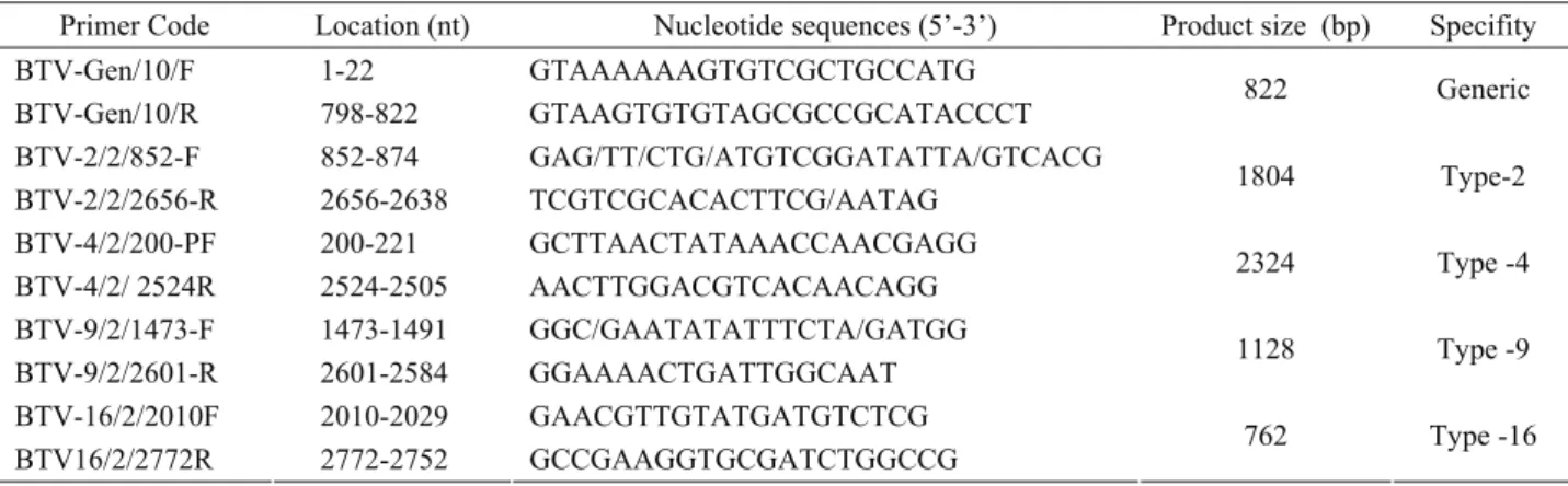
cell-culture supernatant from BTV infected Vero cells using the “Viral RNA extraction kit” (Roche, Germany) as described in the manual. The follow-up reverse transcription (RT) was performed using the “RevertAid kit” protocol based on random hexamer priming (Fermentas, Lithuania) according to the manufacturer's instructions. For Seg-10 amplification, the generic RT-PCR primers designed previously (17) were used (Table 2). The PCR was initiated by a single denaturation step at 96 °C for 10 min, followed by 40 cycles of 1 min at 58 °C, 1 min at 72 °C and 1 min at 94 °C. The mixture was then incubated, as a single step, for 1 min at 50 °C and 10 min at 72 °C for final extension. Five microliters of each PCR product was analyzed in 1% agarose (Prona, Spain) gels containing ethidium bromide (Sigma, USA). On the other hand, serotype specific RT-PCR was carried out on segment 2 with primer sets, two of which had been previously designed (16) while the remaining two (BTV-9 and BTV-16) were designed within this study (Table 2). The serotype specific RT-PCR was performed in conditions similar to that described for generic RT-PCR.
Hyperimmune Serum Production: In order to obtain
the type-specific hyperimmune serums which are the main component of the PRNA test, rabbits were inoculated with three serotypes of BTV (BTV-4, BTV-9 and BTV-16). The rabbits were divided into 3 inoculation groups, each consisting of two rabbits, and two of the rabbits were kept separate to serve as a control
group. Groups 1 to 3 were inoculated with 4, BTV-9 and BTV-16, respectively, while group 4 was injected with Dulbecco’s modified Eagle’s medium (DMEM) to keep them as negative control animals. The injections were made intradermally (id) every fifteen days at multiple region on the back after shaving the skin (Table 3). After the fourth injection, the animals were premedicated with xylasine HCl (Rompun, Bayer) and bled by cardiac puncture. Heat inactivated serum samples were tested with the microneutralization test (5) in order to not only estimate serotype specific antibody titer, but also to see possible cross-reactions between BTV serotypes.
Plaque reduction neutralization test: PRNA was
performed as previously described (19). Briefly, each BTV isolate with 200 pfu in 200 µL was mixed with an equal volume of undiluted hyperimmune serums raised in rabbits and incubated at 37°C for 1 h., subsequently the mixtures were inoculated onto Vero monolayers grown in 24-well plates. After 1 hour of incubation to provide cellular adsorbtion of free virus particles, the cells were overlaid with 3.2% carboxymethyl cellulose in 2x DMEM for the plaque assay. Plaques were scored on day 6 post-inoculation after fixing cells with a 10% formaldehyde solution and subsequent staining with 0.37% crystal violet. The viruses were serotipically identified based on their counted plaque numbers. Briefly, a seventy percent inhibition in plaque numbers by any anti-serotype hyperimmune serum was used as the identification criteria for the BTV isolates.
Table 2. Generic and serotype specific primers used in the study. Tablo 2. Çalışmada kullanılan jenerik ve serotip spesifik primerler
Primer Code Location (nt) Nucleotide sequences (5’-3’) Product size (bp) Specifity BTV-Gen/10/F 1-22 GTAAAAAAGTGTCGCTGCCATG
BTV-Gen/10/R 798-822 GTAAGTGTGTAGCGCCGCATACCCT 822 Generic
BTV-2/2/852-F 852-874 GAG/TT/CTG/ATGTCGGATATTA/GTCACG BTV-2/2/2656-R 2656-2638 TCGTCGCACACTTCG/AATAG 1804 Type-2 BTV-4/2/200-PF 200-221 GCTTAACTATAAACCAACGAGG BTV-4/2/ 2524R 2524-2505 AACTTGGACGTCACAACAGG 2324 Type -4 BTV-9/2/1473-F 1473-1491 GGC/GAATATATTTCTA/GATGG BTV-9/2/2601-R 2601-2584 GGAAAACTGATTGGCAAT 1128 Type -9 BTV-16/2/2010F 2010-2029 GAACGTTGTATGATGTCTCG BTV16/2/2772R 2772-2752 GCCGAAGGTGCGATCTGGCCG 762 Type -16
Table 3. Group of rabbits and inoculation procedure. Tablo 3. Tavşan grupları ve inokulasyon yöntemi.
Group no Rabbit number BTV Serotype Immunization Procedure
I 2 BTV-4 Concerning serotype, 4 inoc. (id) with 15 days apart II 2 BTV-9 Concerning serotype, 4 inoc. (id) with 15 days apart III 2 BTV-16 Concerning serotype, 4 inoc. (id) with 15 days apart
Results
Identification using generic and serotype-specific RT-PCR: RT-PCR using a generic primer set revealed
specific DNA product (822 bp in length) in all of 26 BTV field isolates, while serotype specific RT-PCR identified 20 BT viruses, of which 19 were BTV-9 and the other one was BTV-16. On the other hand, one of the isolates (TR-23) was not able to be identified by serotype specific RT-PCR. There was no specific DNA amplification as a result of sRT-PCR for serotypes 2 and 4. Serotype distribution of local BTV isolates is given in Table 4.
Antibody production serotyping with PRNA:
Immunization protocols for each rabbit group were performed successfully. VN assay revealed that all of the hyperimmune serums produced had 50% neutralizing antibody titer at their 1:10 dilutions for all respective BTV serotypes. Based on this observation, PRNA was carried out using undiluted hyperimmune serums. Three replicates of the PRNA test revealed that nineteen of the BTV isolates were identified as serotype 9 while the remaining two isolates were identified as serotypes 4 and 16, respectively.
Discussion and Conclusion
This study was carried out to identify local Bluetongue viruses (BTV) isolated between 1998 and 2005. For this purpose, generic (g) and serotype specific (s) RT-PCR were used as molecular diagnostic systems and Plaque Reduction Neutralization Assay (PRNA) as a conventional diagnostic system. Generic RT-PCR was performed on segment 10 using primers designed previously (17). On the other hand, sRT-PCR was carried on segment 2 with primer sets, two of which had been designed previously (16) while the remaining two (BTV-9 and BTV-16) were designed within this study.
Seg-10, the smallest genome segment, encodes NS3 and NS3A proteins (21). NS3 is a membrane protein that plays a role in the egress of the virus from infected cells (10). Other important features of NS3 include the two transmembrane domains which allow it to act as a viroporin, and extracellular portions known to form a loop that are thought to be important in terms of recognition by cells (9). Almost no amino acid substitutions were observed in the extra-cellular and glycosylation sites of the NS3 proteins of isolated BT viruses. Recent studies (8) indicate that NS3 protein
Table 4. Comparative results of BTV isolates with gRT-PCR and sRT-PCR/PRNA
Tablo 4. BTV izolatlarının jenerik RT-PZR ile serotip spesifik RT-PZR/PRNT’nin karşılaştırmalı sonuçları sRT-PCR/ PRNA ISOLATE NO gRT-PCR BTV-2 BTV-4 BTV-9 BTV-16 TR-1 + - / - / - + / + - / - TR-2 + - / - / - + / + - / - TR-3 + - / - / - + / + - / - TR-4 + - / - / - + / + - / - TR-5 + - / - / - + / + - / - TR-6 + - / - / - + / + - / - TR-7 + - / - / - + / + - / - TR-8 + - / - / - + / + - / - TR-9 + - / - / - + / + - / - TR-10 + - / - / - + / + - / - TR-11 + - / - / - + / + - / - TR-12 + - / - / - + / + - / - TR-13 + ND ND ND ND TR-14 + ND ND ND ND TR-15 + ND ND ND ND TR-16 + ND ND ND ND TR-17 + ND ND ND ND TR-18 + - / - / - - / - + / + TR-19 + - / - / - + / + - /- TR-20 + - / - / - + / + - / - TR-21 + - / - / - + / + - / - TR-22 + - / - / - + / + - / - TR-23 + - / - / + - / - - / - TR-24 + - / - / - + / + - / - TR-25 + - / - / - + / + - / - TR-26 + - / - / - + / + - / - ND: not determined
depends on the host cell and is not a particularly suitable target to detect BTV nucleic acid in infected mammalian cells. On the contrary, in our study, it was possible to detect specific DNA product (822 bp in length) in all of the BTV field isolates using generic RT-PCR. This was derived from all of the targeted NS3 sequences.
BTV was reported for the first time in Turkey in 1944. The presence of four BTV serotypes (e.g. BTV-2, BTV-4, BTV-9, BTV-16) has been reported in Turkey since that time (4, 11, 14, 26). In this study, 19 BTV isolates were identified as serotype 9 and one isolate was determined to be serotype 16 based on sRT-PCR. DNA amplification was not observed as a result of sRT-PCR for serotypes 2 and 4. Based on these results, it is concluded that BT infection in Turkey was caused dominantly by BTV-9 between 1995 and 2005. These assessments thoroughly conform with BTV epidemics which have been circulating in neighboring countries to the west in the same period. By considering the geographical locations of BTV isolates and the date of occurrence, we assume that BTV-2 does not exist in Turkey, although BTV-9 was isolated predominantly in western Anatolia and a few isolates of BTV-4 and 16 were detected in southern and southwestern Anatolia.
In this study, a total of 26 BTV isolates were inoculated onto the Vero AFFSA cells and 21 of them reached satisfying infectivity titer as calculated by native plaque titration assay. However, Vero AFFSA cell culture adaptation of 5 BTV isolates (TR13-17) was failed and no titer was observed by native plague titration assay performed in case. This situation was thought to be due to Vero AFFSA cell-virus incompatibility or adaptation problem. Consequently, these isolates were not subjected to serotypic identifications by sRT-PCR and PRNA. It is pointed out in this study that following isolation and adaptation to cell culture must be fulfill before diagnostic differentiation.
The isolate TR-23 was identified as serotype 4 by PRNA, but not by RT-PCR. This is most probably caused by incompatibility between primer and target gene sequences as designated primers belonged to Mediterranean Basin isolates, and so BTV-4 primers did not match the target cDNA of isolate TR-23. This observation was found in concordance to several studies in which serotypic discrimination of BTV-4 European isolates and vaccine strains has been performed (16, 28).
Hiperimmunization of rabbits with concentrated viruses for up to 8 weeks did not result in excessive neutralizing of the antibody titer against the serotypes. However, neutralization antibodies can be obtained by immunizations against active infection after inoculations in sheep with similar concentrations (20). When the results obtained after hyperimmunization were evaluated, it was concluded that the antibody titer obtained from
rabbits was low and that it might be possible to obtain a higher antibody titer from animals other than rabbits.
Although PRNA is a labor intensive and time-consuming technique that requires a high level of skill, it has long been recognized as one of the most accurate methods of detecting BTV antibodies (23). The commonly used method of constant serum-varying virus combined with a quantal assay has been so severely criticized elsewhere as to make discussion of it unnecessary (23). While the plaque reduction test is extremely accurate and quantitative, its limitations due to antigenic variation among isolates are as yet unknown. It appears to be a simple and very useful procedure for work with BTV. In this study, hyperimmune serums specific to three serotypes (BTV-4, BTV-9 and BTV-16) produced in rabbits were used in PRNA. The test showed that 19 of the isolates were BTV-9, and the remaining two isolates were identified as BTV-4 and BTV-16. Similarly, PRNA is regarded as the ‘gold standard’ method for identification of some arboviruses (e.g. Bunya-, Flavi- and Togavirus) (7, 19, 24).
In conclusion, in view of BT epidemic’s seasonality and that fact that it is rapidly spread via insects, BTV must be identified by accurate and rapid methods without viral isolation in samples from infected animals. Field screening of BTV infections in endemic countries, and the diagnostic value of RT-PCR applications must be enhanced. Therefore, real-time RT-PCR that is extremely accurate and advanced in-vitro amplification techniques must be developed and utilized for rapid, yet accurate identification of BTV serotypes. On the other hand, as RT-PCR performance is directly influenced by the laboratory or user profile and is an expensive technique, PRNA can be used for serotypic identification of BTV isolates in diagnostic laboratories that do not have molecular diagnostic systems based on similar performance in identifying BTV serotypes found in this study.
References
1. Breard E, Sailleau C, Nomikou K, Hamblin C, Mertens PP, Mellor PS, El Harrak M, Zientara S (2007): Molecular epidemiology of bluetongue virus serotype 4 isolated in Mediterranean Basin between 1979 and 2004. Virus Res, 125, 191–197.
2. Brewer AW, MacLachlan NJ (1992): Ultrastructural characterization of the interaction of bluetongue virus with bovine erythrocytes in vitro. Vet Pathol, 29, 356–359. 3. DeMaula CD, Leutenegger CM, Jutila MA, MacLachlan
NJ (2002): Bluetongue virus-induced activation of primary bovine lung microvascular endothelial cells. Vet Immunol Immunopathol, 86, 147–157.
4. Ertürk A (1994): Çeşitli serumlarda (koyun-keçi-sığır) mavi dil antikorlarının agar-jel presipitasyon testi ile araştırılması. Etlik Vet Mikrobiol Derg, 7, 1-19.
5. Frey HR, Liess B (1971): Vermehrungskinetik und verwendbarkeit eines stark zytopathogenen VD-MD virusstammes für diagnostische untersuchungen mit der mikrotiter-methode. Zbl Vet B, 18, 61-71.
6. Gibbs EP, Greiner EC (1994): The epidemiology of bluetongue. Immun Microbiol Infect Dis, 17, 207–220. 7. Gordon SW, Tammariello RF, Linthicum JK, Dohm
JD, Digoutte JP, Calvo-Wilson MA (1992): Arbovirus ısolations from mosquitoes collected during 1988 in the Senegal River Basin. Am J Trop Med Hyg, 47, 742-748. 8. Guirakhoo F, Catalan JA, Monath TP (1995):
Adaptation of bluetongue virus in mosquito cells results in overexpression of NS3 proteins and release of virus particles. Arch Virol, 140, 967–974.
9. Han Z, Harty RN (2004): The NS3 protein of bluetongue virus exhibits Viroporin-like properties. J Biol Chem, 279, 43092–43097.
10. Hyatt AD, Gould AR, Coupar B, Eaton BT (1991): Localization of the non-structural protein NS3 in bluetongue virus-infected cells. J Gen Virol, 72, 2263– 2267.
11. Karaoğlu T, Özgünlük İ, Demir B, Özkul A, Burgu İ (2007): Seroprevalence of Culicoides-borne disease in cattle in European Turkey. Ankara Üniv Vet Fak Derg, 54, 121-125.
12. Koumbati M, Mangana O, Nomikou K, Mellor PS, Papadopoulos O (1999): Duration of bluetongue viraemia and serological responses in experimentally infected European breeds of sheep and goats. Vet Microbiol, 64, 277–285.
13. Mellor PS, Boorman J (1995): The transmission and geographical spread of African horse sickness and bluetongue viruses. Ann Trop Med Parasitol, 89, 1–15. 14. Mellor PS, Wittmann EJ (2002): Bluetongue virus in the
Mediterranean Basin 1998–2001. Vet J, 164, 20–37. 15. Mertens PPC (2004): dsRNA viruses. Virus Res, 101, 3–
13.
16. Mertens PPC, Maan NS, Prasad G, Samuel AR, Shaw AE, Potgieter AC, Anthony SJ, Maan S (2007): Design of primers and use of RT-PCR assays for typing European bluetongue virus isolates: differentiation of field and vaccine strains. J Gen Virol, 88, 2811–2823.
17. Nikolakaki SV, Nomikou K, Koumbati M, Mangana O, Papanastassopoulou M, Mertens PPC, Papadopoulos O (2005): Molecular analysis of the NS3/NS3A gene of Bluetongue virus isolates from the 1979 and 1998–2001 epizootics in Greece and their segregation into two distinct groups. Virus Res, 114, 6–14.
18. OIE (1996): Bluetongue. In: OIE Standards Commission (Ed.), Manual of Standards for Diagnostic Tests and Vaccines. Paris, 109–118.
19. Ozkul A, Yildirim Y, Pinar D, Akçali A, Yilmaz V, Colak D (2006): Serological evidence of West Nile Virus (WNV) in mammalian species in Turkey. Epidemiol Infect, 134, 826-829.
20. Ramakrishnan MA, Pandey AB, Singh KP, Nandi S, Mehrotra ML (2006): Immune responses and protective efficacy of binary ethylenimine (BEI)-Inactivated Bluetongue Virus vaccines in sheep. Vet Rec, 30, 873-880. 21. Roy P (1989): Bluetongue virus proteins. J Gen Virol, 73,
3051-3064.
22. Roy P (2002): Orbivirus. Ed: C. D. Tidona, G. Darai. The Springer Index of Viruses. Springer-Verlag. Berlin, Germany, pp: 957-963.
23. Russel PK, Nisalak A, Sukhavachan P, Vivona S (1967): A plaque reduction test for dengue virus neutralizing antibodies. J Immunol, 99, 285-290.
24. Ulloa A, Langevin SA, Mendez-Sanchez JD, Arredondo-Jimenez JI, Raetz JL, Powers M, Villarreal-Trevino C, Gubler DJ, Komar N (2003): Serologic survey of domestic animals for zoonotic Arbovirus infections in the Lacandón Forest Region of Chiapas, Mexico. Vector-Borne Zoonotic Dis, 3, 3-9. 25. Urman HK, Milli U, Mert N, Berkin S, Kahraman
MM, Yüce H, Avvuran H (1979): Türkiye’de buzağılarda konjenital epizootik arthrogryposis ve hydranencephaly olayları. Ankara Üniv Vet Fak Derg, 26, 287-292.
26. Yıldırım Y, Burgu İ (2005): Kuzeydoğu Anadolu Bölgesindeki sığırlarda mavidil (BT), IBR, PI-3, EBL ve BVD enfeksiyonlarının seroprevalansı. Ankara Üniv Vet Fak Derg, 52, 113-117.
27. Yonguç AD, Taylor WP, Csonton L, Worrall E (1982): Bluetongue in western Turkey. Vet Rec, 111, 144-146. 28. Zientara S, Breard E, Sailleau C (2006): Bluetongue:
characterization of virus types by reverse transcription-polymerase chain reaction. Dev Biol (Basel), 126, 187– 196.
Geliş tarihi: 24.11.2010 / Kabul tarihi: 31.05.2011 Adress for correspondence
Dr. Volkan Yilmaz
Kafkas Üniversitesi Veteriner Fakültesi Viroloji Anabilim Dalı, Paşaçayır, Kars,
Tel: +90 474 2426807/1107, Faks: +90 474 2426847 E-mail: [email protected]

![Assessment of the cytotoxic and genotoxic potential of pillar[5]arene derivatives by Allium cepa roots and Drosophila melanogaster haemocytes](data:image/gif;base64,R0lGODlhAQABAIAAAP///wAAACH5BAEAAAAALAAAAAABAAEAAAICRAEAOw==)