Case Report: The Formation Variation Of The
Median Nerve
OLGU SUNUMU: NERVUS MEDIANUS’UN OLUŞUM VARYASYONU
Çiğdem İÇKE, Hale KİZANOGLU TEMİZKAN
Department of Anatomy, Dokuz Eylül University, Medical SchoolÇiğdem İÇKE
Dokuz Eylül Universitesi Tıp FakültesiAnatomi AD 35340 İnciraltı, İZMİR
eposta: [email protected]
Tel: (232) 4124357
SUMMARY
Variations in the formation, course and distrubution of the brachial plexus (BP) have been reported in the literature. The anatomy of the BP should be known well for understanding of these variations. The BP is formed by union of the ventral rami of the lower four cervical (C5–C8) and first thoracic (T1) spinal nerves, and supplies motor, sensory, and sympathetic nerve fibers to the upper limb. The fourth cervical ventral ramus frequently gives a branch to the fifth and the first thoracic ventral ramus often receives a contribution from the second thoracic ventral ramus. Classically, the median nerve (MN) (6–C8 and T1) s formed by fusion of its medial and lateral roots, derived from the respective cords of the BP where they oppose the second part of the axillary artery. The objective of this study was the present the case, showing variation in the formation of the median nerve (MN), by the aid of the lineer measurements considering anatomic landmarks.
This study was performed in the laboratory of the Department of Anatomy, Dokuz Eylül University Faculty of Medicine. During the dissections we observed that a formation variation of the MN in the left axillary region of the formaldehid fixed adult male cadaver. The MN was formed by the fusion of three roots, two from the lateral and one from the medial cord of the BP. In the right axillary region of the cadaver formation of the MN was observed as normal. The relationship of the roots, constituting the MN, with the neighbourhood structures was determined by linear measurements.
The variations in the formation of the MN is of great importance for the relationship of the nerve both itself and neighbourhood structures. The roots constituting the MN can be caused to pressure symptoms on axillary artery depending on their locations. Also the structures located in the neighbourhood area of the MN can make pressure to the roots of the MN. Variations in the formation of the MN should be remembered during the surgical approaches and evaluation of the clinical symptoms.
Key words: Brachial plexus, median nerve, medial root, lateral root, variation ÖZET
Literatürde plexus brachialis’in (PB) oluşumu, ilerleyişi ve dağılımı ile ilgili varyasyonlar bildirilmektedir. Bu varyasyonların anlaşılabilmesi için PB anatomisi iyi bilinmelidir. PB alt dört servikal spinal sinir (C5 – C8) ile birinci torakal spinal sinirin (T1) ön dallarının birleşmesi ile oluşur ve üst ekstremiteye giden motor, duyu, sempatik lifleri taşır. Dördüncü servikal spinal sinirin ön dalı sıklıkla beşinci servikal spinal sinire giden bir dal verirken; birinci torakal spinal sinir de ikinci torakal spinal sinirin ön dalından gelen bir dal alır. Klasik olarak, nervus medianus (NM) C6–C8 ve T1 spinal sinirlerin ön dallarından oluşan radix medialis ve radix lateralis’in arteria axillaris’in ikinci bölümüne uyan bölgede birleşmesi ile meydana gelir. Çalışmamızın amacı nervus medianus (NM) oluşum varyasyonu gösteren olgumuzu, anatomik landmarklar dikkate alınarak yapılan
lerin yardımı ile sunmaktır.
Bu çalışma Dokuz Eylül Üniversitesi Tıp Fakültesi Anatomi Anabilim Dalı laboratuvarında gerçekleştirildi. Disseksiyonlar sırasında formaldehid ile fikse edilmiş bir erkek kadavranın sol axilla bölgesinde NM’nin oluşum varyasyonu gözlendi. NM, PB’nin radix lateralis’inden gelen iki, radix medialis’inden gelen bir olmak üzere toplam üç dalın birleşmesi ile oluşmuştu. Olgunun sağ axilla bölgesinde NM’nin oluşumu normal olarak gözlendi. NM’yi oluşturan dalların komşu yapılarla olan ilişkileri yapılan ölçümlerle değerlendirildi.
NM formasyonundaki farklılıklar hem sinirin kendisi hem de çevre dokular ile olan ilişki-sinde önem taşır. NM’nin yapısına katılan dallar yerleşimleri nedeniyle arteria axillaris’de bası bulgularının ortaya çıkmasına neden olabilirler. Ayrıca NM’a komşu bölgelerde yer alan yapılar da siniri oluşturan dallar üzerine bası yapabilir. NM’un oluşum varyasyonları cerrahi uygulamalar sırasında ve klinik bulguların değerlendirilmesi sürecinde hatırda tutulmalıdır.
Anahtar sözcükler: Plexus brachialis, nervus medianus, radix medialis, radix lateralis, varyasyon
Variations in the formation, course and distrubution of the brachial plexus (BP) have been reported in the literature (1). Understanding of these variations need good knowledge about the BP anatomy.
The BP, carrying motor, sensory, symphathetic fibers to the upper extremity, is formed by the union of the anterior divisions of the lower four cervical nerves (C5 – C8) and the greater part of the anterior division of the first thoracic nerve (T1); the fourth cervical usually gives a branch to the fifth cervical, and the first thoracic frequently receives one from the second thoracic (2).
The median nerve (MN) arises by two roots, one from the lateral (LC) and one from the medial cord (MC) of the BP. Its fibers are derived from the sixth, seventh, and eighth cervical and first thoracic nerves. The roots of the MN embrace the lower part of the axillary artery (AA), uniting either in front of or lateral to that vessel (2). Varia-tions in the distribution of the MN in the arm are not com-mon (1).
The objective of this study was the present the case, showing variation in the formation of the MN, by the aid of the lineer measurements considering anatomic landmarks.
MATERIAL AND METHODS
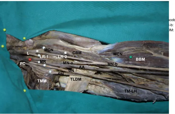
This study was performed in the laboratory of the Department of Anatomy, Dokuz Eylül University Faculty of medicine. During the dissections we observed that a formation variation of the MN in the left axillary region of the formaldehid fixed adult male cadaver. The MN was formed by the fusion of three roots, two from the LC called as LR-1 (First lateral root = Proximal lateral root = Variant lateral root ) and LR-2 (Second lateral root = Distal lateral root = Main lateral root) and one from the MC called as MR (Medial root) of the BP. The union of LR-1 and MR was named as MN-a, after the LR-2 has joined to MN-a then it was called as MN-b.
We measured the thickness of the roots, constituted the MN, at their beginning points. These measurements were described as presenting below:
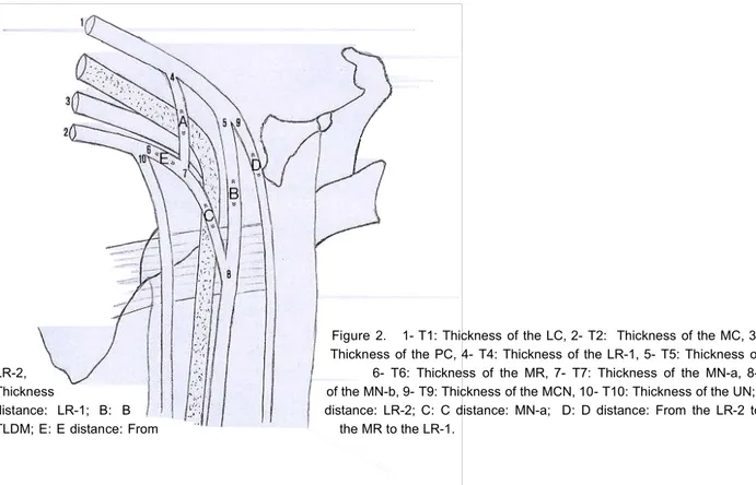
T1: Thickness of the LC at its beginning point T2: Thickness of the MC at its beginning point
T3: Thickness of the posterior cord (PC) at its beginning point
T4: Thickness of the LR-1 at its beginning point T5: Thickness of the LR-2 at its beginning point T6: Thickness of the MR at its beginning point T7: Thickness of the MN-a at its beginning point T8: Thickness of the MN-b at its beginning point
The relationship of the roots, constituting the MN, with the neighbourhood structures was determined by linear measurements considering anatomic landmarks. Axillary artery (AA), tendon of the latissimus dorsi muscle (TLDM), pectoralis minor muscle (PMM) and BP were determined as anatomic landmarks in this study. Measurements were performed when the arm was placed in 90º abduction position with 0.1mm sensitive compass.
A distance: From the beginning point of the LR-1 to the joining point of it to the MR (LR-1)
B distance: From the beginning point of the LR-2 to the joining point of it to the MN-a (LR-2)
C distance: Length of the MN-a (MN-a)
D distance: From the beginning point of the LR-2 to the crossing point of it with the TLDM
E distance: From the beginning point of the MR to the joining point of it to the LR-1.
In the right axillary region of the cadaver the formation of the MN was observed as normal.
RESULTS
In the right axillary region of the case the MN was
formed by the fusion of three roots, two from the LC (named as LR-1 and LR-2) and one from the MC (named as MR) of the BP. MN-a is formed by LR-1 and MR, and both the LR-1 and MN-a crosses the axillary artery (AA) anteriorly (Fig 1). One third proximal part of MN-a has a course adjacent to AA (Fig 1). In our study thickness of the LC, MC and PC at their beginning points were 5.0 mm (K1), 3.6 mm (K2) and 5.7 mm (K3) respectively and the thickness of the branches forming MN were 2.1 mm for LR-1 (K4) and 2.6 mm for LR-2 (K5) and 2.9 mm for MR (K6) at the beginning point (Fig 2). Due to its location and thickness, LR-1 was accepted as variant and LR-2 as normal lateral branch. MR was thicker than 1 and LR-2. MN-a’s thickness at beginning point was 5.6 mm (K7), and MN-b’s was 3.6 mm (K8) (Fig 2). Both the MN-a and MR had significant thickness and they were very close to the AA, so it was thought that they can compress the artery. MCN and UN separated from LC and MC respectively with MN neighbourhood (Fig 1). The thicknesses of them at their beginning points were measured as 3.2 mm (K9) for MCN and 2.7 mm (K10) for UN (Fig 2).
Figure 1. LC: Lateral cord, LR-1: First lateral root, LR-2: Second lateral root, CBM: Coracobrachialis muscle, MCN: Musculocutaneous nevre, BBM: Biceps brachii muscle, MR: Medial root, MN-a: Median nerve-a part, MN-b: Median nerve-b part, UN: Ulnar nevre, RN: Radial nevre, AA: Axillary artery, PC: Posterior cord, TMM: Teres major muscle, TLDM: Tendon of the latissimus dorsi muscle, TM-LH: Triceps muscle-Long head
© 2006
DEÜ
TIP FAKÜLTESİ DERGİSİ CİLT 20, SAYI 2, (MAYIS) 2006, S: 103 - 107L LR-2 LR-1 MN-a MN-b UN RN TLDM AA MR BBM CBM MCN TM-LH TMM MCN PC
Figure 2. 1- T1: Thickness of the LC, 2- T2: Thickness of the MC, 3- T3: Thickness of the PC, 4- T4: Thickness of the LR-1, 5- T5: Thickness of the LR-2, 6- T6: Thickness of the MR, 7- T7: Thickness of the MN-a, 8- T8: Thickness of the MN-b, 9- T9: Thickness of the MCN, 10- T10: Thickness of the UN; A: A distance: LR-1; B: B distance: LR-2; C: C distance: MN-a; D: D distance: From the LR-2 to the TLDM; E: E distance: From the MR to the LR-1.
DISCUSSION
Anatomical BP variations are important because variant nerves with abnormal origins, course and distributions are more susceptible to trap neuropathies and injuries during neck dissections (1,3-6 ). During breast cancer surgery and BP blockage with local anesthetics, and repairing of BP lesions these variations should be remembered (4,7,8)
In the literature some cases had MN’s formed with two LC and one MC branches (1,3,4,9). Sarsilmaz et al. studied 71 adult human cadavers and found 4 of them had BP variation (4). Their third case was similar with our case. Also the variation cases of Sargon and Borman, and Sargon et al’s had similar features with our case (3,9). In the MN variation case, which was defined by Saeed and Rufai beside the similar futures, there was an anastomotic branch connecting MN and MCN (1). In our
For our case lengths of proximal and distal lateral branches were 2.58 cm and 8.08 cm respectively. These lengths were 2.7 cm and 4.6 cm in Sarsilmaz et al’s case (4).
In our case the distance between the connection points of normal and lateral branches with MR was 6.76 cm. It was 1.6 cm in Sarsilmaz et al’s study and 2.5 cm in Saeed and Rufai’s case (1,4).
As in Sarsilmaz et al’s, and Saeed and Rufai’s cases, variant lateral branch crosses AA anteriorly and unites with MR (1,4). Close neighbourhood of variant lateral branch and AA can cause a compressive effect. This can lead to ischemic pain or different levels of arterial insufficiency with shoulder joint’s postural motions and to complications with anterior surgical procedures at the joint. In addition, the axillary lymphadenopathies and AA aneurysms can press the variant lateral branch and can effect the total innervation of MN.
Anatomic BP variation knowledge is helpful for sho-ulder joint traumatology, axilla and shosho-ulder repair operations, radical neck dissections and for the therapy of humerus collum chirurgicum fracture displacement (1,6,-10,11).
Anatomic variations of periferic nerve system can be used to explain the extraordinary clinical symptoms and to interprete the clinical neurophysiology correctly.
LITERATURES
1.
Saeed M, Rufai AA. Median and Musculocutaneous nerves: Variant formation and distribution. Clinical Ana-tomy 2003; 16:453-457.2.
Williams PL, Bannister LH, Berry MM, Collins P, Dyson M, Dussek JE, Ferguson MWJ, editors. 1995. Gray’s anatomy. 38th Ed. Philadelphia: WB-Saunders. 1266-1272.3.
Sargon MF, Uslu SS, Celik HH, Aksit D. A variation of the median nerve at the level of brachial plexus. Bulletin de I' Association des Anatomistes 1995; 79:25-26.4.
Sarsilmaz M, Sendemir E, Celik HH, Gumusalan Y, Sim-sek C. Some variations of the brachial plexus in man.Turkish Journal of Medical Research 1993; 11: 161-165.