The immunohistochemical localization of the fibronectin receptor
(Integrin β1) and laminin in the bitch uterus during the estrous cycle
Güner KÜÇÜK BAYRAM, Narin LİMAN, Feyzullah BEYAZ, Emel ALAN
University of Erciyes, Faculty of Veterinary Medicine, Department of Histology and Embryology, Kayseri, Turkey.Summary: In mammals, the purpose of the estrous cycle is the successful progression of implantation and pregnancy. The endometrium undergoes several changes during the estrous cycle. The extracellular matrix proteins have a likely role in the maintenance of a healthy endometrium. In this study, we investigated the localization of the fibronectin receptor (Integrin β1), a member of the integrin family, and laminin, an extracellular matrix protein, in the bitch uterus throughout the estrous cycle. In the bitch uterus, immunoreactions of similar intensity were observed for the fibronectin receptor (Integrin β1) in the myometrium, perimetrium, peritoneal mesothelial cells and uterine vasculature, and for laminin in the peritoneal mesothelial cells, throughout the estrous cycle. Laminin and the fibronectin receptor (Integrin β1) were found to be present at high intensity in the endometrium of the bitch uterus at the luteal stage.
Keywords: Bitch, estrous cycle, fibronectin, laminin, uterus.
Köpek uterusunda östrus siklusunda fibronektin reseptörü (İntegrin β
1) ve lamininin
immunohistokimyasal lokalizasyonu
Özet: Memelilerde östrus siklusunun amacı implantasyon ve gebeliğin başarıyla sonuçlandırılmasıdır. Endometrium östrus siklusunda birçok değişikliğe uğrar. Ekstrasellüler matriks proteinleri, sağlıklı endometrium yapısının korunmasında önemli bir rol oynar. Bu çalışmada köpek uterusunda, östrus siklusunda integrin ailesinin bir üyesi olan fibronektin reseptörü (Integrin β1) ve ekstrasellüler matriks proteini lamininin lokalizasyonu araştırıldı. Köpek uterusunda, siklus süresince fibronektin reseptör (Integrin β1) miyometriyum, perimetriyum, peritenoal mezotel hücreleri, uterus damarları ve laminin peritenoal mezotel hücrelerinde aynı şiddette reaksiyon gösterdi. Köpek uterus endometriyumunda laminin ve fibronektin reseptör (Integrin β1)’ün siklusun luteal döneminde çok yoğun olduğu belirlendi.
Anahtar sözcükler: Fibronektin, köpek, laminin, östrus siklusu, uterus.
Introduction
The mammalian endometrium shows significant developmental changes during the sexual cycle in preparation for implantation, and these changes continue in early pregnancy (13, 15). The cyclic restructuring of the endometrium implantation and gestation require specific cell-to-stromal matrix and cell-to-cell interactions. The interaction between the cell and the basement layer is mediated by cell adhesion molecules of various classes, of which the most important is integrins. Integrins are heterodimeric (α- and β- chain) cell surface receptors that bind to the extracellular matrix by interacting with cells and extracellular matrix proteins such as collagen, laminin and fibronectin receptor (23, 29). It has been determined that laminin and integrins through dynamic changes in the endometrium during both the menstrual cycle and early pregnancy (22). Although integrins are known to be different in human decidual cells and endometrial stromal
cells, there is no information on the distribution of the fibronectin receptor (Integrin β1) and laminin protein in
the reproductive tract of the bitch during the estrous cycle. The estrous cycle of the bitch is longer than that of many other domestic animals. In the bitch, the follicular stage and ovulation are followed by a luteal stage with an approximate duration of 75 days.The particularities of the estrous cycle of the bitch, such as the luteinization of follicles before ovulation and the cyclic events that take place in the endometrium are quite interesting (6, 7, 20). This study was aimed at demonstrating the localization of the fibronectin receptor (Integrin β1) and laminin in the
uterus of the bitch during the estrous cycle. On the basis of the comparison of the results obtained in this study to the pattern observed in women, it is aimed to gain a better understanding of the role of the fibronectin receptor (Integrin β1) and laminin in the estrous cycle.
Materials and Methods
Tissue preparation and histology: Uterus samples
were taken from 15 healthy bitches, comprising 3 groups of 5 animals, each in different stages of the estrous cycle. Ovarian and uterine tissues, which are normally disposed of as medical waste after ovariohysterectomies performed for the sterilization of female dogs, are preserved in the archives of our department for many years, once having undergone routine histological processing and been embedded in paraffin, and can be used in research whenever needed (Meeting date of Ethics Board of Erciyes University: 15.03.2017, Decision No:17/035). The uterus samples used in this study belonged to 3 to 7- year-old dogs of different breeds, which had been admitted to private veterinary clinics in the Kayseri province for routine ovariohysterectomy. Shortly after ovariohysterectomy, the right and left horns of the uterus were cut into small pieces for histological and immunohistochemical examinations. Fixation was performed in 10% formalin solution for 24 h at room temperature. After being fixed, tissues samples were washed in tap water and dehydrated in a graded ethyl alcohol, cleared in methyl benzoate, and lastly embedded in paraplast. The 5 µm thick cross-sections were stained with Crossman’s staining method to determine the histological structure of the bitch uterus (8).
The determination of the estrous cycle stage: The sections were examined under a light microscope. It was observed that, the histological structure of the bitch uterus varied with the different stages of the estrous cycle. The stage of the estrous cycle, to which the uterine tissue samples belonged, was determined as either the follicular, luteal or anestrous stage, on the basis of the structural changes observed in the surface epithelium of the endometrium, the crypts and glands, as described by Beijerink (1), Butinar et al. (6), Van Cruchten et al. (26), and Vermeirsch et al. (27).
Immunohistochemistry (IHC): Immunohistochemical
analyses were performed at room temperature, according to the immunostaining protocol of Thermo Scientific using the streptavidin-biotin-peroxidase complex method. After being mounted on 3-aminopropyl-ethoxysilane (APES)-coated glass slides, the tissue sections were dried overnight at 37°C in an incubator. Subsequently, the sections were deparaffinized in xylene, hydrated and then followed by incubation in 3% H2O2 in methanol for 20
min. After the sections were thoroughly washed in phosphate - buffered saline (PBS). Next, the sections were washed in PBS and immersed in a blocking serum (Ultra V Block; Thermo Fisher Scientific Lab Vision Corporation, Fremont, CA, USA; TA-125UB) for 5 min. at room temperature to avoid non-specific binding. Afterwards, the sections were applied with rabbit polyclonal laminin AB-11575P (1/200 Abcam)
(Cambridge, Massachusetts, USA) and monoclonal anti-human integrin β1, fibronectin receptor, clone FNR31
(1/10000 Takara, Japan) primary antibodies to specific dog, and incubated in a humidity chamber at room temperature for 1 h. Subsequently, the sections were washed in 0.01M PBS four times, each time for 5 min, treated with biotinylated secondary antibodies (Thermo Scientific, Ultravision kit, TM-125-HL, USA) for 20 min at room temperature, and washed another four times in 0.01M PBS for 5 min. After that, the sections were treated with enzyme-conjugated streptavidin (Thermo Scientific, Ultravision kit, TM-125-HL, USA) for 20 min. To visualize the peroxidase activity, the sections were reacted with 3,3-diaminobenzidine (DAB; Thermo Fisher Scientific Lab Vision Corporation). Later, the sections were counterstained with Gill’s haematoxylin, dehydrated through a graded series of alcohol, cleared in xylene, and mounted with entellan. The specificity of the IHC procedures was controlled using negative control sections. The negative control sections were incubated only with PBS, without any primary antibody. The sections of human spleen were used as a positive control for both antibodies (The blocks of human spleen were supplied from block archive of Medicine Faculty of Erciyes University).
The evaluation of the staining intensity: The
immunohistochemical sections were examined by light microscopy and imaged using an Olympus DP72 digital camera (Olympus, Japan). To evaluate the strength of the immunohistochemical reactions for the fibronectin receptor and laminin in the bitch uterus, a semiquantitative analysis was conducted, such that an arbitrary visual scale was used with a grading score that ranged from (-) to (+++). When observed at high light-microscopic magnification (X40), the absence of stained cells in the reaction site was graded as no reaction (-), the observation of reaction only at high magnification (X40) was graded as weak reaction (+), the clear observation of reaction at low magnification (X10) was graded as moderate reaction (++), and the observation of intense reaction at low magnification (X10) was graded as strong reaction (+++). The immunostaining reactions in the cells were observed by two independent researchers (G.B, N.L) and the mean score of the two observers was calculated.
Results
Histological classification of phase of the oestrus cycle in bitch uterine;
Follicular stage: Endometrium was lined by a single layer of cuboidal or low prismatic epithelial cells. Epithelial cells of crypts were high prismatic. The glandular epithelium was composed of prismatic cells, but the height of the epithelial cells in the deep uterine glands was greater than those in the superficial uterine glands.
Luteal stage: Uterine endometrium and myometrium thickened in luteal phase. Endometrium was lined by low prismatic epithelial cells with long oval nuclei. The endometrial glands were branched and dilated long glands and extended into the basal part of the endometrium. The height of the epithelial cells in the deep uterine glands was more than the height of the superficial uterine gland cells and luminal epithelium of the endometrium.
Anoestrus: The layers of uterine endometrium and myometrium were quite thin during anestrous stage. The endometrium was lined by a single layer of cuboidal cells. A large number of stromal cells existed in the stroma of the endometrium. The luminal epithelium had a few crypts. It was observed that the uterine glands decreased during anoestrous stage.
Findings for Immunohistochemistry;
The localization of the fibronectin receptor (integrin β1): In the bitch uterus, the fibronectin receptor (Integrin β1) displayed a positive immunoreaction at all stages of the
estrous cycle in the endometrium, myometrium and perimetrium. It was determined that the fibronectin receptor (Integrin β1) was localized in the surface, crypt
and glandular epithelial cells of the endometrium, the stromal cells, myometrium, perimetrium and peritoneal mesothelial cells in the bitch uterus. Fibronectin receptor
(Integrin β1) immunoreaction was observed in the
capillaries and the intima, media and adventitia of the large blood vessels in all layers of the uterus. In the bitch uterus, the fibronectin receptor (Integrin β1)
immunoreactions in the cytoplasm of cells insurface and crypt epithelium of the endometrium was moderate at the follicular (Fig. 1A) and anestrous stages (Fig. 3A), whereas reactivity was strong at the luteal stage (Fig. 2A). At the luteal and anestrous stages, a strong immunoreaction was determined for the fibronectin receptor (integrin β1) in glandular epithelium of the
endometrium (Fig. 2B and 3B). But, at the follicular stage, a moderate immunoreaction was observed for this receptor in same region (Fig. 1B). In the stromal cells, the fibronectin receptor (Integrin β1) displayed a moderate
immunoreaction at the follicular stage (Fig. 1A and C), and a strong immunoreaction at the luteal (Fig. 2A and B) and anestrous stages (Fig. 3A). In the uterus, the fibronectin receptor (Integrin β1) displayed a strong
immunoreaction in the myometrium and perimetrium (Fig. 1D and 2D), in the mesothelial (Fig. 1D) and endothelial cells (Fig. 2C and 3C), and media layer of blood vessels (Fig. 1C, 2C and 3C) throughout the estrous cycle.
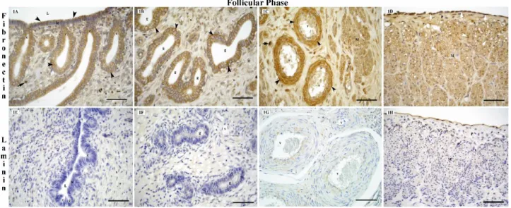
Figure 1. Localization of fibronectin receptor (integrin β1), (1A, 1B, 1C, 1D) and laminin (1E, 1F, 1G, 1H) in the bitch uterus during the follicular phase of the oestrus cycle.
L: lumen, c: crypt, le: luminal epithelium, g: endometrial gland, v: blood vessel, s: stroma, M: myometrium, P: perimetrium, black arrow heads: fibronectin receptor (integrin β1) positive cytoplasmic staining, black arrows: integrin β1 positive stromal cells, white arrow heads: mesothelial cells and white arrows: fibronectin receptor (integrin β1) positive nuclear staining, bars: 50 μm.
Şekil 1. Köpek uterusunda östrus siklusunun folliküler fazında fibronektin reseptör (integrin β1) (1A, 1B, 1C, 1D) ve laminin (1E, 1F, 1G, 1H) lokalizasyonu.
L: lümen, c: kript, le: lüminal epitel, g: endometriyal bez, v: kan damarı, s: stroma, M: miyometriyum, P: perimetriyum, siyah ok başları: fibronektin reseptör (integrin β1) pozitif sitoplazmik boyanma, siyah oklar: integrin β1 pozitif stromal hücreler, beyaz ok başları: mezoteliyal hücreler ve beyaz oklar: fibronektin reseptör (integrin β1) pozitif çekirdek boyanması, bars: 50 μm.
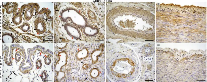
Figure 2. Localization of fibronectin receptor (integrin β1) (2A, 2B, 2C, 2D) and laminin (2E, 2F, 2G, 2H) in the bitch uterus during the luteal phase of the oestrus cycle.
L: lumen, c: crypt, le: luminal epithelium, g: endometrial gland, v: blood vessel, s: stroma, e: endothelial cells, m: media, a: adventitia, M: myometrium, P: perimetrium, black arrow heads: fibronectin receptor (integrin β1) and laminin positive nuclear staining, red arrow heads: fibronectin receptor (integrin β1) and laminin positive cytoplasmic staining, black arrows: fibronectin receptor (integrin β1) and laminin positive stromal cells, red arrows: laminin positive basal membrane, bars: 50 μm.
Şekil 2. Köpek uterusunda östrus siklusunun luteal fazında fibronektin reseptör (integrin β1) (2A, 2B, 2C, 2D) ve laminin (2E, 2F, 2G, 2H) lokalizasyonu.
L: lümen, c: kript, le: lüminal epitel, g: endometriyal bez, v: kan damarı, s: stroma, e: endoteliyal hücreler, m: mediya, a: adventisya, M: miyometriyum, P: perimetriyum, siyah ok başları: fibronektin reseptör (integrin β1) ve laminin pozitif çekirdek boyaması, kırmızı ok başları: fibronektin reseptör (integrin β1) ve laminin pozitif sitoplazmik boyanma, siyah oklar: fibronektin reseptör (integrin β1) ve laminin pozitif stromal hücreler, kırmızı oklar: laminin pozitif bazal membran, bars: 50 μm.
Figure 3. Localization of fibronectin receptor (integrin β1) (3A, 3B, 3C) and laminin (3D, 3E, 3F) in the bitch uterus during the anestrous phase of the oestrus cycle.
L: lumen, c: crypt, le: luminal epithelium, g: endometrial gland, v: blood vessel, s: stroma, e: endothelial cells, m: media, a: adventitia, M: myometrium, black arrow heads: fibronectin receptor (integrin β1) positive nuclear staining, red arrow heads: laminin positive cytoplasmic staining, black arrows: fibronectin receptor (integrin β1) and laminin positive stromal cells, bars: 50 μm.
Şekil 3. Köpek uterusunda östrus siklusunun anöstrus fazında fibronektin reseptör (integrin β1) (3A, 3B, 3C) ve laminin (3D, 3E, 3F) lokalizasyonu.
L: lümen, c: kript, le: lüminal epithel, g: endometriyal bez, v: kan damarı, s: stroma, e: endoteliyal hücreler, m: mediya, a: adventisya, M: miyometriyum, siyah ok başları: fibronektin reseptör (integrin β1) pozitif çekirdek boyanması, kırmızı ok başları: laminin pozitif sitoplazmik boyanma, siyah oklar: fibronektin reseptör (integrin β1) ve laminin pozitif stromal hücreler, bars: 50 μm.
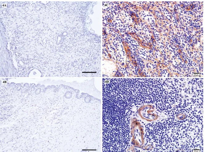
Figure 4. No immunostaining in the bitch uterus sections incubated without the primary antibody fibronectin receptor (integrin β1) (4A) and laminin (4B), Scale bars: 50 µm. Positive immunostaining for fibronectin receptor (integrin β1) in human spleen (4C) and positive immunostaining for laminin in human spleen (4D), bars: 20 µm.
Şekil 4. Köpek uterus kesitlerinde negatif kontrol için primer antikor fibronektin reseptör (integrin β1) (4A) ve laminin (4B) kullanılmadan yapılan immunboyama. İnsan dalağında fibronektin reseptörü (integrin β1) için pozitif immun boyanma (4C). İnsan dalağında laminin için pozitif immun boyanma (4D),bars: 20 µm.
The localization of laminin: It was observed that, in
general, laminin was localized to the basal membranes. Laminin immunoreactions were not observed in the basal membranes of the surface and crypt epithelial cells of the endometrium at the follicular (Fig. 1E) and anoestrus stages (Fig. 3D). In the glandular epithelium, the laminin immunoreaction, which was absent at the follicular stage (Fig. 1F), was determined to be moderate at the anestrous stage (Fig. 3E). At the luteal stage, laminin was present at a high level in the basal membranes of the surface, crypt and glandular epithelium of the endometrium (Fig. 2E and F). While the presence of laminin in the stromal cells was weak at the anestrous stage (Fig. 3E) and strong at the luteal stage (Fig. 2E and F), laminin was absent in the follicular stage (Fig. 1E and F). In the bitch uterus, the laminin immunoreaction, localized to the cytoplasm of the smooth muscle cells of the myometrium, was weak at the follicular stage (Fig. 1H) and strong at the luteal (Fig. 2H) and anestrous stages (Fig. 3F). The laminin immunoreaction in the endothelial and subendothelial cells of the blood vessels was strong, particularly at the
luteal stage (Fig. 2G). Immunreactions in the smooth muscle cells of the blood vessels were weak at the follicular stage (Fig. 1G), moderate at the anestrous stage (Fig. 3F), and strong at the luteal stage (Fig. 2G). It was determined that while the laminin immunoreaction in the adventitia of the blood vessels was weak at the follicular and anestrous stages, it was strong at the luteal phase. The mesothelial cells displayed a strong laminin positive reaction at all stages of the estrous cycle (Fig. 1H). Results obtained for the fibronectin receptor (integrin β1) and
laminin immmunoreactions in the endometrium (surface epithelium, crypt epithelium, glandular epithelium, stromal cells), myometrium, perimetrium, mesothelial cells and blood vessels (tunica intima: endothelial cells, subendothelial cells, tunica media, tunica adventitia) are presented in Tables I and II. No immunostaining was observed in the uterus sections, which were incubated with PBS instead of primary antibody (Fig. 4A-B). Positive immunostainings were detected for fibronectin receptor (integrin β1) (4C) and for laminin in human spleen sections
Table 1. Scoring for specific staining of the fibronectin receptor (Integrin β1) in the bitch uterus during the follicular, luteal and anestrous phases of the estrous cycle.
Tablo 1. Köpek uterusunda östrus siklusunun folliküler, luteal ve anöstrus fazlarında fibronektin reseptör (İntegrin β1)’ün spesifik boyanma skorları.
Portions of the uterus Follicular phase Luteal phase Anestrous phase
Endometrium: Surface epithelium ++ +++ ++ Crypt epithelium ++ +++ ++ Glandular epithelium ++ +++ +++ Stromal cells ++ +++ +++ Myometrium +++ +++ +++ Perimetrium +++ +++ +++ Mesothelial cells +++ +++ +++ Blood vessels: Endothelial cells +++ +++ +++ Subendothelial cells ++ ++ ++ Tunica media +++ +++ +++ Tunica adventitia ++ ++ ++
Staining intensity; (-), no reaction; (+), weak reaction; (++), moderate reaction; (+++), strong reaction.
Boyanma yoğunluğu; (-), reaksiyon yok; (+), zayıf reaksiyon; (++), orta derece reaksiyon; (+++), kuvvetli reaksiyon.
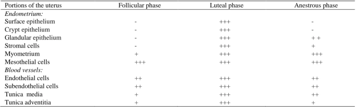
Table 2. Scoring for specific staining of the laminin in the bitch uterus during the follicular, luteal and anestrous phases of the estrous cycle. Tablo 2. Köpek uterusunda östrus siklusunun folliküler, luteal ve anöstrus fazlarında lamininin spesifik boyanma skorları.
Portions of the uterus Follicular phase Luteal phase Anestrous phase
Endometrium: Surface epithelium - +++ - Crypt epithelium - +++ - Glandular epithelium - +++ + + Stromal cells - +++ + Myometrium + +++ +++ Mesothelial cells +++ +++ +++ Blood vessels: Endothelial cells ++ +++ ++ Subendothelial cells ++ +++ ++ Tunica media + +++ ++ Tunica adventitia + +++ +
Staining intensity: (-), no reaction; (+), weak reaction; (++), moderate reaction; (+++), strong reaction.
Boyanma yoğunluğu; (-), reaksiyon yok; (+), zayıf reaksiyon; (++), orta derece reaksiyon; (+++), kuvvetli reaksiyon.
Discussion and Conclusion
In several studies, the pattern of expression of the integrin family of cell adhesion molecules has been described in the human, (13, 15, 23) rat, (11, 14, 18) mouse, (4, 28) cow, (12) monkey, (9) mare, (17) and sow (19, 21) endometrium.However, most of these studies are limited to the investigation of implantation and gestation, and only very few research is available on the estrous cycle. In this study, the presence of the fibronectin receptor (Integrin β1) and laminin, and the cyclic changes
observed in their distribution in the bitch uterus were determined for the first time during the estrous cycle, which is a preparatory period for implantation. Today, integrins serve as markers of uterine receptivity. At least 7 integrin subunits are found in the sow endometrium, (19, 21) 10 in the baboon endometrium, (9) and 14 in the human endometrium (16).Klentzeris et al. (16) and Bilalis et al. (3) suggested that, at the luteal stage, the β1integrins
localized to the endometrium positively affected both
uterine receptivity and the endometrial compartments, and aided in cell-ECM and cell-cell recognition as well as in the attachment of the blastocyst to the endometrium. Taylor et al. (24) determined strong integrin β1 reaction in
the glands, stroma and vasculature of the healthy human uterus during the proliferative and secretory phases. Reports indicate that, in the human uterus, the expression of the β1integrins varies throughout the estrous cycle and
predominant expression is reached in the secretory phase. Furthermore, it has been indicated that β1integrins are
located in the glandular epithelium during the early proliferative phase and in the stromal cells during the mid-secretory phase (13, 23, 29). In the present study, it was determined that in the bitch uterus, the expression of the fibronectin receptor (Integrin β1) reached its peak level at
the luteal stage. Beliard et al. (2) reported that, the absence of the fibronectin receptor α4β1 in the epithelial cells of the
endometrium during the proliferative phase, reduced the number of cell-matrix connections, and resulted in the
localization of flattened and elongated glandular structures to the deep layers of the stroma, while the increase in the level of α4β1 after ovulation, at the luteal
stage, strengthened stromal adhesion and enabled the maintenance of the undulated appearance of the glands. In the present study, it was ascertained that in the bitch uterus, the fibronectin receptor (integrin β1) reaction in the
surface epithelium, crypt epithelium and glandular epithelium was moderate at the follicular stage, and strong at the luteal stage. At the follicular stage, it was observed that glands with a narrow lumen were few in the surface of the stroma and extended to the deeper layers, whereas at the luteal stage, as a result of the increased level of the fibronectin receptor (integrin β1) in the endometrium,
glands with a wide lumen and strengthened stromal connections predominated in the basal region. The integrins α4β1 (13, 23) and αvβ3 (16) which are highly
expressed by the epithelial cells of the fertile human endometrium at the luteal stage of the estrous cycle, were found not to exist in the infertile human endometrium. It has been reported that as these molecules enable the attachment of the trophoblast to the endometrium, their absence results in implantation failure (30). In the glandular epithelium of the baboon uterus, the fibronectin receptor α4β1 is expressed at the luteal stage and the
integrin αvβ3 during gestation, whilst the vitronectin and
osteopontin receptor α5β3 is expressed by stromal cells
throughout the estrous cycle (9).In the bovine uterus, the integrin αvβ3 is expressed strongly in the basal membrane
of the intercaruncular luminal epithelium during metestrus, proestrus, and estrus, and moderately in the blood vessels, myometrium and the basal membrane of the shallow glands at all stages of the estrous cycle (12).The expression of the subunits α4, α5 and β1 in the uterine
luminal epithelium depends on the stage of the estrous cycle with the highest level of expression being observed between days 10-15 of the cycle in sow (5). In the porcine uterus, it was determined that, between days 15-35 of gestation, the integrin α5β1 was expressed in the surface
and glandular epithelium. Furthermore, it was ascertained that the expression of the integrin α5β1 increased with the
advance of gestation, as a result of increased sex steroid levels (21). Nishida et al. (18) reported that the fibronectin receptor (integrin β1) was expressed in the epithelial and
vascular endothelial cells of the endometrium in the rat uterus throughout the estrous cycle, and indicated that at the proestrus stage, during which the active migration of epithelial cells occurs, the fibronectin receptor (integrin β1) was localized to the basal and lateral surfaces of the
epithelial cells, but began to disappear at the estrus stage. Researchers (18) have indicated that the binding of fibronectin to the fibronectin receptor protects and restores the epithelium. As it is not known whether the commercially available antibodies used to identify the subtypes of integrins react with canine tissues, in the
present study, only the human placental fibronectin receptor (Integrin β1) (Clone FNR31) antibody was used.
At all stages of the estrous cycle, a positive immunoreaction was detected for the fibronectin receptor (integrin β1). Basement membranes, which are structures
composed of extracellular matrix separate different tissues from each other.Laminins are the predominating proteins of the basal lamina, which serves as a protein network foundation for the majority of cells and organs. Laminins are an active and fundamental component of the basal lamina, which are involved in cell adhesion, migration, and differentiation (25).The changes observed for laminin in relation to the sexual cycle have been demonstrated in several animal species. It has been reported that, in the uterus of healthy women and women suffering from endometriosis, laminin is found in the glandular epithelial cells and vascular basal membrane at all stages of the sexual cycle, and is expressed in the stromal cells of the endometrium only during the secretory phase (2). Reports indicate that, in the uterus of the baboon, (9) rat, (10) and mare (17) laminin is located in the basal membrane of both the endometrial glands and the vascular endothelium, and that the expression of laminin does not change during the estrous cycle. It was determined that laminin was localized to the glandular and endometrial epithelial cells and the vascular basal laminae in the involution period in the porcine uterus (19), throughout the estrous cycle in the bovine uterus (12) and at the anestrous stage in the bitch uterus (1). The results of the present study demonstrated that, similar to the human, baboon, rat and mare uterus, in the bitch uterus laminin was located in the basal membranes. The presence of laminin in the basal membrane of the surface and glandular epithelial cells was strong only at the luteal stage of the estrous cycle and weak at the anoestrus stage. On the other hand, laminin was absent at the follicular stage. It has been reported that during the decidualization process, while the level of laminin increases in the decidual cells, the levels of fibronectin and collagen types I and IV decrease. It has also been indicated that laminin stimulates implantation (10). In the present study the detection of an increased laminin immunoreaction in the bitch uterus at the luteal stage is in support of the view that laminin stimulates implantation. Research has shown that in rats, laminin is expressed by the smooth muscle cells of the myometrium at all stages of the sexual cycle, as well as during gestation (18). Despite being weak at the follicular stage, laminin immunoreaction was detected in the myometrial smooth muscle cells of the bitch uterus at all stages of the estrous cycle, in the present study. The dense muscular localization of laminin at the luteal stage, which is the preparatory period for implantation, and during gestation, is considered to be an indicator of the contribution of laminin to the maintenance of the strength of the myometrial structure, which is required for the continuation
of gestation. In the healthy bitch uterus, laminin and fibronectin receptor (Integrin β1) immunoreactions are
very strong at the luteal stage, during which both ovulation and implantation occur. The results of the present study are expected to provide a better understanding of the causes of ovulation, implantation and embryonic development problems encountered in bitches, the clinical use of laminin and fibronectin for the treatment of reproductive disorders, and the implantation period, which is considered the most important stage of reproduction.
Acknowledgements
This study received financial support under Project No: VA-07-06, as approved by the Scientific Research Council of Erciyes University.
References
1. Beijerink NJ (2007): Endocrinology of physiology and
progestin - induced canine anoestrus. PhD thesis, Faculty
of Veterinary Medicine, Utrecht University, The Netherlands. 2. Beliard A, Dönmez J, Nisolle M, et al. (1997):
Localization of laminin, fibronectin, E-cadherin and integrins in endometrium and endometriosis. Fertil Steril,
67, 266-272.
3. Bilalis DA, Klentzeris LD, Fleming S (1996):
Immunohistochemical localization of extracellular matrix proteins in luteal phase endometrium of fertile and infertile patients. Hum Reprod, 11, 2713-2718.
4. Bowen JA, Hunt JS (1999): Expression of cell adhesion
molecules in murine placentas and a placental cell line. Biol
Reprod, 60, 428-434.
5. Burghardt RC, Bowen JA, Newton GR, et al. (1997):
Extracellular matrix and the implantation cascade in pigs.
J Reprod Fertil, 52,151-164.
6. Butinar J, Mujagic E, Galac S. (2004): The oestrus cycle
in the bitch. Slov Vet Res, 41, 5-11.
7. Chandra SA, Adler RR (2008): Frequency of different
estrous stages in purpose - bred beagles: A retrospective study. Toxicol Pathol, 36, 944-949.
8. Crossman GA (1937): A Modification of Mallory’s
connective tissue stain with a discussion of the principles involved. Anat Rec, 69, 33-38.
9. Fazleabas AT, Bell SC, Fleming S, et al. (1997):
Distribution of integrins and the extracellular matrix proteins in the baboon endometrium during the menstrual cycle and early pregnancy. Biol Reprod, 56, 348-356.
10. Glasser SR, Lampelo S, Munir MI, et al. (1987):
Expression of desmin, laminin and fibronectin during in situ differentiation (decidualization) of rat uterine stromal cells.
Differentiation, 35, 132-142.
11. Kayışlı ÜA, Asar M, Demir R (2000): Ratlarda
desidualizasyon süresince ekstrasellüler matriksin yeniden modellenmesinde laminin ve fibronektin ile reseptör alt birimleri integrin β4 ve α5’in dağılımları ve muhtemel
rolleri. Turk J Biol, 24, 379 - 395.
12. Kimmins S, Maclaren LA (1999): Cyclic modulation of
integrin expression in bovine endometrium. Biol Reprod,
61, 1267-1274.
13. Klentzeris LD, Bulmer JN, Trejdosiewicz LK, et al. (1993): Beta-1 integrin cell adhesion molecules in the
endometrium of fertile and infertile women. Hum Reprod, 8,
1223-1230.
14. Korgun ET, Çayli S, Asar M,et al. (2007): Distribution
of laminin, vimentin and desmin in the rat uterus during initial stages of implantation. J Mol Hist, 38, 253-260.
15. Lessey BA, Damjanovich L,Coutifaris C, et al. (1992):
Integrin adhesion molecules in the human endometrium-correlation with the normal and abnormal menstrual cycle.
J Clin Invest, 90, 188-195.
16. Lessey BA, Ilesanmi AO, Lessey MA, et al. (1996):
Luminal and glandular endometrial epithelium express integrins differently throughout the menstrual cycle: implications for implantation, contraception, and infertility.
Am J Reprod Immunol, 35, 195-204.
17. Mansour GD, Henry M, Ferreira MR (2003):
Immunohistochemical study of equine endometrial extracellular matrix during the oestrous cycle. J Comp Path,
129, 316-319.
18. Nishida T, Murakami J, Otori T (1991): Expression of
fibronectin receptor (integrin) in the uterus of rats in relation to the estrous cycle. Histochemistry, 96, 279-283. 19. Ogawa H, Takahashi M, Takahashi H, et al. (2001):
Histochemical observations during uterine involution in meishan pigs. J Reprod Develop, 47, 83-89.
20. Okkens AC, Kooistra HS (2006): Anoestrus in the dog: A
fascinating story. Reprod Dom Anim, 41, 291-296.
21. Rashev P, Georgieva R, Rees D (2005): Expression of
alpha 5 beta 1integrin and fibronectin during early pregnancy in pigs. Folia Biol, 51, 121-125.
22. Ruoslahti E (1991): Integrins. J Clin Invest, 87, 1-5. 23. Tabibzadeh S (1992): Patterns of expression of integrin
molecules in human endometrium throughout the menstrual cycle. Hum Reprod, 7, 876-882.
24. Taylor CV, Letarte M, Lye SJ (1996): The expression of
integrins and cadherins in normal human uterus and uterine leiomyomas. Am J of Obstet Gynecol, 175, 411-419.
25. Timpl R, Rohde H, Robey PG, et al. (1979): Laminin - a
glycoprotein from basement membranes. J Biol Chem, 254,
9933-9937.
26. Van Cruchten S, Van den Broeck W,D’haeseleer M,et al. (2004): Proliferation patterns in the canine endometrium
during the estrous cycle. Theriogenology, 62, 631-64130.
27. Vermeirsch H, Simoens P, Hellemans A, et al. (2000):
Immunohistochemical detection of progesterone receptors in the canine uterus and their relation to sex steroid hormone levels. Theriogenology, 53, 773 -788.
28. Wewer UM, Damjanov A,Weiss J,et al. (1986): Mouse
endometrial stromal cells produce basement-membrane components. Differentiation, 32, 49-58.
29. Yoshimura Y, Shiokawa S, Nagamatsu S,et al. (1995):
Effects of beta-1 integrins in the process of implantation.
Horm Res, 44, 36-41.
30. Yoshimura Y (2002): The role of integrin in the human
reproductive process. J Reprod Develop, 48, 215-232. Geliş tarihi: 28.03.2017 / Kabul tarihi: 03.07.2017
Address for correspondence:
Prof. Dr. Güner KÜÇÜK BAYRAM
Erciyes University, Faculty of Veterinary Medicine, Department of Histology and Embryology, 38039 Talas, Kayseri, Turkey