Online available at: www.entupdates.org
1Zeliha KAPUSUZ, 2Mahmut ÖZKIRIŞ, 3Muzaffer GENCER, 4Ayşe Yeşim GÖÇMEN, 5Yusuf Kenan DAĞLIOĞLU
1Department of Otolaryngology, Head and Neck Surgery, İstinye University Medical Faculty, Associate Professor 2Department of Otolaryngology, Head and Neck Surgery, Acıbadem Hospital, Kayseri, Associate Professor
3Department of Anesthesia and Resuscitation İstinye University Liv Hospital, Assistant Professor 4Department of Biochemistry, Bozok University Medical Faculty, Associate Professor 5Medical Sciences Research and Application Center, Cukurova University, Associate Professor
Abstract
Objectives: We aimed to look into potential associations between
specific biomarkers and trauma to Cranial Nerve VII (CNVII) in a rabbit model, focusing on whether endocrine studies have potential as bio-markers in this context.
Methods: 30 adult New Zealand rabbits with intact facial muscles
were used for the research. Each animal underwent identical surgery by the same surgeon. The facial nerve divisions were exposed by incis-ing below the level of the mandible. After dissection of the skin and subcutaneous layers, the buccal division of CNVII was located with the nerve stimulator and microscopic dissection and a section of nerve 10mm long was excised in each case from the buccal branch of CNVII. Blood samples were drawn 8 weeks and 12 weeks after nerve injury had been surgically induced. The samples from day 1, week 4 and week 8 were tested for the following levels: Testosterone, oestrogen, progesterone, free T3 and T4, Cancer antigen 19-9 (Ca19-9), folate, TPSA, FPSA, FSH, LH, CA15.3, CAE, AFP and prolactin.
Results: The levels of free T3 and T4 as well as testosterone, were
down at 4th week, but at 8 weeks each had increased. Ca19-9 levels were also above the baseline. At 4 weeks, whilst oestrogen had mark-edly risen, progesterone had fallen. The statistical significance of the change in levels of free T3 and T4, testosterone, oestrogen, progester-one and FPSA was evaluated. For the group of animals with induced paralysis, the association between the lesion and testosterone, oes-trogen, progesterone, free T3 and T4, Ca19-9, and folate levels were strong and at the level of statistical significance.
Conclusion: There were statistically significant alterations in the
se-rum levels of free T3 and T4, testosterone, oestrogen, progesterone and FPSA at the 4 and 8 week intervals post surgically-induced CNVII injury. It is likely that rabbit pathophysiology resembles human patho-physiology in nervous injury, hence these six biomarkers may be of value in managing trauma or idiopathic degeneration of CNVII in hu-mans. The authors hope this study will pave the way for future re-search in this area.
Keywords: Injury, facial nerve, hormones
Changes In Various Hormone Levels In The Rabbit
Traumatic Facial Nerve Injury Model
Correspondence: Zeliha KAPUSUZ
İstinte unv. Liv Hospital Esenkent/Istanbul E-mail: [email protected]
Received: July 10, 2018; Accepted: July 25, 2018
Introduction
The facial musculature is principally innervated by crani-al nerve VII (CNVII), thus damage to this supply causes complete muscular paralysis of the face on the same side
as the injury.[1] Trauma to the skull and facial skeleton
of-ten result in damage to the peripheral branches of CNVII
and the resulting loss of function may be both severe and
ongoing.[2] Whilst there are now better outcomes in
inju-ries involving complete transection of the nerve thanks to microsurgery, for a subgroup of cases with CNVII injury, the residual functional deficits are still a cause for concern.
[3] Potential problems that may remain include undue
ex-posure of the cornea, which may develop ulcers and lead to visual impairment, hypersalivation, dysarthric speech and leakage from the oral cavity. These deficits are not in-frequently accompanied by a loss of self-confidence and a
tendency to withdraw socially.[2]
There is a need for deeper knowledge concerning the relationship between neurological trauma and biochemical markers (biomarkers) to assist with the discovery of novel agents and methods to promote neuronal regrowth.
Biomarkers are molecules of endogenous origin, the lev-els of which are taken to reflect disease onset, progression and treatment response. They are of growing significance in nervous system trauma to augment other investigato-ry diagnostic techniques such as electroencephalography, evoked sensorimotor responses, transcranial vascular ul-trasonography, near-infrared spectroscopic techniques or other imaging modalities. Several proteins that originate from the nervous system are now known and they are being evaluated at present for their ability to elucidate how severe injury is, what the likely prognosis will be, and to see if they can cast light on the level of cell injury and the molecular
pathways involved when neurones are damaged.[4] At the
time of writing, a biomarker that can be used routinely in the context of diagnosing and managing CNVII damage is lacking. This research therefore aimed to look into poten-tial associations between specific biomarkers and trauma to CNVII in a rabbit model, focusing on whether endocrine studies have potential as biomarkers in this context.
Materials and Methods
Animals
30 adult rabbits, male and female, of New Zealand White type and with a weight between 2.5 and 3.2 kg each were utilized for the research. Only those animals whose facial muscles were intact were used. Ethical approval for this study was obtained from the Ethics Committee of the Çukurova University Faculty of Medicine. Careful precau-tions were taken to keep suffering by the animals at the lowest level and the minimum number of animals con-sistent with obtaining a statistically significant result were used. The animals were held under fixed environmental conditions – ambient temperature of 25°C with relative humidity of between 10% and 55%. There was an unlim-ited access to standard feed material and water.
Each animal underwent identical surgery by the same
in-dividual. Anesthesia consisted of 10 mg/kg xylasine hydro-chloride (Rompun®, Bayer Pharmaceuticals, Turkey) and 50 mg/kg ketamine hydrochloride (Ketalar®, Eczacibasi Pharmaceuticals, Turkey). Prior to administering anesthe-sia, atropine was given subcutaneously to provide antispas-modic and anticholinergic cover. The area to be operated upon was first sterilized using iodine-povidine (Batticon® solution, Adeka Pharmaceuticals, Turkey).
Facial Nerve Surgery
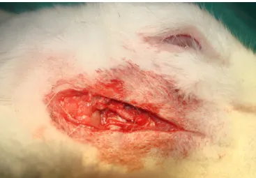
Surgery was undertaken using sterile technique and the operating microscope. The rabbit was lain on its side and the area anterior to the auricle and the left side of the neck were shaved. The facial nerve divisions (dorsal buccal, ventral buccal and marginal mandibular) were exposed by incising below the level of the mandible. After dissection of the skin and subcutaneous layers, the buccal division of CNVII was located with the facial nerve stimulator and microscopic dissection and a section of nerve 10 mm long was excised in each case from the buccal branch of CNVII (n=25) (Figures 1-3). Postoperatively the rabbits were kept on heated pads and then placed in separate cages where they could eat and drink water ad libitum. Recovery from surgery was in all cases uncomplicated.
Blood samples and biochemical measurements
Blood samples were drawn 8 weeks and 12 weeks after nerve injury had been surgically induced. First, the animals were fasted for 12 hours. The samples were centrifuged at 4°C, then divided into no more than three aliquots to be deep frozen at -80°C prior to analysis. The samples from day 1, week 4 and week 8 were tested for the following levels: Testosterone, oestrogen, progesterone, free T3 and T4, Ca19-9, folate, TPSA, FPSA, FSH, LH, CA15.3, CAE, AFP and prolactin. The assays were performed by means of the Abbott Architect 1000i analyzer, using the manufacturer’s own kits for CMIA (Chemiluminescent Microparticle Immunoassay).
Statistical analysis
The means plus standard errors were noted for each as-say and the Statistical Package for Social Sciences version 17.0 was used for analysis. One-way ANOVA was used to analyze differences between groups. Comparisons were performed by Tukey procedure post hoc and correlation was analyzed using the Pearson test. A value of p<0.05 was taken to indicate statistical significance.
Results
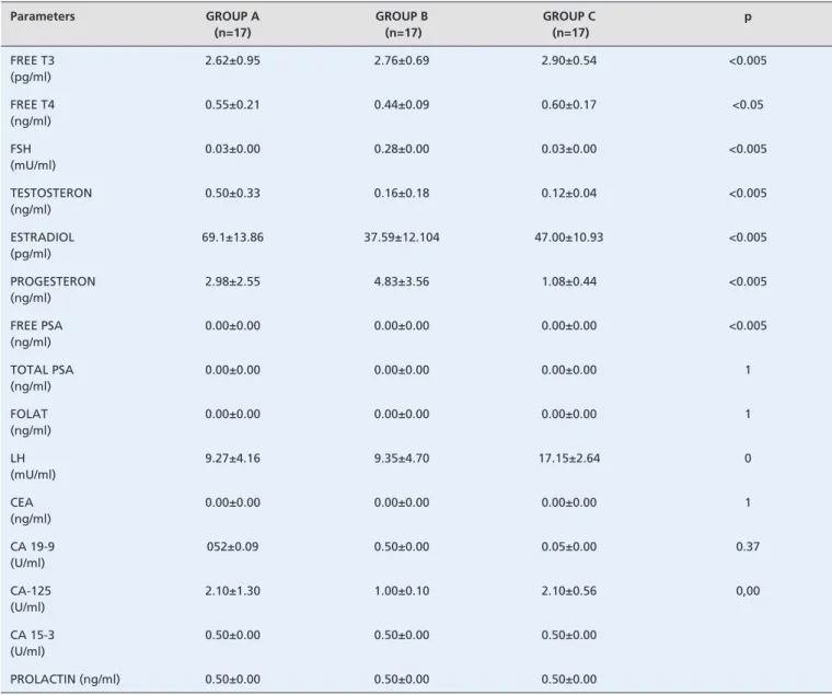
The 3 groups can be compared in Table 1, showing the situation prior to surgery, at 4 and 8 weeks post-surgical-ly and at the point the experiment was terminated. At 4 weeks, the levels of free T3 and T4 as well as testosterone were down, but at 8 weeks each had increased. Ca19-9 lev-els were also above baseline. At 4 weeks, whilst oestrogen had markedly risen, progesterone had fallen. The statisti-cal significance of the change in levels of free T3 and T4, testosterone, oestrogen, progesterone and FPSA was eval-uated. For the group of animals with induced paralysis, the association between the lesion and testosterone, oestrogen, progesterone, free T3 and T4, Ca19-9, and folate levels were strong and at the level of statistical significance.
Discussion
Trauma to the skull and facial skeleton frequently causes damage to the peripheral branches of CNVII and the re-sulting loss of function may be both severe and ongoing. Complete transection of CNVII can be better treated cur-rently as a result of improved methods in microsurgery for remediating nerve injury. The treatment for partial nerve transection, by contrast, is medical, and thus only progress in drug discovery or novel methods aimed at regenerating
neurones can improve the prognosis for such patients.[2]
For this study, rabbits were considered preferable to mice for disease model as their axonal recovery process-es from crush injury are more similar to those of humans (chromatolysis preceding restoration of neuronal function)
than those seen in mice.[5] Anatomically, CNVII in rabbits
resembles that of humans.
“Stress response” refers to a pattern of endocrine and metabolic alterations subsequent upon trauma or insult that forms an element of the larger systemic response, in-volving, as it does, biochemical, endocrine, immune and
hematological adaptations.[6] Ischemia and trauma to
neu-ral tissues (such as CNVII) result in lesions both directly (primary insult) and indirectly (secondary to endogenous mediator activity). Trauma to CNVII may result in a cas-cade of biochemical and pathological adaptations leading to cellular necrosis and loss of function. The first adap-tations include the hydrolytic conversion of membrane components into fatty acids, the synthesis of bioactive ei-cosanoids and production of reactive oxygen species (ROS) through oxidative degradation. ROS are the principal means through which cellular injury occurs. Peroxidation
Figure 1. Exposure of the buccal branch of CNVII
Figure 2. Dissection of the buccal branch of CNVII
of lipid typically occurs by involvement of superoxide, hy-droxyl or ferryl moieties.
Laboratory investigations may help to define how inju-ry occurs as well as delimit a period in which it is beneficial to attempt therapeutic remediation – an issue of clinical as well as economic significance. The availability of suita-ble investigations (i.e. biomarkers) may greatly assist with defining probable outcomes to aim for in planning
medi-cal care.[7] Biomarkers should be isolable from plasma and
their level should reflect the severity of the neural insult. Biomarkers in blood or cerebrospinal fluid might point to what types of cells or what cellular function is being
im-paired. Early results in their use for diagnosis of neoplasia, cardiac failure, infectious disorders and genetic conditions have been encouraging.
Currently the biomarkers with most widespread ac-ceptance are CK-MB (the MB isoform of creatine kinase) and the troponins, which find application as the primary way to confirm a suspected acute myocardial infarction,
replacing ECG in this regard.[8] Neuron-specific enolase
[9-11], S100B [12,13], myelin-basic protein [14], glial fibrillary
acidic protein [10,12] and cleaved tau protein [15] have all been
put forward as potentially of value in predicting prognosis following traumatic brain injury from a moderate to high
Table 1. Comparison of various biochemical markers.
Parameters GROUP A (n=17) GROUP B (n=17) GROUP C (n=17) p FREE T3 (pg/ml) 2.62±0.95 2.76±0.69 2.90±0.54 <0.005 FREE T4 (ng/ml) 0.55±0.21 0.44±0.09 0.60±0.17 <0.05 FSH (mU/ml) 0.03±0.00 0.28±0.00 0.03±0.00 <0.005 TESTOSTERON (ng/ml) 0.50±0.33 0.16±0.18 0.12±0.04 <0.005 ESTRADIOL (pg/ml) 69.1±13.86 37.59±12.104 47.00±10.93 <0.005 PROGESTERON (ng/ml) 2.98±2.55 4.83±3.56 1.08±0.44 <0.005 FREE PSA (ng/ml) 0.00±0.00 0.00±0.00 0.00±0.00 <0.005 TOTAL PSA (ng/ml) 0.00±0.00 0.00±0.00 0.00±0.00 1 FOLAT (ng/ml) 0.00±0.00 0.00±0.00 0.00±0.00 1 LH (mU/ml) 9.27±4.16 9.35±4.70 17.15±2.64 0 CEA (ng/ml) 0.00±0.00 0.00±0.00 0.00±0.00 1 CA 19-9 (U/ml) 052±0.09 0.50±0.00 0.05±0.00 0.37 CA-125 (U/ml) 2.10±1.30 1.00±0.10 2.10±0.56 0,00 CA 15-3 (U/ml) 0.50±0.00 0.50±0.00 0.50±0.00 PROLACTIN (ng/ml) 0.50±0.00 0.50±0.00 0.50±0.00
degree of severity. Of these potential biomarkers, S100B has received the most interest. Studies investigating the outcome in low severity neural injury secondary to trauma have used S100B as a possible marker.
This research investigated biomarkers experimentally. There were statistically significant alterations in the serum levels of free T3 and T4, testosterone, oestrogen,
proges-terone and FPSA at the 4 and 8 week intervals post surgi-cally-induced CNVII injury. It is likely that rabbit patho-physiology resembles human pathopatho-physiology in nervous injury, hence these six biomarkers may be of value in man-aging trauma or idiopathic degeneration of CNVII in hu-mans. The authors hope this study will pave the way for future research in this area.
1. Odebode TO, Ologe FE. Facial nerve palsy after head injury: Case incidence, causes, clinical profile and outcome. J Trauma 2006;61:388-91.
2. Diaz LM, Steele MH, Guerra AB, et al. The role of topically ad-ministered FK506 (tacrolimus) at the time of facial nerve repair using entubulation neurorrhaphy in a rabbit model. Ann Plast Surg 2004;52:407-13.
3. Wu G, Ju L, Jin T, et al. Local delivery of recombinant human bone morphogenetic protein-2 increases axonal regeneration and the expression of tau protein after facial nerve injury. J Int Med Res 2010;38:1682-8.
4. Cata JP, Abdelmalak B, Farag E. Neurological biomarkers in the pe-rioperative period. Br J Anaesth 2011;107:844-58.
5. Costa HJ, Silva CF, Korn GP, Lazarini PR. Posttraumatic facial nerve regeneration in rabbits. Braz J Otorhinolaryngol 2006;72:786-93.
6. Mukhtar AM, Obayah EM, Hassona AM. The use of dexmedetomi-dine in paediatric cardiac surgery. Anesth Analg 2006;103:52-6. 7. Kövesdi E, Lückl J, Bukovics P, et al. Update on protein biomarkers
in traumatic brain injury with emphasis on clinical use in adults and pediatrics. Acta Neurochir (Wien) 2010;152:1-17.
8. Lewandrowski K, Chen A, Januzzi J. Cardiac markers for myocar-dial infarction. A brief review. Am J Clin Pathol 2002;118:S93-9. References
9. Bandyopadhyay S, Hennes H, Gorelick MH, Wells RG, Walsh-Kel-ly CM. Serum neuron-specific enolase as a predictor of short-term outcome in children with closed traumatic brain injury. Acad Emerg Med 2005;12:732-8.
10. Vos PE, Lamers KJ, Hendriks JC, et al. Glial and neuronal proteins in serum predict outcome after severe traumatic brain injury. Neurol-ogy 2004;62:1303-10.
11. Lima JE, Takayanagui OM, Garcia LV, Leite JP. Use of neuron-spe-cific enolase for assessing the severity and outcome in patients with neurological disorders. Braz J Med Biol Res 2004;37:19-26. 12. Pelinka LE, Kroepfl A, Leixnering M, Buchinger W, Raabe A,
Redl H. GFAP versus S100B in serum after traumatic brain inju-ry: relationship to brain damage and outcome. J Neurotrauma. 2004;21:1553-61.
13. Petzold A, Green AJ, Keir G, et al. Role of serum S100B as an early predictor of high intracranial pressure and mortality in brain injury: a pilot study. Crit Care Med 2002;30:2705-10.
14. Thomas DG, Palfreyman JW, Ratcliffe JG. Serum-myelin-basic-pro-tein assay in diagnosis and prognosis of patients with head injury. Lancet. 1978;1:113-5.
15. Shaw GJ, Jauch EC, Zemlan FP. Serum cleaved tau protein levels and clinical outcome in adult patients with closed head injury. Ann Emerg Med 2002;39:254-7.
This is an open access article distributed under the terms of the Creative Commons Attribution-NonCommercial-NoDerivs 3.0 Unported (CC BYNC-ND3.0) Licence (http://creativecommons.org/licenses/by-nc-nd/3.0/) which permits unrestricted noncommercial use, distribution, and reproduction in any medium, provided the original work is properly cited.
Please cite this article as: Kapusuz Z, Özkiriş M, Gencer M, Göçmen A. Y, Dağlioğlu Y. K. Changes in Various Hormone levels in the Rabbit Traumatic Facial