The Evaluation of Clinical Signs in Patients with Suspected
Renovascular Hypertension
Renovasküler Hipertansiyon Şüphesi Olan Hastalarda Klinik
İpuçlarının Değerlendirilmesi
ABSTRACT
OBJECTIVE: Renovascular hypertension (RVH) is the most common yet correctable cause of
secondary hypertension if diagnosed early. There are many clinical signs that can suggest RVH. The aim of this study was to find which clinical or laboratory signs are more indicative in diagnosing RVH and in determining which patients should go through renal angiography.
MATERIAL and METHODS: The study included 184 patients who presented to our clinic due to
hypertension and were under risk of RVH. All patients underwent three-dimensional time-of-flight Magnetic Resonance Angiography with phase-contrast. The patients were divided into two groups as with and without renal artery stenosis, supported by MRA.
RESULTS: Advanced age, low body mass index, high serum creatinine level, presence of proteinuria,
and patients with diabetes mellitus and coronary artery disease were found to be significant risk factors for RVH. Only the presence of renal asymmetry and the history of coronary artery disease were found to be independent risk factors.
CONCLUSION: In conclusion, detailed patient history and the evaluation of renal size are very
important for patients with hypertension. Coronary artery disease and a difference in renal size of more than 1.5 cm could be strong indicators of RVH.
KEY WORDS: Renovascular hypertension, Renal artery stenosis, Magnetic resonance angiography
ÖZ
AMAÇ: Renovasküler hipertansiyon (RVH); erken teşhis edildiğinde sekonder hipertansiyonun
en fazla düzeltilebilme ihtimali olan nedenlerinden biridir. RVH’u destekleyen pek çok klinik ipucu bulunmaktadır. Çalışmanın amacı, RVH tanısında, klinik ve laboratuvar ipuçlarından hangilerinin daha belirleyici olduğunu bulmak ve renal anjiyografi yapılması gereken hastaları belirlemektir.
GEREÇ ve YÖNTEMLER: Çalışmaya hipertansiyon nedeni ile başvuran ve RVH için risk faktörü
taşıyan 184 hasta dahil edildi. Tüm hastalara üç boyutlu kontrastlı manyetik rezonans anjiyografi (MRA) uygulandı. Hastalar MRA sonucuna göre; renal arter stenozu olanlar ve olmayanlar şeklinde iki gruba ayrıldı.
BULGULAR: İleri yaş, düşük vücut kitle indeksi, yüksek serum kreatinin seviyesi, proteinüri, diabetes
mellitus ve koroner arter hastalığı varlığı, RVH için önemli risk faktörleri olarak bulundu. Koroner arter hastalığı öyküsü ile renal asimetri varlığı bağımsız risk faktörü olarak saptandı.
SONUÇ: Sonuç olarak, ayrıntılı öykü alınması ve böbrek boyutunun değerlendirilmesi hipertansiyon
hastaları için büyük önem taşımaktadır. Koroner arter hastalığı ve böbrek boyutları arasında 1.5 cm’den büyük fark, RVH’nun güçlü birer göstergesi olabilir.
ANAHTAR SÖZCÜKLER: Renovasküler hipertansiyon, Renal arter stenozu, Manyetik rezonans
anjiyografi
Correspondence Address:
Nihan TEKKARIŞMAZ
Başkent Üniversitesi Tıp Fakültesi, Nefroloji Bilim Dalı,
Adana, Turkey Phone : + 90 322 344 44 44 E-mail : [email protected] Received : 21.12.2016 Accepted : 27.07.2017 Nihan TEKKARIŞMAZ1 Dilek TORUN1 Uğur ÖZKAN2 Ayşegül ZÜMRÜTDAL3
Fatma Nurhan ÖZDEMIR ACAR4
1 Başkent University, Faculty of Medicine, Department of Nephrology,
Adana, Turkey
2 Private Adana Middle East Hospital, Department of Interventional Radiology, Adana, Turkey
3 Acıbadem Adana Hospital, Department of Nephrology, Adana, Turkey
4 Başkent University, Faculty of Medicine, Department of Nephrology,
INTRODUCTION
Renovascular hypertension (RVH) is a clinical situation characterized by high blood pressure (BP) resulting from renal ischemia in the presence of stenosis of the the renal artery or arteries (RAS) (1). It is the most common yet correctable cause of secondary hypertension and closely associated with chronic renal failure, increased cardiovascular mortality, and end organ damage (1-5). The prevalence of RVH varies with the clinical settings. It is about 1-5% in all the hypertensive population, less than 1% in patients with mild-moderate high blood pressure, and between 10-45% in patients with severe, malignant or resistant hypertension (1-10)
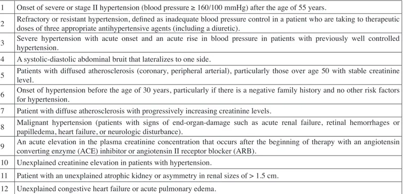
Although renal angiography is the gold standard for the diagnosis of RAS, it is an invasive and costly method. Due to the risks associated with contrast nephropathy, angiography is recommended for patients at high risk for RVH (1, 4). MRA is being increasingly used as the fist line screening test for RVH (10). Compared with DSA, MRA has median sensitivity and specificity of 90-96% and 76-94%, respectively (7,10). There are many clinical signs that can suggest RVH. However, no single sign is useful to determine patients at high risk for RVH. There is a commonly used clinical risk index for RVH (Table I) (5, 10-15). In this study, the aim was to find which clinical or laboratory signs are more indicative in the diagnosis of RVH and to determine which patients should go through renal angiography.
MATERIAL and METHODS
In our clinic, renal MRA examination is routinely required for admitted patients suspected of renovascular hypertension. In addition, we obtain informed consent from each patient before the MRA examination.
This study was approved by the Baskent University Institutional Review Board (Project no: KA 17/170). The study included 184 patients who presented to our clinic due to hypertension and had risk factor(s) for RVH between October 2007 and March 2009. Of these patients, 77 were male (42%) and 107 were female (58%) (mean age ± SD: 53.5 ± 14 years).
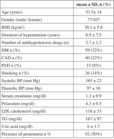
Patient demographics (age, gender, body mass index (BMI), medical history (duration of hypertension, number of antihypertensive drugs, presence of diabetes mellitus (DM), coronary artery disease (CAD) and peripheral arterial disease (PAD), smoking habits, mean BP), laboratory values (serum creatinine, potassium, low density lipoprotein-cholesterol (LDL- cholesterol), triglycerides (TG), and uric acid levels in blood, and presence of proteinuria (Table II), were recorded. All patients were assessed by the clinical risk index (Table I).
Blood pressure was measured three times with at least one week interval by standard sphygmomanometer. Measurements were taken from patients in the sitting position, with the back supported and the arm supported at the heart level, after resting for 5 minutes without speaking. The last two measured blood pressure values were averaged and recorded. Patients with a
Table I: Clinical Risk Index for Renovascular Hypertension (adapted from reference 11: 2005 ACC/AHA practice guidelines).
1 Onset of severe or stage II hypertension (blood pressure ≥ 160/100 mmHg) after the age of 55 years.
2 Refractory or resistant hypertension, defined as inadequate blood pressure control in a patient who are taking to therapeutic doses of three appropriate antihypertensive agents (including a diuretic). 3 Severe hypertension with acute onset and an acute rise in blood pressure in patients with previously well controlled hypertension. 4 A systolic-diastolic abdominal bruit that lateralizes to one side.
5 Patients with diffused atherosclerosis (coronary, peripheral arterial), particularly those over age 50 with stable creatinine level. 6 Onset of hypertension before the age of 30 years, particularly if there is a negative family history and no other risk factors for hypertension. 7 Patient with diffuse atherosclerosis with progressively increasing creatinine levels.
8 Malignant hypertension (patients with signs of end-organ-damage such as acute renal failure, retinal hemorrhages or papilledema, heart failure, or neurologic disturbance). 9 An acute elevation in the plasma creatinine concentration that occurs after the beginning of therapy with an angiotensin converting enzyme (ACE) inhibitor or angiotensin II receptor blocker (ARB). 10 Unexplained creatinine elevation in patients with hypertension.
11 Patient with an unexplained atrophic kidney or asymmetry in renal sizes of > 1.5 cm. 12 Unexplained congestive heart failure or acute pulmonary edema.
mean systolic blood pressure ≥140 mm Hg, mean diastolic blood pressure ≥90 mm Hg or those using antihypertensive drugs were recorded as patients with hypertension. The diagnosis of CAD was determined according to the clinical history of the patients (angina pectoris, acute myocardial infarction, bypass surgery, balloon dilatation or stent implantation in coronary arteries) and the results of the electrocardiography and echocardiography test. Patients with a fasting blood glucose level ≥126 mg/dl or those using antidiabetic drugs were recorded as patients with DM. The diagnosis of PAD was determined based on presence of intermittent claudication, stroke or previous endovascular surgery in the history of the patients and Doppler ultrasound signs in the lower extremity. If the patients had smoked regularly for at least 1 year, we considered them to be habitual smokers.
Technical Data
The levels of glucose, creatinine, LDL- cholesterol, TG, and uric acid were studied by the enzymatic colorimetric method in Beckman Unicel DXC 800 analyzer (Beckman Coulter, Inc., Fullerton, CA, USA). The levels of LDL-cholesterol were calculated by the Friedewald formula. The concentration
of potassium was determined by same analyzer using the ion-selective electrode method. Protein levels in urine were studied by turbidimetric methods in the Hitachi 912 analyzer.
All patients with suspected RVH underwent contrast enhanced renal magnetic resonance angiography (MRA). MRA was performed using 1.5T MR systems (Avanto, Siemens, Erlanger, Germany). Contrast-enhanced breath-hold FISP three-dimensional MRA was applied in the coronal view, centered at the renal arteries. An anterior-posterior phased-array surface coil (torso array coil) was used. We used sagittal, coronal, and axial localizing pulse sequences, followed by image acquisition in the coronal plane. The FLASH 3D gradient echo sequence was employed. The following imaging parameters were used: repetition time 2.8 ms; echo time 1.02 ms; flip angle 25°; field of view 400 mm; matrix size 256 x 512; slice thickness 1.2 mm. For contrast administration, an intravenous cannula was placed in an antecubital arm vein and connected to long extension tubing. We used approximately 15-20 mL (0.2 mmol/kg) of meglumine gadoterate (Dotarem, Guerbet, France). The contrast material was injected with an automatic injector at 2 mL/sec, followed by a 20 mL saline flush. All patients were required to hold their breath during image acquisition. After the extracted images were processed by the ‘maximum intensity projection’ method, they were converted into a three-dimensional image. The two- and three-dimensional images were evaluated separately. The diameter of the renal artery and the size of both kidneys were evaluated.
Patients were divided into two groups as with and without RAS. Patient with RAS were classified into two subgroups according to the degree of RAS as hemodynamically significant (≥60% diameter stenosis) and not hemodynamically significant (1,2,9,14,16).
Statistical Analysis
SPSS for windows 11.0 was used in statistical evaluation of the data (SPSS Inc., Chicago, IL, USA). Descriptive statistics (number, percentage and mean ± SD) were used as the statistical method. Parametric and nonparametric differences between groups were evaluated using the Student T and Mann-Whitney U tests. Chi-square was used to compare categorical values. Linear regression analysis was used to determine the possible risk factors associated with RAS. P value <0.05 was accepted as statistically significant.
RESULTS
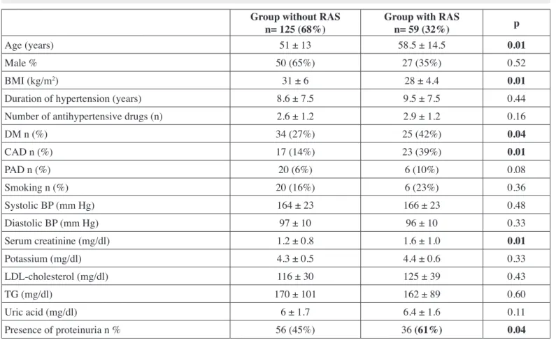
Renal artery stenosis was found in 32% (59/184) of the patients with hypertension. Hemodynamically significant RAS was found in 21% (39/184) of all patients. Age (58.5 ± 14.5 vs. 51 ± 13 years, respectively, p<0.01) and serum creatinine levels (1.6 ± 1.0 vs. 1.2 ± 0.8 mg/dl, respectively, p<0.01) were significantly higher while BMI (28 ± 4.4 vs. 31 ± 6 kg/ m2, respectively, p<0.01) was significantly lower in patients with RAS compared to those without RAS. The prevalence of
Table II: Demographic and basal laboratory data of patients. mean ± SD, n (%)
Age (years) 53.5± 14
Gender (male/ female) 77/107
BMI (kg/m2) 30.1 ± 5.8
Duration of hypertension (years) 8.9 ± 7.5 Number of antihypertensive drugs (n) 2.7 ± 1.2
DM n (%) 59 (32%) CAD n (%) 40 (22%) PAD n (%) 15 (8%) Smoking n (%) 26 (14%) Systolic BP (mm Hg) 165 ± 23 Diastolic BP (mm Hg) 97 ± 10 Serum creatinine (mg/dl) 1.3 ± 0.9 Potassium (mg/dl) 4.3 ± 0.5 LDL-cholesterol (mg/dl) 118 ± 33 TG (mg/dl) 167 ± 97 Uric acid (mg/dl) 6 ± 1.7 Presence of proteinuria n % 92 (50%)
BMI: Body mass index, DM: Diabetes mellitus, CAD: Coronary
artery disease, PAD: Peripheral arterial disease, BP: Blood pressure,
LDL-Cholesterol: Low density lipoprotein-cholesterol, TG: Triglycerides.
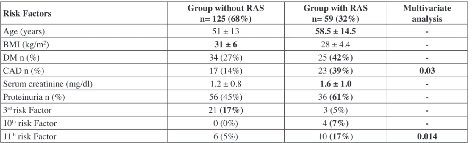
DM (25 (42%) vs. 34 (27%), respectively, p= 0.04), CAD (23 (39%) vs. 17 (14%) respectively, p<0.01), and proteinuria (36 (61%) vs. 56 (45%) respectively, p=0.04) were more significant in patients with RAS. There were no statistically significant difference between the groups with respect to gender, duration of hypertension, number of antihypertensive drugs, PAD, smoking, systolic and diastolic BP, and laboratory parameters including serum potassium, LDL-cholesterol, TG, uric acid levels (Table III). The presence of the clinical risk factors including the 3rd (Severe hypertension with acute onset and an acute rise in BP in patients with previously well controlled hypertension) risk factor was significantly lower, while the 10th (Unexplained creatinine elevation in patients with hypertension) and 11th (Patient with an unexplained atrophic kidney or asymmetry in renal sizes of >1.5 cm) risk factors were significantly higher in patients with RAS (Table IV). CAD and the 11th risk factor of clinical risk index were independent variables that were associated with RAS in a multivariate linear regression analysis (Table V).
Hemodynamically significant RAS with MRA was detected in 39 patients. 33 of these 39 patients underwent DSA. 6 patients did not give consent for the procedure. Significant RAS was
detected in 29 of 33 patients who underwent DSA. Of the 29 patients undergoing revascularization, only 21 had a decrease in the number of antihypertensive drugs.
DISCUSSION
Although the prevalence of RAS is not known exactly, it has been reported to be between 7% and 45% in the literature according to method used for the diagnosis and the characteristic of the patients (1-10). Hemodynamically significant RAS was detected about 21% of our patients with MRA. In the literature, this rate was reported at between 6.3% and 23% in different patients groups (1-3,9,13,17). Compared with DSA, MRA has median sensitivity and specificity of 90-96 and 76-94%, respectively (7,10). We were unable to calculate sensitivity and specificity in our study because we did not perform DSA in patients without RAS according to MRA.
Advanced age has been reported as a significant risk factor for RAS in many studies (1,3-4,17-18). We found similar results.
The association between gender and RAS has varied in different studies. Similar to the studies of Ozkan and Krijnen (2,4), we found that gender was not a risk factor for RAS.
Table III: Distribution of patients according to the presence of renal artery stenosis. Group without RAS
n= 125 (68%) Group with RASn= 59 (32%) p
Age (years) 51 ± 13 58.5 ± 14.5 0.01
Male % 50 (65%) 27 (35%) 0.52
BMI (kg/m2) 31 ± 6 28 ± 4.4 0.01
Duration of hypertension (years) 8.6 ± 7.5 9.5 ± 7.5 0.44
Number of antihypertensive drugs (n) 2.6 ± 1.2 2.9 ± 1.2 0.16
DM n (%) 34 (27%) 25 (42%) 0.04 CAD n (%) 17 (14%) 23 (39%) 0.01 PAD n (%) 20 (6%) 6 (10%) 0.08 Smoking n (%) 20 (16%) 6 (23%) 0.36 Systolic BP (mm Hg) 164 ± 23 166 ± 23 0.48 Diastolic BP (mm Hg) 97 ± 10 96 ± 10 0.33 Serum creatinine (mg/dl) 1.2 ± 0.8 1.6 ± 1.0 0.01 Potassium (mg/dl) 4.3 ± 0.5 4.4 ± 0.6 0.33 LDL-cholesterol (mg/dl) 116 ± 30 125 ± 39 0.43 TG (mg/dl) 170 ± 101 162 ± 89 0.60 Uric acid (mg/dl) 6 ± 1.7 6.4 ± 1.6 0.11 Presence of proteinuria n % 56 (45%) 36 (61%) 0.04
BMI: Body mass index, DM: Diabetes Mellitus, CAD: Coronary artery disease, PAD: Peripheral arterial disease, BP: Blood pressure, LDL-Cholesterol: Low density lipoprotein-cholesterol, TG: Triglycerides.
Table IV: Distribution of Clinical Risk Index for Renovascular Hypertension according to the presence of renal artery stenosis.
Risk Factors without RASGroup
(n=125) n %
Group with RAS
(n=59) n % p
1. Onset of severe or stage II hypertension (blood pressure ≥ 160/100 mm Hg) after the
age of 55 years. 7 (6%) 3 (5%) 0.88
2. Refractory or resistant hypertension, defined as inadequate blood pressure control in a patient who are taking to therapeutic doses of three appropriate antihypertensive
agents (including a diuretic). 40 (32%) 12 (20%) 0.10
3. Severe hypertension with acute onset and an acute rise in blood pressure in patients
with previously well-controlled hypertension. 21 (17%) 3 (5%) 0.02
4. A systolic-diastolic abdominal bruit that lateralizes to one side. 2 (2%) 2 (3%) 0.43 5. Patients with diffused atherosclerosis (coronary, peripheral arterial), particularly
those over age 50 with stable creatinine level. 12 (10%) 9 (15%) 0.26
6. Onset of hypertension before the age of 30 years, particularly if there is a negative
family history and no other risk factors for hypertension. 10 (8%) 3 (5%) 0.47 7. Patient with diffuse atherosclerosis with progressively increasing creatinine levels. 10 (8%) 5 (8%) 0.91 8. Malignant hypertension (patients with signs of end-organ-damage such as acute renal
failure, retinal hemorrhages or papilledema, heart failure, or neurologic disturbance). 4 (3%) 0 (0%) 0.16 9. An acute elevation in the plasma creatinine concentration that occurs after the
beginning of therapy with an angiotensin converting enzyme (ACE) inhibitor or
angiotensin II receptor blocker (ARB). 12 (10%) 8 (14%) 0.42
10. Unexplained creatinine elevation in patients with hypertension. 0 (0%) 4 (7%) 0.003
11. Patient with an unexplained atrophic kidney or asymmetry in renal sizes of > 1.5 cm. 6 (5%) 10 (17%) 0.006
12. Unexplained congestive heart failure or acute pulmonary edema. 0 0
RAS: Renal artery stenosis.
Table V: Distribution of patients with and without renal artery stenosis according to the potential risk factors.
Risk Factors Group without RASn= 125 (68%) Group with RASn= 59 (32%) Multivariate analysis
Age (years) 51 ± 13 58.5 ± 14.5 -BMI (kg/m2) 31 ± 6 28 ± 4.4 -DM n (%) 34 (27%) 25 (42%) -CAD n (%) 17 (14%) 23 (39%) 0.03 Serum creatinine (mg/dl) 1.2 ± 0.8 1.6 ± 1.0 -Proteinuria n (%) 56 (45%) 36 (61%) -3rd risk Factor 21 (17%) 3 (5%) -10th risk Factor 0 (0%) 4 (7%) -11th risk Factor 6 (5%) 10 (17%) 0.014
RAS: Renal artery stenosis, BMI: Body mass index, DM: Diabetes Mellitus, CAD: Coronary artery disease.
Various results have been reported regarding the relationship between BMI and RAS. In one study, obesity was shown as a risk factor for atherosclerotic RAS (9). Other studies have reported that low BMI is a risk factor for RAS (3,4). In our study, BMI was found to be lower in patients with RAS.
In a study it was shown that the prevalence of RAS decreased when the duration of hypertension increased (4). In our study, there was no relation between these two parameters.
Some studies have suggested that use of multiple antihypertensive agents is a risk factor for RAS (1,19). In our study, no correlation was found between the two parameters. Only 23% of the patients whose BP was not adequately controlled (2nd risk factor) despite the use of three appropriate
antihypertensive agents including diuretics had RAS. In our study, the 2nd risk factor was not considered to be a determinant
for RAS.
The prevalence of RAS is high in patients with DM because of increased atherosclerotic vascular disease. While Ozkan et al. did not show any association between DM and RAS, Paraskevas et al. showed the presence of DM to be a risk factor for atherosclerotic RAS (2,9). Similar to Paraskevas, our study showed that the presence of DM increased the risk of RAS.
The presence of various sites of concomitant atherosclerotic vascular disease in hypertensive patients is a clinical marker for RAS. Various studies have shown that CAD in addition to hypertension is a clinical marker (1,3,18,17). Some studies have indicated that the number of vessels affected and degree of coronary artery stenosis are associated with RAS (9,18). Park et al. have determined RAS in 13.5% of their patients with significant CAD (17). In our study, the presence of CAD in addition to hypertension was found to be the strongest independent risk factor for RAS. We have found RAS in 57.5% of patients with CAD.
In some studies, PAD was specified as a risk factor for RAS (2,3,7,17). However, in our study, the presence of PAD in addition to hypertension was not identified as a risk factor. This difference may be due to difficulties in the diagnosis of PAD.
Some studies show that the smoking habit is a risk factor for atherosclerotic RAS (4, 9). Smoking in our patient population was not detected as a risk factor for RAS.
In various studies, an increased level of plasma creatinine has been shown to be a powerful marker for RAS (1,3,9,20). Uzu et al. found that 39% of patients with renal failure had RAS (18). Similarly, it was found in 44% of patients with renal failure in this study. Only 4 patients had the 10th risk factor. RAS was
observed in all patients. The 10th risk factor was also found to be
an important risk factor.
Hyperlipidemia as a risk factor for developing RAS has been challenged in the literature. In different studies by Park, Krijnen and Paraskevas, hyperlipidemia has been shown to be a clinical marker for RAS (4,9,17). Hansen et al. showed low HDL to be an independent risk factor (8). A relationship between hyperlipidemia and RAS was not demonstrated in Ozkan’s study (2). In our study, a correlation was not found between the lipid levels and RAS.
Proteinuria is an indicator of renal parenchyma damage in the presence of atherosclerotic RAS (21). In this study, the presence of proteinuria was seen at higher rates in patients with RAS
compared to those without at 61% to 45% respectively. Similar to this study, Uzu et al. found that the presence of proteinuria was associated with RAS (18).
In previous studies, sudden onset or accelerated hypertension was defined as a risk factor regardless of age (19,20). In this study, the 3rd risk factor was found to have the lowest predictive value. Our patients 55 years or older, with severe or stage II hypertension (our 1st risk factor), had RAS at a rate as low as 30%, which led to a statictically low predictive value.
The presence of an abdominal murmur was shown to be a marker for RAS (4). In literature, the presence of murmur was reported in only 45% of the patients with RAS, and in 9% of the patients with essential hypertension (1). In a study, abdominal bruits have been reported in 77.7% to 86.9% of RAS patients (22). We had four hypertensive patients with a murmur in the abdomen (4th risk factor). RAS was detected in only two of them
with MRA. The presence of an abdominal murmur was not statistically significant in our study. The reason for this result could be that the number of patients with the 4th risk factor was
not adequate for the study.
Diffuse atherosclerosis with stable (5th risk factor) or
progressively increased (7th risk factor) serum creatinine levels is
an important determinant of RAS. In this study, both risk factors showed similar rates for RAS, 43% and 33% respectively. These ratios do have determinative values, even though they are not statistically significant.
In our study, only 3 patients who were 30 years old without a family history of hypertension (6th risk factor) had RAS detected
with MRA.
In the medical literature, RAS is closely associated with malignant hypertension (8th risk factor) (9,19). However, we
were not able to demonstrate a relationship between RAS and the 8th risk factor. The reason for this outcome might be the low
number of patients with the 8th risk factor.
Safian’s study showed that RAS must be suspected in cases of renal failure triggered by ACE inhibitors (20). We detected RAS in 40% of the 20 patients who had undergone renal MRA because of the 9th risk factor. However, this was not considered
statistically meaningful in our study.
In previous reports, a strong correlation was reported between the presence of RAS and renal asymmetry (9,20,23). The 11th risk factor was an independent and strong marker in our
study. Our patients with renal asymmetry had a RAS rate as high as 62.5%. This result was important for early diagnosis of RAS that could cause kidney failure.
There were no patients with the 12th (Unexplained congestive
heart failure or acute pulmonary edema) risk factor among those who presented to the nephrology clinic with hypertension. The reason for not having any patients with the 12th risk factor in this
acute pulmenary edema tend to go directly to emergency care rather than the nephrology clinic.
In conclusion, the kidney sizes and parenchymal structures of patients with hypertension should be evaluated by imaging techniques for the early diagnosis of RVH. If there is more than 1.5 cm difference in size between the two kidneys or renal atrophy is present, the possibility of RVH should be considered. A detailed history should be obtained from hypertensive patients. The existence of additional CAD should be queried. Patients with a positive history of CAD should be noted regarding the possibility of RVH.
REFERENCES
1. Krzesinski JM: Diagnostic criteria for renovascular hypertension. Acta Chir Belg 2002;102:159-166
2. Ozkan U, Oguzkurt L, Tercan F, Nursal TZ: The prevalance and clinical predictors of incidental atherosclerotic renal artery stenosis. Eur J Radiol 2009;69:550-554
3. Przewlocki T, Kablak-Ziembicka A, Tracz W, Kopec G, Rubis P, Pasowicz M, Musialek P, Kostkiewicz M, Kozanecki A, Stompór T, Sulowicz W, Sokolowski A: Prevalence and prediction of renal artery stenosis in patients with coronary and supraaortic artery atherosclerotic disease. Nephrol Dial Transplant 2008;23:580-585 4. Krijnen P, van Jaarsveld BC, Steyerberg EW, Man in ‘t Veld AJ,
Schalekamp MA, Habbema JD: A clinical prediction rule for renal artery stenosis. Ann Intern Med 1998;129:705-711
5. Textor S: Evaluation of secondary hypertension. Uptodate. Literature review current through: [İnternet yayını]. Jan 2015. Erişim:http://www.uptodate.com/contents/evaluation-of-secondary-hypertension.
6. Vasbinder GBC, Nelemans PJ, Kessels AGH: Diagnostic tests for renal artery stenosis in patients suspected of having renovascular hypertension: A meta-analysis. Ann Intern Med 2001;135:401-411 7. Hirsch AT, Haskal ZJ, Hertzer NR, Bakal CW, Creager MA, Halperin
JL, Hiratzka LF, Murphy WR, Olin JW, Puschett JB, Rosenfield KA, Sacks D, Stanley JC, Taylor LM Jr, White CJ, White J, White RA, Antman EM, Smith SC Jr, Adams CD, Anderson JL, Faxon DP, Fuster V, Gibbons RJ, Hunt SA, Jacobs AK, Nishimura R, Ornato JP, Page RL, Riegel B; American Association for Vascular Surgery; Society for Vascular Surgery; Society for Cardiovascular Angiography and Interventions; Society for Vascular Medicine and Biology; Society of Interventional Radiology; ACC/AHA Task Force on Practice Guidelines Writing Committee to Develop Guidelines for the Management of Patients with Peripheral Arterial Disease; American Association of Cardiovascular and Pulmonary Rehabilitation; National Heart, Lung, and Blood Institute; Society for Vascular Nursing; TransAtlantic Inter-Society Consensus; Vascular Disease Foundation. Circulation 2006;113:e463-654 8. Hansen KJ, Edwards MS, Craven TE, Cherr GS, Jackson SA, Appel
RG, Burke GL, Dean RH: Prevalence of renovascular disease in the elderly: A population based study. J Vasc Surg 2002;36:443-451
9. Paraskevas KI, Hamilton G, Cross JM, Mikhailidis DP: Atherosclerotic renal artery stenosis: Association with emerging vascular risk factors. Nephron Clin Pract 2008;108:c56-c66 10. Kallıstratos MS, Giannakopoulos A, German V, Manolis AJ:
Diagnostic modalities of the most common forms of secondary hypertension. Hellenic J Cardiol 2010;51:518-529
11. Safian RD, Textor SC: Renal artery stenosis. N Engl J Med 2001;344:431-442
12. White CJ, Jaff MR, Haskal ZJ, Jones DJ, Olin JW, Rocha-Singh KJ, Rosenfield KA, Rundback JH, Linas SL: Indications for renal arteriography at the time of coronary arteriography: A science advisory from the American Heart Association Committee on Diagnostic and Interventional Cardiac Catheterization, Council on Clinical Cardiology, and the Councils on Cardiovascular Radiology and Intervention and on Kidney in Cardiovascular Disease. Circulation 2006;114:1892-1895
13. Chobanian AV, Bakris GL, Black HR, Cushman WC: The Seventh Report of the Joint National Committee on prevention, detection, evaluation, and treatment of high blood pressure: The JNC 7 Report. JAMA 2003;289:2560-2572
14. Borelli FA, Pinto IM, Amodeo C, Smanio PE, Kambara AM, Petisco AC, Moreira SM, Paiva RC, Lopes HB, Sousa AG: Analysis of the sensitivity and specificity of noninvasive imaging tests for the diagnosis of renal artery stenosis. Arq Bras Cardiol 2013;101:423-433
15. Herrmann SMS, Textor SC: Diagnostic criteria for renovascular disease: Where are we now? Nephrol Dial Transplant 2012;27:2657-2663
16. Baumgartner I, Lerman LO: Renovascular hypertension: Screening and modern management. European Heart Journal 2011;32:1590-1598
17. Park S, Jung JH, Seo HS, Ko YG, Choi D, Jang Y, Chung N, Cho SY, Shim WH: The prevalence and clinical predictors of atherosclerotic renal artery stenosis in patients undergoing coronary angiography. Heart Vessels 2004;19:275-279
18. Uzu T, Inoue T, Fujii T, Nakamura S, Inenaga T, Yutani C, Kimura G: Prevalence and predictors of renal artery stenosis in patients with myocardial infarction. Am J Kidney Dis 1997;29:733-738 19. Smoszna J, Wańkowicz Z: Significance of clinical anamnesis in the
preliminary diagnosis of renovascular hypertension. Pol Merkur Lekarski 1998;5:266-268
20. Safian RD: Aterosklerotic renal artery stenosis. Curr Treat Options Cardiovasc Med 2003;5:91-101
21. Chrysochou C, Cheung CM, Durow M, Middleton RJ, Solomon LR, Craig A, Venning M, Kalra PA: Proteinuria as a predictor of renal functional outcome after revascularization in atherosclerotic renovascular disease. QJM 2009;102:283-288
22. Turnbull JM: Is listening for abdominal bruits useful in the evaluation of hypertension? JAMA 1995;274:1299-301
23. Soluez G, Therasse E, Qanadli SD, Froment D, Léveillé M, Nicolet V, Turpin S, Giroux MF, Guertin MC, Oliva VL: Prediction of clinical response after renal angioplasti; respective value of renal doppler sonography and scintigraphy. AJR Am Roentgenol