Fax +41 61 306 12 34 E-Mail [email protected] www.karger.com
Case Report
Acta Haematol 2012;128:73–76 DOI: 10.1159/000338220Higher Expression of the Novel Gene Upregulated
Gene 4 in Two Acute Lymphoblastic Leukemia
Patients with Poor Prednisolone Response
Yesim Oymak
a
Yavuz Dodurga
b
Aysen Turedi
c
Yontem Yaman
d
Gulcihan Ozek
d
Ozgur Carti
d
Burcak Tatlı Gunes
d
Esin Erbudak
d
Ergul Berber
f
Cıgır Biray Avci
e
Canan Vergin
d
a Department of Pediatric Hematology, Harran University, Sanliurfa , b Department of Medical Biology and
Genetics, Medical Faculty, Pamukkale University, Denizli , c Diyarbakir Children’s Hospital, Diyarbakir ,
d Department of Pediatric Hematology, Behcet Uz Children’s Government Hospital, and e Department of Medical
Biology and Genetics, Medical Faculty, Ege University, Izmir , and f Department of Molecular Biology and
Genetics, Arel University, Istanbul , Turkey
Introduction
Acute lymphoblastic leukemia (ALL) is the most com-mon childhood malignant disease. Variations in the ex-pression level of the cell cycle genes including cyclins, cy-clin-dependent kinases (CDKs) and CDK inhibitors have been associated with leukemia [1–3] .
Recently, upregulated gene 4 (URG4) located on chro-mosome 7p13 has been implicated in tumorigenesis. For example, it was demonstrated that HBxAg-positive hepa-tocellular carcinoma cells have a strong expression of URG4 [4] . Moreover, the high level of URG4 expression in osteosarcoma tissues was correlated with proliferative activity of osteosarcoma cells [5] . It was suggested that URG4 may play a role in the development of carcinoma by regulating the expression of the cyclin D1 gene ( CCND1) [5] . URG4 has also been proposed as a therapeu-tic target in the treatment of carcinomas such as gastric cancer due to its implication in carcinogenesis [6] .
Although research in the understanding of the mo-lecular basis of ALL has been going on, the momo-lecular pathogenesis of leukemia has not yet been elucidated. However, more precise risk factors have been identified
Key Words
Acute lymphoblastic leukemia ⴢ Leukemogenesis ⴢ Poor prednisolone response ⴢ URG4
Abstract
Elucidation of the molecular mechanisms of leukemogene-sis is important for a better understanding of the prognoleukemogene-sis of acute lymphoblastic leukemia (ALL). Studies have shown that the expression of upregulated gene 4 (URG4), which pro-motes cell growth and survival, is increased in different types of carcinomas including hepatocellular carcinoma, gastric cancer and osteosarcoma. Similarly, higher expression of URG4 and cyclin D1 gene might promote proliferation of the blast cells by causing escape from the G1 checkpoint and entry into the S phase. This study reports the high expression level of URG4 in 2 high-risk ALL patients for the first time in the literature. In conclusion, the higher expression of URG4 in our 2 patients suggests that URG4 might be involved in leukemogenesis. Future studies with a large number of high-risk ALL patients and cell culture studies are needed to dem-onstrate the exact role of URG4 in leukemogenesis.
Copyright © 2012 S. Karger AG, Basel
Received: January 9, 2012
Accepted after revision: March 8, 2012 Published online: June 6, 2012
Dr. Yavuz Dodurga
Pamukkale University Department of Medical Biology and Genetics Kinikli Kampusu Morfoloji Binasi Kat: 3
TR–20070 Denizli (Turkey)
Tel. +90 258 296 2534, E-Mail yavuzdodurga @ gmail.com
© 2012 S. Karger AG, Basel 0001–5792/12/1282–0073$38.00/0 Accessible online at:
www.karger.com/aha
Oymak et al.
Acta Haematol 2012;128:73–76
74
in childhood leukemia. Prednisolone response on the 8th day of induction therapy is the earliest one for the Berlin-Frankfurt-Munich (BFM) group. Patients with a poor prednisolone response on the 8th day of treatment are classified as high-risk patients in the BMF group.
In the present study, we report 2 cases of high-risk ALL patients according to the BFM criteria with a higher ex-pression of CCND1 and URG4 . This is the first report showing URG4 upregulation in 2 ALL patients with poor prednisolone response.
Case Reports
Case 1
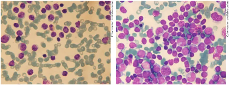
A 12-year-old girl presented to the Hematology Clinic of Dr. Behcet Uz Children’s Hospital because of high fever and weight loss. She had splenomegaly and her peripheral blood sample pro-file showed 12.7 g/dl hemoglobin, 146.0 ! 10 9 /l platelets and 6.4
! 10 9 /l leukocytes with 35% of blast cells. Moreover, L1 type
blas-tic infiltration was observed in the bone marrow aspiration smear ( fig. 1 ). The patient was diagnosed as a pre-B-cell ALL patient ac-cording to flow cytometry analysis. Her central nervous system was affected by dural enhancement in the supratentorial com-partments, which was isointense on T 1 -weighted and
hyperin-tense on T 2 -weighted MR images. There were four lymphocytes
in the cerebrospinal fluid count (by the Nageotte chamber meth-od). Cytogenetic analysis, fluorescence in situ hybridization and RT-PCR analysis of bone marrow did not show any abnormality.
Initially, chemotherapy of the BFM-2000 protocol with inter-mediate-risk group was applied. However, she had a poor pred-nisolone response with 1.178 ! 10 9 /l absolute blast count on the
8th day of treatment. Therefore, she was reclassified as a high-risk
group patient. Although 98% of the blast cells were observed on the 15th day of bone marrow treatment, she was in remission at the end of the remission induction therapy. She underwent hema-topoietic stem cell transplantation from a human leukocyte anti-gen-identical older sister after the first three high-risk blocks and she has been leukemia free for 2 years.
Case 2
Case 2 was an 8-year-old boy with a 2-week history of increas-ing fatigue, weakness and pallor. Physical examination revealed hepatosplenomegaly. His blood counts indicated leukocytosis (172.0 ! 10 9 /l), anemia (hemoglobin 3 g/dl) and
thrombocytope-nia (22.0 ! 10 9 /l). The peripheral blood analysis and bone
mar-row analysis showed L1 type blasts (55 and 65% blast cells, respec-tively) which is the characteristic feature of ALL patients ( fig. 2 ). The blast cells coexpressed T-cell markers and CD13. Although hypodiploidy was observed during cytogenetic analysis, fluores-cence in situ hybridization and RT-PCR analysis did not show any abnormality. The patient was classified as an intermediate-risk patient due to his age, his high leukocyte count and his T-cell im-mune phenotype, and thus, the BFM-2000 protocol was applied. The absolute blast count of the patient on day 8 was 70.40 ! 10 9 /l
and bone marrow on day 15 showed 3% blast cells. At bone mar-row evaluation on day 33, the patient was in remission. His risk group changed to high risk due to the poor prednisolone response on the 8th day. The patient is still alive and receiving chemother-apy due to the absence of a matching donor.
Method
Quantitative RT-PCR
In order to evaluate the molecular pathogenesis of leukemia in our 2 patients, the expression level of URG4 was analyzed. Bone marrow samples from the 2 patients and 6 controls without
Fig. 1. Bone marrow aspirate smear of case 1. Bone marrow
aspi-ration shows diffuse L1 type blastic infiltaspi-ration. ! 100.
Co lo r v e rs io n av a il a b le o n li n e
Fig. 2. Bone marrow aspirate smear of case 2. The bone marrow
shows blast cells that are characteristic of ALL. ! 100.
Co lo r v e rs io n av a il a b le o n li n e
URG4 in ALL Patients with Poor Prednisolone Response
Acta Haematol 2012;128:73–76 75
lignancy were obtained for molecular analysis. Control bone mar-row samples were taken during routine screening for diagnostic purposes, and the informed consent of the patients was obtained. The expression levels of URG4 and CCND1 were analyzed by quantitative RT-PCR method with the Light Cycler 480 Real Time PCR system. Fifty microliters (50–200 ng) of total RNA was iso-lated from the bone marrow samples by using the RT 2 First Strand
Kit (Roche Diagnostics, Germany) according to the manufactur-er’s manual. Glyceraldehyde-3-phosphate dehydrogenase (‘house-keeping’ gene) was used as an internal control in the RT-PCR. The primer sequences are given in table 1 . The results were analyzed by using the ⌬⌬CT method and quantitated with Light Cycler 쏐 480 Quantification Software.
Results
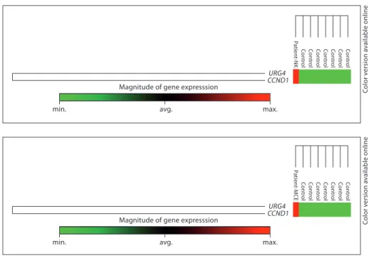
Previous studies have implicated a functional role for URG4 in cell cycle control and tumor development. In the present study, increased expression of URG4 was demon-strated in 2 patients diagnosed with childhood ALL. The expression level of URG4 and CCND1 in the patients was analyzed by quantitative RT-PCR method and compared with the expression level in the control patients without malignancy. The expression of URG4 ( fig. 3 ) and CCND1 ( fig. 4 ) was increased in both patients. Case 1 had a 6,623.3- and a 14,050.5-fold increase in URG4 and CCND1 expression, respectively. Similarly, case 2 showed a 544.3- and a 3,133.0-fold increase in URG4 and CCND1 expres-sion, respectively ( table 2 ).
Discussion
Genome-wide approaches have contributed to a better understanding of tumorigenesis by identifying the onco-genes and have also delineated novel genetic alterations
Table 1. Primers used for RT-PCR
Primer name Sequence URG4 F: 5ⴕ-CGGGAGATGGGACAGTTTTA-3ⴕ URG4 R: 5ⴕ-CATGGTGTTGAGGAGTGTGG-3ⴕ CCND1 F: 5ⴕ-AGCTCCTGTGCTGCGAAGTGGAAAC-3ⴕ CCND1 R: 5ⴕ-AGTGTTCAATGAAATCGTGCGGGGT-3ⴕ GAPDH F: 5ⴕ-CCCCACACACATGCACTTACC-3ⴕ GAPDH R: 5ⴕ-CCTAGTCCCAGGGCTTTGATT-3ⴕ
F = Forward; R = reverse; GAPDH = glyceraldehyde-3-phos-phate dehydrogenase.
Magnitude of gene expresssion
min. avg. max.
Co n tro l Co n tro l Co n tro l Co n tro l Co n tro l Co n tro l Co n tro l P a tient-NK URG4 CCND1
Magnitude of gene expresssion
min. avg. max.
Co n tro l Co n tro l Co n tro l Co n tro l Co n tro l Co n tro l Co n tro l P a tient-MCE URG4 CCND1
Fig. 3. Expression levels of URG4 and
CCND1 for case 1. Min. = Minimum; avg. = average; max. = maximum; NK = patient name ID.
Fig. 4. Expression levels of URG4 and
CCND1 for case 2. Min. = Minimum; avg. = average; max. = maximum; MCE = patient name ID.
Co lo r v e rs io n av a il a b le o n li n e Co lo r v e rs io n av a il a b le o n li n e
Table 2. Change in URG4 and CCND1 expression in 2 patients
(according to the comparison of cases and control patients)
Case 1 Case 2
Gene symbol URG4 CCND1 URG4 CCND1
Fold change 6,623.3 14,050.5 544.3 3,133.0
Oymak et al.
Acta Haematol 2012;128:73–76
76
that are associated with prognosis. Recent studies have implicated that the novel oncogene URG4 has a role in tumorigenesis.
We used the BFM study group protocol to classify the patients. According to the BFM study group protocol, poor prednisolone response on the 8th day of therapy characterizes patients as high-risk patients. Accordingly, case 1 and case 2 are classified as high-risk patients with
1.178 ! 10 9 /l and 70.400 ! 10 9 /l peripheral blast on the
8th day of therapy, respectively.
In the present study, higher expression of URG4 was demonstrated in 2 high-risk ALL patients for the first time. A 6,623.3- and a 544.3-fold increase in URG4 ex-pression was observed together with an increased expres-sion of CCDN1 .
CDKs and cyclins are important components of the eukaryotic cell division cycle. D-type cyclins are regula-tory partners of G1-CDKs which are required for entry into the S phase of the cell cycle. The cyclin protein level is important for cell proliferation since the activity of CDKs depends on the availability of cyclins [7] . Studies have shown that an abnormal increase in the cellular
CCND1 level can cause malignant cell transformation and resistance to apoptosis, thus contributing to the re-sistance of several tumor cells to chemotherapeutic agents [8–11] . In addition, studies have indicated that expression of URG4 promotes growth factor-independent survival [12] and URG4 has a role in tumorigenesis by affecting the CCND1 level through bypassing the checkpoint dur-ing the G1 to S phase transition [5, 12] . Therefore, we sug-gest that URG4 might act via CCND1 and contribute to uncontrolled proliferation of blasts in the 2 leukemia cas-es prcas-esented here, since overexprcas-ession of both CCND1 and URG4 were observed.
Another distinguishing feature of our 2 patients was poor prednisolone response which is a well-known prog-nostic feature for childhood ALL. The high level of URG4 and CCND1 in addition to the high blast count on the 8th day might implicate the presence of an ALL subtype.
In conclusion, this study provides the first clue that URG4 could be implicated in the molecular pathogenesis of leukemia. Future studies with a large number of pa-tients are needed to reveal the exact role of URG4 in the molecular pathogenesis of leukemia.
References
1 Clarke MF, Dick JE, Dirks PB, Eaves CJ, Ja-mieson CH, Jones DL, Visvader J, Weissman IL, Wahl GM: Cancer stem cells – perspec-tives on current status and future directions: AACR workshop on cancer stem cells. Can-cer Res 2006; 66: 9339–9344.
2 Benekli M, Baer MR, Baumann H, Wetzler M: Signal transducer and activator of tran-scription proteins in leukemias. Blood 2003; 101: 2940–2954.
3 Gesbert F, Griffin JD: Bcr/Abl activates tran-scription of the Bcl-X gene through STAT5. Blood 2000; 96: 2269–2276.
4 Satiroglu-Tufan NL, Dodurga Y, Gok D, Ce-tinkaya A, Feitelson MA: RNA interference mediated URG4 gene silencing diminishes cyclin D1 mRNA expression in HepG2 cells. Genet Mol Res 2010; 9: 1557–1567.
5 Huang J, Zhu B, Lu L, Lian Z, Wang Y, Yang X, Satiroglu-Tufan NL, Liu J, Luo Z: The ex-pression of novel gene URG4 in osteosarco-ma: correlation with patients’ prognosis. Pa-thology 2009; 41: 149–154.
6 Song J, Xie H, Lian Z, Yang G, Du R, Du Y, Zou X, Jin H, Gao J, Liu J, Fan D: Enhanced cell survival of gastric cancer cells by a novel gene URG4. Neoplasia 2006; 8: 995–1002. 7 Pardee AB: A restriction point for control of
normal animal cell proliferation. Proc Natl Acad Sci USA 1974; 71: 1286–1290.
8 Shintani M, Okazaki A, Masuda T, Kawada M, Ishizuka M, Doki Y, Weinstein IB, Imoto M: Overexpression of cyclin DI contributes to malignant properties of esophageal tumor cells by increasing VEGF production and decreasing Fas expression. Anticancer Res 2002; 22: 639–647.
9 Harada H, Nakagawa K, Iwata S, Saito M, Kumon Y, Sakaki S, Sato K, Hamada K: Res-toration of wild-type p16 down-regulates vascular endothelial growth factor expres-sion and inhibits angiogenesis in human gli-omas. Cancer Res 1999; 59: 3783–3789.
10 Kornmann M, Arber N, Korc M: Inhibition of basal and mitogen-stimulated pancreatic cancer cell growth by CCND1 antisense is associated with loss of tumorigenicity and potentiation of cytotoxicity to cisplatinum. J Clin Invest 1998; 101: 344–352.
11 Warenius HM, Seabra LA, Maw P: Sensitiv-ity to cisdiamminedichloroplatinum in hu-man cancer cells is related to expression of CCND1 but not c-raf-1 protein. Int J Cancer 1996; 67: 224–231.
12 Tufan NL, Lian Z, Liu J, Pan J, Arbuthnot P, Kew M, Clayton MM, Zhu M, Feitelson MA: Hepatitis Bx antigen stimulates expression of a novel cellular gene, URG4, that promotes hepatocellular growth and survival. Neopla-sia 2002; 4: 355–368.