BIOTINYLATED PEPTIDE NANOFIBERS FOR
MODULATING THE IMMUNE RESPONSE
A THESIS SUBMITTED TO
THE GRADUATE SCHOOL OF ENGINEERING AND SCIENCE OF BILKENT UNIVERSITY
IN PARTIAL FULFILLMENT OF THE REQUIREMENTS FOR THE DEGREE OF
MASTER OF SCIENCE IN
MATERIALS SCIENCE AND NANOTECHNOLOGY
By
Şehmus Tohumeken June 2016
BIOTINYLATED PEPTIDE NANOFIBERS FOR
MODULATING THE IMMUNE RESPONSE
By Şehmus Tohumeken June 2016
We certify that we have read this thesis and that in our opinion it is fully adequate, in scope and in quality, as a dissertation for the degree of Master of Science.
Ayşe Begüm Tekinay (Advisor)
Mustafa Ö. Güler
Melih Ö. Babaoğlu
Approved for the Graduate School of Engineering and Science
Levent Onural
i
ABSTRACT
BIOTINYLATED PEPTIDE NANOFIBERS FOR
MODULATING THE IMMUNE RESPONSE
Şehmus Tohumeken
M.Sc. in Materials Science and Nanotechnology Advisor: Ayşe Begüm Tekinay
Co-Advisor: Mustafa Özgür Güler June 2016
Despite the fact that vaccination eradicates many diseases, a broad variety of disorders cannot be treated using current vaccine development methods. In addition, it is difficult to rapidly develop new vaccines following the sudden onset of a new pandemic, as the production and transport of vaccines to impoverished areas is still a major issue. The lack of sufficient vaccine production, for example, enabled the spread of swine flu in 2009, while HIV, Zika and malaria viruses currently lack effective vaccinations. In addition, while cancer vaccines represent a promising area of research, their clinical implementation is also limited by the absence of rapid and effective vaccine development methods. The development of new and effective vaccines is therefore quite vital. Moreover, recently used vaccines promote either humoral or cellular immune responses, while effective treatment requires the induction of both systems. Consequently, there is an urgent need for effective and easy-to-prepare vaccines that are capable of eliciting immune action through multiple channels.
Peptide amphiphiles are small molecules that are able to self-assemble into nanoscale fibrous networks. These nanofibers are biodegradable, biocompatible and do not
ii
generate toxic byproducts, making them ideal for designing biomaterials. As such, nanofibers are a promising class of materials for inducing an effective immune response and overcoming some of the problems faced by current vaccine development methods.
In this thesis, I detail the use of biotinylated peptide nanofiber systems as immunomodulatory materials that are capable of incorporating a broad variety of antigens in a modular manner. Briefly, biotinylated and non-biotinylated peptide amphiphiles (PA) were first synthesized, purified and characterized to determine their material properties. PAs were then induced to self-assemble in the presence of CpG oligonucleotide (ODN) adjuvants, and ovalbumin was conjugated to self-assembled biotinylated-PA (B-PA) nanofibers by streptavidin linkers. Splenocytes were isolated from the mouse spleen and treated with bioactive nanofibers to investigate the effect of bioactive nanofibers on the immune response. Following the confirmation of an effective combined immune response, live mice were exposed to the nanofiber adjuvants as a proof-of-concept demonstration of in vivo PA-vaccine efficiency. Both in vivo and in vitro studies demonstrated that B-PA nanofibers are able to effectively modulate the immune response.
Given these observations, I suggest that the B-PA nanofiber can be used as an immunomodulatory material for promoting effective immune response against extracellular and intracellular pathogens, and especially for the vaccine-based treatment of cancer. As the antigen presented by the PA system can be changed in a modular manner, B-PA nanofibers can also be employed to rapidly develop new vaccines against sudden outbreaks of new viral strains.
Keywords: peptide amphiphile, self-assembly, nanofiber, adjuvant, Toll-like receptors, immune cell activation
iii
ÖZET
İMMÜN CEVABIN DÜZENLENMESİ İÇİN BİYOTİNLENMİŞ
PEPTİT NANOYAPILAR
Şehmus Tohumeken
Malzeme Bilimi ve Nanoteknoloji Programı, Yüksek Lisans Tez Danışmanı: Ayşe Begüm Tekinay
Tez Eşdanışmanı: Mustafa Özgür Güler Haziran 2016
Günümüz aşıları birçok hastalığı ortadan kaldırabildiği halde, bazı hastalıkların tedavisi için hala yetersiz kalmaktadır. Özellikle aniden yayılan hastalıklarda, aşının üretilme kapasitesi ve gerekli yerlere hızlıca nakil edilmesi, ya da nakil sırasında bozulmadan kalabilmesi sınırlıdır. Bu yüzden yeni aşıların geliştirilmesi hayati önem taşımaktadır. 2009 domuz gribi vakası sırasında, günümüz aşı sisteminde ne kadar eksiklik olduğu gün yüzüne çıkmıştır. HIV, malarya, zika gibi hastalık yapıcı patojenler için henüz etkili bir tedavi bulunamadığı gibi, kanserden ölen insanların sayısı da gün geçtikçe artmaktadır. Ayrıca, kullanılan çoğu aşılar, ya hücresel ya da humoral immün cevap oluşmasına neden olmaktadır. Etkili bir immun cevabın oluşması için hem hücresel hem de humoral immün cevabın etkili ve dengeli bir şekilde oluşması gerekmektedir. Bu yüzden yeni, etkili ve kolay hazırlanabilir bir aşı modelinin geliştirilmesine ihtiyaç duyulmaktadır.
Peptit amfifil denilen küçük moleküller, kendiliğinden bir araya gelip nano boyutlarda nanofiberler oluşturabilmektedir. Bu yapılar kolayca biyobozunabildiğinden, vücuda uyumlu olduğundan ve bozunduktan sonra zehirli ürün oluşturmadığından dolayı organizmalar için biyouyumluluk göstermekte ve uygulanabilir bir materyal olmaktadır. Bu yüzden bu yapılar, immünolojik cevabın etkin bir şekilde oluşturulması ve aşıların eksikliklerini kapatması için etkili bir yol olabilir.
iv
Bu tezde, immün cevabın düzenlenmesi için kullanılan biyotinlenmiş peptit nanofiber sistemleri anlatılmaktadır. İlk olarak bu peptidin biyotinli ve biyotinsiz hali sentezlenmiş ve karakterize edilmiştir. Bir adjuvan olan CpG-oligonükleotid (ODN) ile karıştırılarak nanofiber yapılar oluşturulmuştur. Streptavidin bağlayıcısı kullanılarak model antijen olan ovalbumin, biotinli nanofibere bağlanmıştır. Daha sonra bu yapıların fareden alınan dalak hücrelerine etkisi ve farelere verildiğinde nasıl bir reaksiyon oluşturulduğu incelenmiştir. Bütün deneyler sonucunda, biyotinli nanofiberler immün hücreleri etkin ve düzenlenebilir bir immün cevap oluşturması için uyarmaktadır. Bu sonuçlar ışığında, biyotinli peptit nanofiberlerin etkili bir immün cevap tetikleyicisi olarak kullanılarak, hücre içi, hücreler arası patojenlere ve kansere karşı etkili bir çözüm sunabileceği kanısına varılmıştır. Kolay bir şekilde hazırlanabildiğinden ve taşıdığı antijeni ortaya çıkmış salgına göre ayarlanabileceğinden dolayı, bu sistem uygulanabilirlik açısından umut vadetmektedir.
Anahtar Kelimeler: peptit amfifil, kendiliğinden bir araya gelme, nanofiber, adjuvan, Toll benzeri reseptör, bağışıklık hücrelerinin aktivasyonu
v
Kuzenim Ali Durğun’a
(1989-2013)
vi
ACKNOWLEDGEMENTS
I would like to express my appreciations to all people who contribute my MS thesis either by their experiences, knowledge, help or their mental and emotional support. I would like thank to my advisors, Prof. Tekinay and Prof. Güler for their guidance and support to my research. Their encouragement pushed me to perform at my best throughout my Master. They also contributed to my personal development and gave me a different perspective about social issues.
I would like to thank the National Nanotechnology Research Center (UNAM) and Bilkent University for providing pleasant facilities and required equipment. Bilkent University has been my home with all the unforgettable memories for the past eight years, including my undergraduate education I would like to thank to The Scientific and Technological Research Council of Turkey (TÜBİTAK) for BİDEB 2210 E master fellowship and TÜBİTAK grant number 114Z562.
I would like to indicate my greatest thanks and most sincere gratitude to my entire family’s members. My father Aşem, my mother Halime, my brother İlhan, and my sisters Sevgül, Birgül who always believe in me and support me to become who I am now. My youngest brothers and sister deserves a lot more than acknowledgement for their emotional support. I have always felt the love and endless support of my family with me. They always show respect to my decisions and never turn their backs. They always encourage me and be with me when I feel hopeless to overcome struggles. I am proud of them and I feel very lucky to have such a family.
I would love to express my special thanks to Merve Tohumeken, my wife, my friend, my ewinamin and mother of Said Musa, our son. She was always with me and always support me either in good or bad times, her supports has been priceless. I am proud of her, because he is doing in both maternity and master. I would like to express my gratitude to The Kunt family for their support to my carrier and allowing me to get married with their daughter.
I would like to express my special thanks to our old PhD Dr. Rashad Mammadov and His wife Dr.Büşra Mammadov for sharing their knowledge, and valuable collaboration. I would like to express my special thanks to Nuray Gündüz, M. Burak
vii
Demircan, Zeynep Orhan, Alper Devrim Özkan, Nurcan Haştar, Berna Şentürk, Fatih Yergöz, İdil Uyan and Merve Şen for their help in my hard time for their collaboration and friendship, which would not have been possible without their sincere efforts. I would like to thank to Ahmet Emin Topal, Aref Khalily, Gökhan Günay, Elif Arslan, Özüm Sehnaz Günel, Çağla Eren, Yasin Tümtaş, Seher Yaylacı(Üstün) and Melike Sever for their sharing of lots of knowledge, experiences in AFM,CD, flow cytometer, microscopy etc. and for the fruitful scientific discussions and for their friendship. I would also like to thank Gülistan Tansık, Dr. Ruslan Garifullah, Dr. Gülcihan Gülseren, Dr. Gözde Uzunallı, Meryem Hatip, Sever, Dr. Melis Şardan, Göksu Çinar, Mevhibe Yakut, Dr. Özlem Erol, Dr. Hakan Ceylan, Hatice Kübra Kara, Seren Hamsici, Egemen Deniz Eren, Oya İlke Şentürk, Begüm Kocatürk, Dr. Aslı Çelebioğlu, Zeynep Aytaç, Sude Selin Su Yirmibeşoğlu, Hepi Hari Susapto, İ. Ceren Garip, Dilek Sezer, İkra Gizem Yıldız, Elif Ergül, Aygül Zengin, Mustafa Beter, Canelif Yılmaz, Oğuz Tuncay, İbrahim Çelik, Özge Uysal, Zelal Yavuz, Ebru Şahin Kehribar, Ebuzer Kalyoncu, Aybegüm Samast, Kerem Emre Ercan and Tuğçe Önür for creating such a warm working environment.
I would like to thank to Mustafa Güler and Serhat, for technical assistance and helpful discussion. I especially thank to Suna Abla for spreading their happiness with her sincere smile. My special thanks to Zeynep Ergül Ülger and Zeynep Erdoğan for their technical help and friendship.
viii
TABLE OF CONTENTS
TABLE OF CONTENTS ... viii
CHAPTER 1 ... 1
1.1 Introduction ... 1
1.2 Self-assembled peptide nanostructures ... 2
1.3 An overview of the immune system ... 8
1.3.1 Innate immune system ... 9
1.3.2 Adaptive immune system ... 15
1.4 Toll like receptors and adjuvants ... 21
CHAPTER 2 ... 30
2.1 Introduction ... 30
2.2 Experimental ... 34
2.2.1 Material ... 34
2.2.2 Peptide synthesis ... 34
2.2.3 Preparation of fiber nanostructures ... 35
2.2.4 Preparation of antigen bearing fiber nanostructures ... 35
2.2.5 Transmission electron microscopy (TEM) imaging ... 36
2.2.6 Atomic force microscopy (AFM) imaging ... 36
ix
2.2.8 Polyacrylamide gel electrophoresis (PAGE) ... 37
2.2.9 Immunogold staining and transmission electron microscopy imaging .. 38
2.2.10 Binding assay by ELISA ... 39
2.2.11 Animals ... 40
2.2.12 Splenocyte culture and stimulation experiment ... 40
2.2.13 ELISA ... 41
2.2.14 The effect of nanostructures on cell viability ... 42
2.2.15 Staining of surface markers, SIINFEKL and flow cytometry ... 42
2.2.16 Evaluation of effects of PA/ODN nanofibers on immunized mice ... 43
2.2.17 Effects of PA/ODN nanostructure on splenocytes proliferation ... 43
2.2.18 Uptake of ODNs into splenocytes ... 44
2.3 Results and Discussion ... 45
2.3.1 Design and characterization of the PA/ODN nanostructure ... 45
2.3.2 Biotinylated ovalbumin binds to B-PA nanofibers ... 54
2.3.3 Nanofiber size and epitope density can be controlled by dilution and non-OVA binding peptide incorporation ... 56
2.3.4 B-PA/ODN nanostructure complex promotes cytokine secretion ... 60
2.3.5 PA/ODN nanofibers enhance expression of the maturation markers and cross-presentation of antigens and uptake of ODN... 62
2.3.6 Effect of PA/ODN nanostructures on humoral immunity and splenocytes proliferation ... 66
x
CHAPTER 3 ... 73 3. Conclusions and future perspectives ... 73 Bibliography ... 76
xi
LIST OF FIGURES
Figure 1.1 A peptide amphiphile molecule structure. ... 3
Figure 1.2 Fluorescent βtail fusion proteins stably integrate into self-assembling peptide nanofibers. ... 4
Figure 1.3 Enzymatic degradation profile of ODNs bound to self-assembled PA. ... 7
Figure 1.4 Haematopoietic stem cells differentiation... 11
Figure 1.5 Relationship between innate immunity, adaptive immunity and invaders. ... 14
Figure 1.6 General mechanism of the adaptive immune responses after antigen recognition by antigen presenting cells. ... 18
Figure 1.7 CD4+ helper cells induce cells by secreting distinct but overlapping sets of cytokines. ... 20
Figure 1.8 Simple demonstration of PRRs and PAMPs. ... 23
Figure 1.9 Pattern recognition receptors and their downstream pathways. ... 24
Figure 2.1 Chemical representation of PAs.………...………46
Figure 2.2 Characterization of K-PA. ... 47
Figure 2.3 Characterization of B-PA. ... 48
Figure 2.4 Structural characterization of PA/ODN nanostructures. ... 49 Figure 2.5 Morphological characterization of PA/ODN nanostructures.51
xii
Figure 2.6 Evaluation of K-PA/B-PA mixture on formation of self-assembled nanostructure with PAGE. ... 52 Figure 2.7 Cytotoxic effect of materials. ... 53 Figure 2.8 Antigen-bearing B-PA/ODN nanostructures. ... 55 Figure 2.9 Morphological characterizations of diluted self-assembled B-PA/ODN nanostructures. ... 57 Figure 2.10 Control of antigen binding to PA/ODN nanostructure via ELISA. ... 59 Figure 2.11 Cytokine secretion profile of splenocytes triggered by PA/ODN nanostructures and ODN. ... 61 Figure 2.12 Effect of PA/ODN and OVA on maturation marker of splenocytes. ... 63 Figure 2.13 In vitro stimulation of immune responses by PA/ODN and OVA. ... 65 Figure 2.14 In vivo immunization with PA/ODN versus soluble antigen or antigen with ODN. ... 67 Figure 2.15 IgG isotopes in sera of immunized mice with PA/ODN versus soluble antigen or antigen with ODN. ... 68 Figure 2.16 Splenocytes proliferation of immunized mice with PA/ODN versus soluble antigen or antigen with ODN. ... 70 Figure 2.17 Schematic illustration of B-PA nanofiber formation and associated transmission electron microscope images. ... 71 Figure 2.18 Effect of PAs on the cellular uptake of FITC-ODN in dendritic cells. ... 72
xiii
LIST OF TABLES
Table 1-1 TLR recognition of microbial components ... 25
Table 1-2 Characteristics of the ideal adjuvants... 27
Table 1-3 Major adjuvants and clinically approved adjuvants ... 27
xiv
LIST OF ABBREVIATION
PA : Peptide Amphiphile
AFM : Atomic Force Microscope
CD : Circular Dichroism
BrdU : Bromodeoxyuridine
FACS : Fluorescence Activated Cell Sorter
MHC : Major Histocompatibility Complex
ODN : Oligodeoxynucleotide
LC-MS : Liquid Chromatography Mass Spectroscopy
HPLC : High Pressure Liquid Chromatography
PAGE : Polyacrylamide Gel Electrophoresis
SEM : Scanning Electron Microscopy
TEM : Transmission Electron Microscopy
FBS : Fetal Bovine Serum
DMEM : Dulbecco’s Modified Eagle Medium
TCP : Tissue Culture Plate
TLR : Toll Like Receptor
1
CHAPTER 1
1.1 Introduction
Vaccination has been used as a preventive treatment for more than 200 years, and the eradication of smallpox and rinderpest stands as a testament to its impact in saving human and animal life. Nevertheless, the development of effective vaccines is still necessary due to the constant evolution of newer viral strains and transmission of zoonoses to human hosts. No vaccine has so far been successful against highly threatening diseases such as Zika, Ebola, HIV and malaria. In addition, new vaccine development methods may allow the selective targeting of cancer cells by the immune system, which would be of major importance due to the fact that cancer is expected to claim more than 11 million lives worldwide by 2030. Autoimmune diseases, neurodegenerative diseases and organ failures are other unsolved problems of the modern era, and vaccine development efforts may likewise assist in their treatment. Rationally designed biomaterials allow us to solve a wide range of health problems. Recent developments in material science and biology have greatly broadened the scope of intelligent biomaterial design for therapeutic applications, since these materials can now be synthesized, functionalized, characterized, controlled and mass produced with considerable ease. Liposomes, self-assembled nanofibers, gold nanoparticles and other materials can now be synthesized and functionalized in the nanoscale with different sizes, and subsequently used for different purposes. Liposomes, for example, are commonly employed as drug delivery vesicles for cancer therapy. In this thesis, we have used self-assembled biotinylated peptide amphiphile nanofibers for tuning the
2
immune response against a specific antigen that is noncovalently attached to the peptide matrix.
1.2 Self-assembled peptide nanostructures
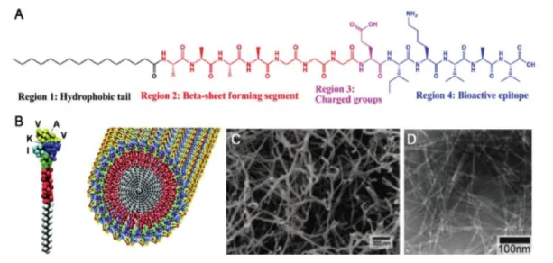
Self-assembed systems are prevalent in nature, and self-assembly is an attractive mechanism to construct materials exhibiting desired combinations of physical, chemical and biological properties. For example, the cell membrane is one of the best-known self-assembled structures. Self-assembled peptide nanostructures can be formed by mixing oppositely charged peptide amphiphiles (PAs) in aqueous environments [1-3]. PA molecules are mainly composed of hydrophilic and hydrophobic regions that are covalently bound. A typical PA includes four key regions as shown in Figure 1.1:
i) Hydrophobic domain: Generally formed by an alkyl tail
ii) β-sheet forming domain: Covalently binds to the alkyl tail and is composed of a short peptide sequence. This domain has the ability to form intermolecular hydrogen bonds.
iii) Charged domain: This domain is a versatile region. It enables PAs to be dissolved in water by providing charges, and is formed by either basic or acidic amino acids. Self-assembly can be controlled by neutralizing the charge of a PA with oppositely charged ions, pH change, an oppositely charged PA, or other materials such as ODNs.
iv) Bioactive domain: Similar to the charged domain this region is also a versatile region. This part can present bioactive epitopes, binding sites, differentiation signals etc. [3-7].
3
PAs can self-assemble into a range of novel nanostructures, which makes them attractive for various purposes in biomaterial engineering. Over the past decade, by changing the bioactive epitopes of PAs, a considerable number of studies have been performed for understanding the effects of PAs on biological systems, such as cell signal transduction, cell differentiation, cell adhesion in extra-cellular matrix (ECM), cell growth and cell mobility [3, 8].
Figure 1.1 A peptide amphiphile molecule structure. A) Chemical structure of a PA molecule. B) Molecular demonstration of PA molecule and their self-assembled structure,(IKVAV was used as bioactive epitope). C) Scanning electron micrograph of the PA nanostructure in aqueous solution. D) Transmission electron micrograph of the PA nanostructure (Copyright © 2010 Wiley Periodicals, Inc. Reproduced with permission from [4])
4
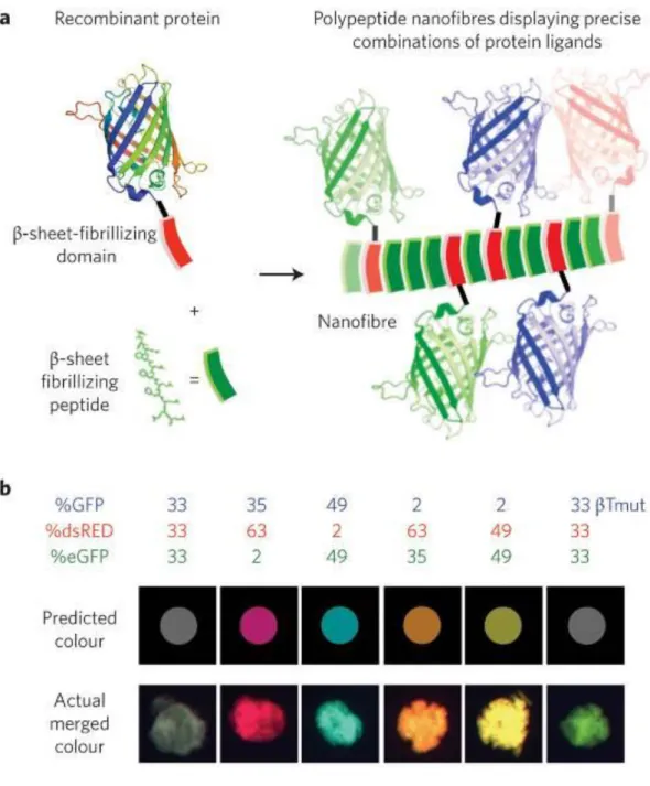
Figure 1.2 Fluorescent βtail fusion proteins stably integrate into self-assembling peptide nanofibers. a) A schematic representation of engineered fusion β-sheet fibrillating domain integrating into self-assembling peptide. b) Different fluorescent βTail proteins co-integrated into self-assembling peptide microgels at a different dose, and the correlation between actual gel colour and the predicted colour. (Green fluorescent protein, GFP; red fluorescent protein, dsRed; enhanced GFP, eGFP, mutated (βTmut)) (Copyright © 2010 NPG. Reproduced with permission from ref. [9])
5
Nanostructure formulation by peptide self-assembly is achieved through noncovalent interactions. Self-assembled peptides are commonly utilized in biomaterial research because of their ease of design, biocompatibility, defined functional motifs, fast response to the external environment, biodegradability, low immunogenicity as well as bioactivity [10, 11]. Active domains of self-assembled peptide nanofibers can also be designed for different purposes.
In one study, heparan-sulfate-mimicking PAs (HSM-PA) were synthesized and the effect of HSM-PA in combination with laminin-mimicking PAs on neurite outgrowth was analyzed. It was revealed that hydrogels made of self-assembling laminin-derived PA and HSM-PA increase the efficiency of neurite outgrowth compared to laminin-derived PAs alone. It was also shown that the scaffold in question is able to reduce the inhibitory effect of chondroitin sulfate proteoglycans (CSPGs), which makes it a promising therapeutic agent for central nervous system damages [12].
Another other commonly used bioactive epitope is IKVAV, which is a neurite-promoting laminin epitope that upregulates the proliferation of neural cells. Moreover, IKVAV reduces astrocyte formation and minimizes the risk of glial scars. In addition, it also prevents cells from undergoing apoptosis and mitigates astrogliosis. It was observed that self-assembling IKVAV-PA is more effective in modulating neuronal differentiation compared to laminin [13, 14].
In a series of interesting studies, Dr. Collier and coworkers approached self-assembled PAs in a different perspective. Instead of changing the active domain of PAs, they integrated β-sheet fibrillating domains into self-assembling peptides (Figure 1.2). This was accomplished by genetically synthesizing a GFP fusion β-sheet fibrillating
6
domain and mixing this protein with PAs. After characterization studies, they observed that PA bound to large proteins can nevertheless self-assemble into well-organized nanostructures [9]. Dr. Stupp and coworkers demonstrated a similar but harder approach for functionalized PAs. They first formed self-assembled peptide nanofibers and subsequently conjugated yellow fluorescent protein, cyan fluorescent protein or both to these self-assembled PA nanofibers using covalent linkage. Chemoselective native chemical ligation (NCL) reactions were utilized to synthesize such “hybrid” materials [15].
In recent studies, it was shown that PAs can be used for gene delivery as well [16, 17]. We have previously designed PAs that form nanostructures in the presence of CpG-ODN. We have shown that when ODN and PA/ODN nanostructures were treated with DNase1 enzyme, ODN alone gets degraded faster than the PA/ODN nanostructure (Figure 1.3). Additionally, it was shown that the PA/ODN nanostructure can prevent half of carried ODN from degradation for more than 24 h [18]. This system is very important for gene and adjuvant delivery systems, since effective delivery systems must protect their DNA or RNA cargo from endonucleases.
7
Figure 1.3 Enzymatic degradation profile of ODNs bound to self-assembled PA. (a) ODN alone, (b) nanofiber forming PA/ODN and (c) micell forming PA/ODN. Each group had been treated with DNAse I for different time periods. Remained ODN was analyzed by PAGE. Lane 1 is Marker, Lane 2 is non-treated ODN, Lane3-Lane7 = 10 min, 30 min, 1 h, 4 h and 24 h treatment with DNAse. (d) Graph of remaining ODN after degradation. Plotted according to calculated band intensities (Copyright © 2015 NPG. Reproduced with permission from ref. [18])
As a result, all information given from literature shows that rationally designed, self-assembled PAs serve as an interesting new tool for designing and developing powerful, controllable and functional materials for biological applications.
8
1.3 An overview of the immune system
Immunology was long known as the study of the body’s defense against infection, but with the improvement in technology and diagnosis systems, it is found that immunology is not only the body’s defense against infections but also body’s defense against itself and other things. The foundation of immunology was laid in 1796 by Edward Jenner. He showed that inoculation with cowpox could prevent smallpox infection. He named this procedure as vaccination and this term is still used. Recently, vaccine are composed of weakened, inactivated or live-attenuated disease-causing agents. Basically, this strategy aims to teach immune cells about unknown diseases to be recognized by immune systems. Although, this strategy gave rise to successful results in eradication of some diseases such as smallpox, there is still some limitations. i) Effective vaccines are still not available in developing countries because of their high costs. ii) There is still no effective vaccine against HIV, malaria, zika viruses, tuberculosis and cancers. iii) Vaccine technology is old and not suitable for a rapid response to emerging outbreaks. For example, in the 2009 influenza A (H1N1) pandemic, it was concluded that there is limitation for producing more vaccine in short time, as well as distribution of them to other countries. iv) Traditional vaccines are produced by using live-attenuated viruses, inactivated viruses, attenuated bacteria or non-replicating recombinant viruses, however, there are safety concerns related to these vaccines. For instance, attenuated organism in the vaccine could gain virulent properties and cause disease.
To develop effective vaccines with minimum side effects, basic concepts in immunology should be taken into account.
9
1.3.1 Innate immune system
The innate immune system is the network of cellular and molecular elements that protect body from infections or diseases. The innate immune system responds to infection or damage rapidly and non-specifically. It is present at birth and is not gained after encountering infections, so it is not antigen specific [19]. The elements of the innate immune system include anatomical barriers, humoral molecules and cellular components.
Anatomical barriers are first innate immune system organs that infection agents encounter. It is composed of physical, chemical and biological barriers. Epithelial surfaces covering the skin, lungs and gut provide a physical barrier between the inside of the body and the invaders. The interior epithelial surfaces covered with a mucus layer prevented the entrance of microbial organisms into the body. Tight junctions between neighboring cells are other barriers that protect cells from potential pathogens. Movement of cilia or peristalsis protect air passages and the gastrointestinal tract from microorganisms. Tears and saliva including protective chemicals prevent infection of pathogens in the eyes and mouth. Fatty acids, lysozyme and phospholipase, which are abundantly found in the first line of immune system help clearing of microorganisms. Lysozyme and phospholipase are some of the material found in tears, saliva and nasal secretions. They can breakdown the cell walls of bacteria. Fatty acids in sweat, and some chemicals in stomach reduce pH of sweat and gastric secretions, preventing growth of bacteria. Gut and skin micro flora also help body to prevent colonization of microorganisms by secreting some toxic materials to other organisms or by competing them for nutrition [20]. When anatomical barriers are breached due to damages or
10
diseases, infection may occurs and humoral molecules are secreted for preventing infection. These humoral factors are complement systems proteins, cytokines, chemokines, acute-phase protein, interferons, interleukin1, lactoferrin and transferrin. Humoral factors play important roles in inflammation, recruitment of cells, promoting antibody formation, neutralizing viruses, attracting macrophages and neutrophils to a troubled spot, or killing bacteria directly [21]. Injured cells release specific chemical factors to stimulate inflammation. This mechanism generates a physical barrier to stop the spreading of infection, and also to promote healing of damaged tissues [22]. In response to tissue injury, a multifactorial network of chemical signals initiate a host response in the afflicted tissue. This response includes activation and migration of leukocytes (neutrophils, monocytes and eosinophils) from the venous system to the sites of damage [23]. Cellular barriers are formed after innate immune cells take their places. As shown in Figure 1.4, immune cells originated from hematopoietic stem cells. White blood cells (WBCs) which are also called leukocytes, make up 1% of total blood volume, and red blood cells constitute 40% to 50% of all blood volume. WBCs can be divided into two subgroups: granulocytes and agranulocytes [24]. Granulocytes are neutrophils, eosinophils, basophils, and mast cells; whereas agranulocytes are lymphocytes and monocytes. WBCs also can be separated into five main types: neutrophils, eosinophils, basophils, lymphocytes, and monocytes. Lymphocytes are composed of B cells, T cells and natural killer (NK) cells, and
11
monocytes include dendritic cell and macrophages. Additionally, dendritic cells, macrophages and neutrophils are known as phagocytic.
Figure 1.4 Haematopoietic stem cells differentiation. Haematopoietic stem cells (HSCs), are divided into at least two subsets: long-term reconstituting HSCs (LT-HSCs) and short-term reconstituting HSCs (ST-(LT-HSCs). LT-HSCs have self-renewal and multi-lineage differentiation potential throughout life. ST-HSCs have multipotency, but have more-limited self-renewal potential. Further differentiation of ST-HSCs generates multipotent progenitors (MPPs). Haematopoietic progenitor cells lose their differentiation potential in a stepwise fashion until they eventually generate all of the mature cells of the blood system. Lineage-committed oligopotent progenitors derived from MPPs include the common lymphoid progenitor (CLP), common myeloid progenitor (CMP), megakaryocyte-erythrocyte progenitor (MEP) and granulocyte-monocyte progenitor (GMP) populations. NK, natural killer. (Copyright © 2011 NPG. Reproduced with permission from ref.[25])
12
Innate immune cells include macrophages (MQ), dendritic cells (DC), neutrophils, basophils, eosinophils, mast cells and natural killer cells (NK). Each cell type in immune system has a different function. Neutrophils are specialized in engulfing microorganisms at infection sites. Basophils and eosinophils secrete toxic proteins and free radicals at the site of infection to kill parasites. Mast cells reside in connective tissues and mucosal membranes and take role in wound healing by releasing histamine and heparin. NK cells are quite special cells that can kill infected cells. Instead of engulfing pathogens, they are specialized in killing infected cells which do not express little expressions of self-antigens, which is a signal used to save normal cells from killing. Moreover, NK cells do not need further activation for killing any cells. Macrophages and dendritic cells are special cells that can take up pathogens, process, and present them to adaptive immune cells.
NK cells are responses to viral infections and tumor development. They are an important member of lymphocytes, their main function overlaps with T cells, however they take role in the first line of defense systems. NK cells were originally recognized by their ability to mediate lysis of certain susceptible tumor cell lines and infected cells. They have large granular lymphocyte morphology, but do not express T cell antigen receptors and the CD3 complex which are specific for T cells recognition. In addition, they can produce antimicrobial and immunoregulatory cytokines. These cells can secrete gamma interferon (IFNγ), tumor necrosis factor (TNF), and granulocyte/macrophage colony stimulation factor (GM-CSF). Their regulation can be controlled by innate cytokines which can mediate biological functions of NK cell
13
responses of cytotoxicity, proliferation, and interferon gamma (IFNγ) production [26-28].
Dendritic cells are also important cells of the innate immune system and they can be differentiated from common lymphoid progenitors and common myeloid progenitors. Dendritic cells can capture and process antigens, present antigen to adaptive immune cells, express lymphocyte co-stimulatory molecules, migrate to lymphoid organs and secrete cytokines to initiate immune responses. Moreover, besides activation of lymphocytes, they can also tolerate T cells to self-antigens, and other antigens, thereby minimizing autoimmune reactions. Like macrophages and B cells, dendritic cells are also known as antigen presenting cells (APC). They are efficient stimulators of B and T cells. After presenting antigens on their peptide-binding proteins, which are MHC class I and MHC class II, they stimulate cytotoxic T cells (CTLs) and helper T cells respectively. Without this engagement, T cells cannot be activated. They can produce IL-12, interferon and tumor necrosis factor alpha (TNF alpha) which have roles in T cell activation and activation of other immune cells. Therefore, these cells are powerful in manipulating the immune system. [29-31]
Monocytes are another class of innate immune system cells. They are the largest cells of the innate immune system. They become mature in bone marrow. They can circulate through the blood. Half of them are found in the spleen and the other half is located throughout the body. Monocytes differentiate into macrophages and dendritic cells. After crossing into tissue, monocytes differentiate into macrophages. They are very crucial for removing excess, damaged, or dead, and phagocytosis of bacteria, viruses and other pathogens. Macrophages express different types of pattern recognition
14
receptor (PRR) on their surface which are important for recognition of danger signal. Toll like receptor ones (TLRs) are the most important ones. When TLRs are activated by pathogens or other signals, this stimulates the macrophages to phagocytes pathogens or damaged cells. Additionally, it induces macrophages to secrete cytokines which can activate or recruit additional immune cells.
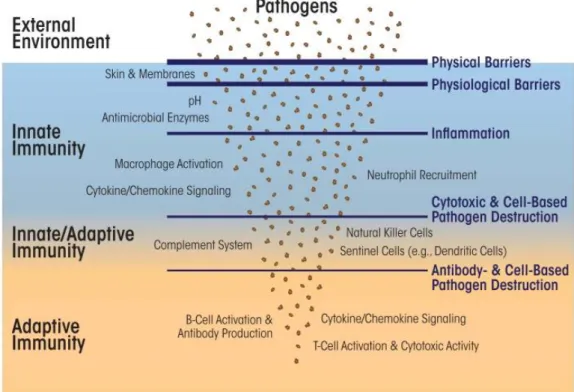
Figure 1.5 Relationship between innate immunity, adaptive immunity and invaders. Innate immunity consist of non-specific protective mechanisms against infection. The barriers including physical, physiological, chemical barriers, are cells and small proteins. The adaptive immune system comprises specialized cells and proteins. Several cells in adaptive immunity can store immune memory of a pathogenic invasion. The natural killer cells and dendritic cells have roles in both innate and adaptive immunity. (Copyright © 2015 ARCR. Reproduced with permission from Ref. [32])
15
Macrophages are members of antigen presenting cells, like B cells and dendritic cells. Another important role of macrophages is to induce inflammation. They are also known as scavenger cells in the body because they can clear dead cells and debris. Tumor necrosis factor α (TNFα) is mainly produced by macrophages. Macrophages also have important roles in healing process [32-34]. As it is shown in Figure1.5, innate immune system works as a supplementary system of adaptive immune system. They have common cells and also common cytokines which are important for regulating immune cell activity.
1.3.2 Adaptive immune system
Adaptive immune response, also called acquired immune response, is mediated by B and T cells. This system includes specialized cells and unique processes to eliminate pathogens from the body or prevent infections. It is acquired after birth. Adaptive immune cells are specialized to learn and remember, forming an immunological memory after encountering pathogens. The adaptive immune system is highly specific because the response is formed for specific antigens or toxins. If a patient is able to recover from an initial infection, they will not be affected by the same pathogen again, because adaptive immune response remains in reserve and responds quickly in the case of a second infection. Similar to the innate immune response, the adaptive immune response functions through antigen-specific humoral and cellular immune responses. T and B cells bear clonally-distributed antigen receptors. T-cell receptors (TCRs) and immunoglobulin are formed by somatic rearrangement. Once T cells are activated, they can directly attack infected cells, stimulate B cells to produce antibodies against pathogens, or enhance inflammation in the area of infection [35-37]. The somatic rearrangement of immunoglobulin is responsible for antibody production by B cells.
16
Activation of B cells by pathogens initiates a process of hypermutation in genes associated with the production of immunoglobulins, which increases the diversity of the resulting antibodies and increases the likelihood that one such protein will be specific against the antigen in question [38]. In some cases, B cells produce antibodies without undergoing hypermutation and secrete low-avidity antibodies [38].
T cells are specialized to eliminate cells infected with viruses, destroy detected cancer cells, or orchestrate the functions of innate and adaptive cells. T cells are further divided into different categories, including naive T cells (which further differentiate into T helpers and CTLs), memory T cells and regulatory T cells. An effective T cell-mediated immune response occurs after the activation of T lymphocytes by APCs in secondary lymphoid organs (the spleen, lymph nodes (LNs), Peyer’s patches (PPs), tonsils, adenoids) [39].
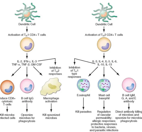
Naive T cells are located in the thymus and differentiate from bone marrow. Following positive and negative clonal selection, they become mature non-effector cells that are individually specific towards a given antigen, but cannot recognize these antigens directly [40]. Helper T cells, known as CD4+ cells, are key elements of adaptive immunity. T helper cells can activate immune system cells, including B cells, killer T cells, and macrophages. Main T helper cells are Th1 and Th2. They serve as an important line of defense against intra- and extracellular pathogens and autoimmune and allergic responses. Th17 is a subgroup of CD4+ cells, and plays a role in chronic inflammation, autoimmune disease and the elimination of extracellular microorganisms. Regulatory T (Treg) cells are T cells that modulate the responses of effector T cells and suppress pro-inflammatory pathways. Due to this self-correcting mechanism, they also prevent immune cells from attacking normal body cells. T helper cells are activated by APCs, which express antigen fragments on their MHC class II molecules. This complex binds to T cell receptors (TCR) on T cells. Following this interaction, a second activation signal is provided through the effects of co-stimulatory molecules, which are either CD80 or CD86 on APCs. These co-stimulatory molecules bind to CD28 on T cells. A third signaling molecule, CD4+, then binds to MHC-CD80/CD86 complexes and enhances their binding affinity. Following MHC-TCR
17
binding, the helper T cell is activated and begins to proliferate. Activated helper T cells differentiate into Th1 or Th2 according to the milieu that T cells are found within. For example, IL-12 induces naïve T cells to differentiate into Th1 cells, while IL-4-stimulated cells form a Th2 response. In addition, Th1 cells produce IL-2, IFN-γ, TNF-α, and LT-α; while Th2 cells produce IL-4, IL-5, IL-6, IL-10, and IL-13. Some proliferating T cells may also become memory helper T cells. Those cells contribute to immunological memory and respond quickly when the body encounters the same pathogen again. The activated T cells are known as effector T cells, which release cytokines and induce apoptosis. Cytokines produced by T helper cells attract macrophages, B cells, and cytotoxic T cells, and/or regulate the activity of these cells [41-48].
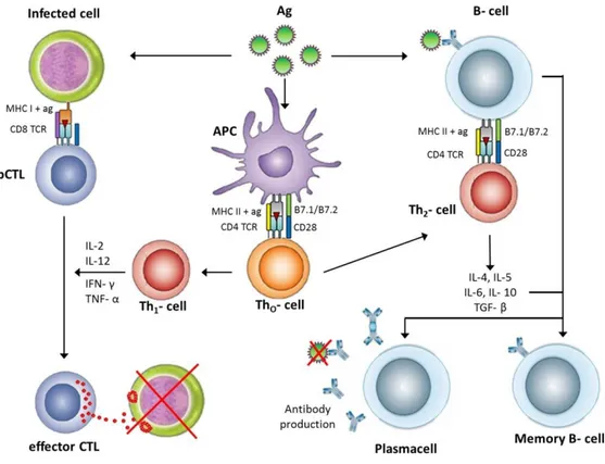
Concentration of antigenic peptides, affinity of TCR toward the Ag/MHC complex, cytokines, inflammatory, and co-stimulatory signals in lymphoid organs affect the efficiency of T cell activation. CD8+ naive T cells differentiate into effector cytotoxic T lymphocytes (CTL) after encountering the above-mentioned requisite signals. As shown in Figure 1.6, CTL is activated after encountering an antigen segment presented on MHC I, and induces apoptosis. This mechanism is also important for clearing cancerous cells, which express tumorous antigens. Cluster of differentiation 28 (CD28) and cytotoxic T-lymphocyte-associated protein 4 (CTLA-4) are expressed on T cells. The ligation of CD28 with CD80/86 on APCs plays a co-stimulatory role for T cell activation, while the ligation of CTLA-4 with CD80/86 acts as a negative regulator. Conventional anticancer antibodies such as ipilimumab or tremelimumab were used to block the binding of CTLA-4 with CD80/86, suppressing the inactivation of T cells. The programmed death 1 (PD-1) inhibitory receptor is also expressed by T cells and negatively regulates their activity following its ligation with L1 and PD-L2 [49-51].
18
Figure 1.6 General mechanism of the adaptive immune responses after antigen recognition by antigen presenting cells. Antigens can be presented by either dendritic cells or B cells to T cells. APCs bearing the peptide-MHC I complex are recognized by precursor cytotoxic CD8+ T lymphocytes (CTLs). Moreover, infected cells can express pathogen specific antigens on their surface with MHC I or MHC II. Activated Th1 cells produce IL-2, IFN-γ, and TNF-α cytokines to induce activation and differentiation of the precursor CTLs into memory or effector CTLs. Effector CTLs can directly recognize and kill infected cells by the production of perforines and granzymes. Activated Th2 cells produce B cell stimulating cytokines such as 4, IL-5, IL-6, IL-10, TGF−β; which activate naive B cells. After activation, B cells differentiate into memory B cells and plasma cells that produce large amounts of IgG, IgA, IgE antibodies to prevent further virus infection. Ab: antibody, Ag: antigen, APC: Antigen Presenting Cell, DC: Dendritic Cell, IL: Interleukin, TCR: T Cell Receptor, Th: CD4+ T helper cell. (Copyright © 2012 Wiley Periodicals, Inc. Reproduced with permission from ref [47])
19
B cells are APCs and have the ability to recover antigens through their clonally formed antigen receptors, which are called membrane immunoglobulins (Ig). The B cell first recognizes antigens via its antigen receptor, and subsequently internalizes and degrades the antigen. After processing, antigens bound to MHC class II molecules are presented on the cell surface. Similar to other APCs, T cells recognize the processed antigens on the B cell surface and bind through the activity of TCR, CD4, CD40-L and/or CD28. CD40 is a co-stimulator expressed by B cells, and its interactions with CD40-L on T cells result in the differentiation and activation of B cells. Following binding, T helper cells recognize peptide fragments on MHC II molecules on the B-cells, allowing the activation of both cells against the same antigen. This interaction also triggers the expression of CD40-L on the T cell surface, which binds to CD40 on the B cell. This binding results in the stimulation of B cell clonal expansion, isotype switching, affinity maturation and differentiation into memory cells [52]. In addition to T cell-mediated activation, there is another B cell activation pathway that does not require T helper cells. This type of activation occurs in response to non-protein antigens such as polysaccharides, glycolipids, and nucleic acids. These antigens are not processed and presented along with MHC proteins, and cannot be recognized by helper T cells. Figure 1.7 shows that both Th1 and Th2 responses are able to activate B cells, and that cytokine exposure is able to determine the type of immunoglobulin secreted by the cells. Th1 helper cells preferentially induce B cells for IgG2a secretion, while Th2 helpers induce B cells for the synthesis of IgG1 and IgE [53]. Immunoglobulins are divided into five primary classes: IgG, IgM, IgA, IgD and IgE. The type of heavy chain in the molecule makes them distinguishable. IgGs are
20
abundantly found in serum: around 75% of serum IGs belong to subgroups such as IgG1 and IgG2a. IgG is the only antibody that can penetrate through the umbilical cord and impart passive immunity to the fetus.
Figure 1.7 CD4+ helper cells induce cells by secreting distinct but overlapping sets of cytokines. TH1 CD4+ cells activate immune cells for inflammatory and cellular
reactions against intracellular bacteria or viruses. TH2 CD4+ cells activate immune
cells for specific types of antibody against parasites and extracellular encapsulated
bacteria. TH2 CD4+ cells play a role in allergic diseases. GM-CSF,
granulocyte-macrophage colony stimulating factor; IFN, interferon; IL, interleukin; TNF, tumor
necrosis factor. (Copyright © 2007 Elsevier, Reproduced with permission from ref
21
1.4 Toll like receptors and adjuvants
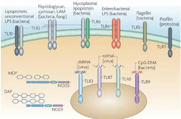
Pattern-recognition receptors (PRRs) are innate immune system proteins that are used to recognize microorganisms or their characteristic antigens. They are encoded in the germline. PRRs recognize microbial components such as lipoteichoic acids, lipoarabinomannan (LAM), lipopolysaccharides (LPS), CpG-ODNs and lipoproteins. These components are known as pathogen-associated molecular patterns (PAMPs). PAMPs are essential for the survival of many microorganisms; consequently, the evolutionary pressures associated with their synthesis are too strict to drive their change, making them ideal targets for the innate immune system. For example, LPS are found in the cell wall of Gram-negative bacteria and protect them from chemical factors. PRRs are expressed constitutively in the host, allowing the recognition of pathogens at all stages of life. Different PRRs recognize specific PAMPs and activate specific signaling pathways, resulting in distinct anti-pathogen responses. For instance, LPS are recognized by Toll like receptor 4 (TLR4) and induce interferon secretions [35, 36]. PRRs are found on the surface of the cell membrane, within the endosome and in cytosol (Figure 1.8). Basic classes of PRRs are shown in Figure 1.8. However, these only represent a small part of the diversity exhibited by PRRs, because a broad range of PRRs are required to recognize common pathogens and pathogenic particles (Figure 1.9)
Toll like receptors are important members of PRRs. Toll was initially identified in Drosophila, and was demonstrated to be essential for the development of embryonic dorsoventral polarity in this organism [37]. Later, it was also shown to play a critical role in the antifungal response of flies [38]. To date, 12 members of Toll like receptors
22
have been identified. Macrophages, dendritic cells (DCs), B cells, specific types of T cells, fibroblasts and epithelial cells are known to express TLRs intracellularly or extracellularly. Environmental stresses and cytokines can affect the expression of TLRs. While TLRs 1, 2, 4, 5, and 6 are expressed on the cell surface, TLRs 3, 7, 8, and 9 are found in intracellular compartments such as endosomes. As shown in Figure 1.8, intracellular TLRs generally recognize bacterial DNA or viral RNA or DNA. As such, they are important for recognition of nucleotide sequences associated with intracellular pathogens [36, 39, 40].
TLR9 recognizes bacterial genomic DNA or virus DNA. Both bacterial and viral DNA are immunostimulants because of the presence of unmethylated CpG dinucleotide motifs [41]. Compared to mammalian genomes, bacterial genomes have a greater tendency to feature the unmethylated CpG motif. The methylated CpG motif, in contrast, is characteristic of vertebrate genomes and cannot activate mammalian immune cells. The presence of CpG-DNA can activate immune cells by upregulating the production of inflammatory cytokines that support the Th1 response. It was shown that synthetically designed DNA containing the CpG motif can induce immune cells in a manner comparable to bacterial DNA. TLR9 is found in the endosome, so bacterial DNA must first be delivered to this intracellular compartment prior to its recognition. Bacteria or viruses that are engulfed by the endosome are exposed to acidic and reducing conditions, which lead to the degradation of DNA into multiple
single-23
stranded CpG-motifs. After the degradation of envelope or coat of bacteria or viruses, TLR9 can interact with CpG-containing regions [42].
Figure 1.8 Simple demonstration of PRRs and PAMPs. Nod-like receptor (NLR) family. DAP, diaminopimelic acid; ds, double-stranded; MDP, muramyl dipeptide; LPS, lipopolysaccharides; LAM, lipoarabinomannan; ss, single-stranded. (Copyright © 2007 NPG. Reproduced with permission from ref.[43])
24
Figure 1.9 Pattern recognition receptors and their downstream pathways. The soluble lectin receptor mannose-binding lectin (MBL) can bind to mannose-rich structures. The membrane-bound C-type lectin receptors, macrophage mannose receptor 1 (MMR), dendritic cell-specific ICAM3-grabbing non-integrin (DC-SIGN) and macrophage-inducible C-type lectin (MINCLE) can recognize mannose-rich structures. Dectin 1 can recognize and bind to β-glucans, and dectin 2. Toll-like receptor 4 (TLR4) recognizes O-linked mannans, and TLR2 can recognize phospholipomannans or, together with galectin 3, recognizes β-mannosides. TLR9 is located in the cytosol and recognizes DNA. Furthermore, the NOD-like receptor (NOD-, LRR), apoptosis-associated speck (ASC). IFNs, interferons; IL-1β, interleukin-1β and NF-κB, nuclear factor-κB also assist in the recognition process. (Copyright © 2007 NPG. Reproduced with permission from ref.[44])
25
Table 1-1 TLR recognition of microbial components (taken from ref. [36])
As demonstrated in Table 1-1, TLRs and their ligands may differ considerably between organisms. Additionally, particles that drive different types of TLR’ immunity may
26
originate from the same organism. This mechanism enhances the recognition of pathogens by TLR.
Vaccination is an effective way to prevent infectious diseases. The main aim of vaccination is to induce a strong immune response to pathogen-derived antigens and provide long-term protection against infection. Purified or recombinant antigens exhibit poor immunogenicity and cannot raise a substantial immune response. Materials that are used to enhance the specific immune response against co-inoculated antigens are called adjuvants. The word adjuvant originates from the Latin word adjuvare, which means to help or to enhance. As previously mentioned, immune cells recognize organisms by binding to PAMPs through their PRRs. As such, the former category of molecules can be used as adjuvants. However, if they are not purified sufficiently, they can be toxic. Therefore, less toxic derivatives of PAMPs are particularly attractive as potential adjuvants. For example, bacterial components are often potent immune activators. Bacterial DNA sections including CpG motifs are one of the most potent cellular adjuvants. CpG motifs are unmethylated cytosine-guanine dinucleotide sequences that are found in bacterial DNA and are absent in the mammalian genome. The CpG motif is recognized by TLR9 and induces the proliferation, activation and differentiation of immune cells Additionally, it directs the immune response toward the Th1 pathway [45, 46].
Adjuvants are used for different purposes: to enhance the immunogenicity of highly purified or recombinant antigens; to reduce the amount of antigens used for immunization; to reduce the number of immunizations; to improve the efficacy of
27
vaccines in newborns, the elderly or immunocompromised persons; or to enhance the uptake of antigens by the mucosa [47, 48].
Table 1-2 Characteristics of the ideal adjuvants (taken from ref.[49])
28
The biological pathways by which adjuvants perform their functions are subject to debate. However, there is some evidence that adjuvants may utilize a variety of mechanisms to elicit immune responses. These mechanisms include the depot effect (through which the adjuvant sustains the release of antigens at the site of injection); up-regulation of cytokines and chemokines; cellular recruitment at the site of injection; increasing antigen uptake of APCs and presentation of antigens; activation and maturation of APCs (e.g. by promoting the expression of MHC class I, MHC class II or co-stimulatory molecules); inducing APCs to migrate to the draining lymph nodes; and/or the activation of the inflammasome [50]. A great number of adjuvants have been characterized to date, but few have been approved by Food and Drug Administration (FDA) (Table1.3.3). Approved adjuvants are used for different treatments and can induce different immune responses, as shown in Table 1.3.4. Consequently, the choice and dose of adjuvants can be optimized to efficiently induce the immune system towards a desired response mechanism.
29
30
CHAPTER 2
2.1 Introduction
Despite improvements in vaccine technology, effective vaccines have yet to be developed for diseases such as cancer, human immunodeficiency virus (HIV) infection, tuberculosis and malaria, which together cause the deaths of more than 4 million people worldwide each year [51, 52].Modern vaccines also require an extended development process, and cannot respond immediately to emerging outbreaks that follow the transmission of new viral strains from animal reservoirs to the human population [51]. For example, in the recent outbreak of Ebola and Zika viruses, it is reported that 13,562 people were infected by Ebola in West Africa [53] and 32,000 people were infected by Zika in French Polynesia [54]. It is also predicted that in 2030, cancers will claim the lives of more than 11 million people worldwide [55, 56]. Currently, clinically approved vaccine adjuvants such as aluminum hydroxide and the oil-in-water emulsion MF59 can promote humoral immune responses but not cellular immune responses [46, 57]. The activation of CD8+ T cells (CD8T) in particular is critical for the development of vaccines against cancer or intracellular pathogens such as human immunodeficiency virus (HIV), malaria and hepatitis C. As such, materials modulating humoral and cellular responses in a synchronized manner are preferred in vaccine development [58, 59]. Accordingly, inactivated virus-, live-attenuated virus- and viral vector-based subunit vaccines have been developed for inducing effective humoral and cellular responses [60]. However, these approaches have so far faced problems involving incomplete viral inactivation, regain of virulence and unfavorable host responses to viral vectors, which have limited their clinical utility [61].
31
Safety concerns associated with inactivated and live-attenuated virus vaccines have led to the development of recombinant protein-based subunit vaccines, which consist of one or more recombinant proteins or polysaccharides and do not utilize the active viral agent in any form. However, subunit vaccines induce poor or non-existent CD8T responses and exhibit low cross-presentation efficiencies, as they are not uptaken, processed and/or presented to naïve CD8T cells to the same extent as the entire viral unit [60-63]. The cross-presentation and immunogenicity of antigens can be improved through the administration of immunostimulatory molecules or adjuvants [64, 65], including aluminum, MF59, MPL, AS04, AS01B and AS02A, toll-like receptor (TLR) ligands, CpG-ODN1826, oil based adjuvants and other factors [49, 66-68]. In addition, antigens may also be delivered in particulate form in order to generate specific T cell responses, such as the Th1-type response [69]. Encapsulated or conjugated antigens have likewise been shown to induce greater humoral responses than soluble antigens [69-72]. To induce an effective immune response, adjuvant and antigens should be in close proximity [18]. The size of carrier materials is another important parameter for the generation of an effective immune response. Immunization with nanoparticles in the 200–600 nm range are known to induce higher levels of IFN-γ production, upregulation of MHC class I molecules and the production of antibody isotypes favoring the Th1-type immune response. Immunization with 2 – 8 μm microparticles, in contrast, enhance IL-4 secretion, upregulate MHC class II expression, and direct the immune response towards the Th2-type. On the other hand, nanoparticles in the 20−200 nm range can efficiently enter the lymphatic system through lymph drainage [73-75]. Previous studies have also shown that epitope density displayed on the
32
nanoparticles are crucial for effectively eliciting humoral immune responses, and especially the IgG response [76]. As such, materials to be used for antigen delivery applications should have controllable sizes and exhibit the ability to present epitopes efficiently to produce concerted T-cell responses. Highly effective T-cell vaccines fulfilling these criteria are still being sought, and a broad variety of methods have been employed for their development [63, 73]. As virus mimetic materials exhibit the potential to elicit strong immune responses while posing little to no health risks, we hypothesized that a rationally designed network of self-assembled peptide nanofibers can be used as an effective vaccine candidate to meet the stringent demands of the vaccine industry.
Self-assembly has been used as a powerful strategy for the construction of functional materials. Mediated by electrostatic forces rather than covalent binding, nanofiber formation by peptide self-assembly has attracted intensive research interest due to the ease of design, biocompatibility, defined functional motifs, fast response to external environment, biodegradability and low immunogenicity associated with these materials [11]. Self-assembled nanomaterial formation has been shown to exhibit immense potential for regenerative medicine, drug delivery, cell culture, analytical detection, biosensors and immunotherapy applications [4, 9, 77, 78]. Recently, Collier and co-workers have reported that self-assembling peptides can be used as powerful immune adjuvants [71, 79]. In another study, it was shown that peptide amphiphile (PA)-based micelles could serve as an effective antigen delivery system [77, 80]. Although the strategies developed by these groups are powerful and effective for vaccine production, they also suffer from limitations with regards to their synthesis
33
and assembly methods. The preparation of these materials involves the covalent conjugation of a protein or protein fragment to the self-assembling peptide, which is not readily applicable to all antigen fragments. However, the streptavidin (SA)-biotin system can be used in situations where direct covalent attachment is problematic, as it is known that the SA-biotin interaction is among the strongest non-covalent interactions in nature [81]. In a previous study, it was demonstrated that streptavidin binds strongly to biotinylated self-assembled nanostructures, and it was also shown that biotinylated insulin-like growth factor 1 (IGF-1) can bind to the peptide nanostructure through an SA linker [82].
In this work, we describe the development of a modular delivery system using a biotin-linked approach that allows a broad diversity of antigens to be presented on the surface of peptide nanofibers. Nanostructure formation by the ODN1826 adjuvant (a TLR9 agonist) [83] and biotinylated self-assembling peptides allowed highly specific binding of antigens to the nanostructure and positioned the antigens in close proximity with the adjuvant through the SA linker. We also demonstrate that the designed self-assembled (PAs/ODN+SA+OVA) nanostructure is able to induce humoral and cellular responses, and detail the mechanism of action of the adjuvant system using in vitro and in vivo models.
34
2.2 Experimental
2.2.1 Material
9-Fluorenylmethoxycarbonyl (Fmoc) and tert-butoxycarbonyl (Boc) protected amino acids, [4-[α-(20,40-dimethoxyphenyl) Fmoc-aminomethyl] phenoxy] acetamidonorleucyl-MBHA resin (Rink amide MBHA resin), and 2-(1H-benzotriazol-1-yl)-1,1,3,3-tetramethyluronium hexafluorophosphate (HBTU) were purchased from NovaBiochem and ABCR. Other chemicals for peptide synthesis were purchased from Fisher, Merck, AlfaAesar, or Aldrich. CpG ODNs were purchased from Invivogen. Paired antibodies and recombinant proteins of IFNγ and IL-12 were obtained from R&D systems, that of IL-6 from eBioscience. Reagents for polyacrylamide gel electrophoresis were obtained from Sigma Aldrich. Horseradish peroxidase-conjugated goat anti-mouse IgG (sigma), IgG1(eBioscience), IgG2a (abcam), or IgG2b (eBioscience) were obtained from Southern Biotechnologies (AL, USA). All cell culture and ELISA reagents were purchased from Life Technologies, except non-essential amino acid solution (Sigma Aldrich). Fluorescently labelled antibodies were obtained from eBioscience (CD40, CD86, Anti-Mouse OVA257-264 (SIINFEKL) peptide).
2.2.2 Peptide synthesis
Lauryl-VVAGK-Am (K-PA) and Lauryl- VVAGKK(Bio)-Am (B-PA) were synthesized on Rink Amide MBHA resin. Amino acid couplings were performed with 2 equivalents (equiv) of Fmoc-protected amino acid, 1.95 equiv of HBTU and 3 equiv of N,N-diisopropylethylamine (DIEA) for 2 h. After removal of Mtt group of
Fmoc-35
Lys(Mtt)-OH by %5 TFA solution in DCM, biotin was conjugated in the same manner as amino acid couplings. Removal of Fmoc group was achieved by 20% (v/v) piperidine in DMF for 20 minutes. To block the remaining free amine groups after amino acid coupling, 10% (v/v) acetic anhydride solution in DMF was used (30 min). After each step, the resin was washed using DMF, dichloromethane (DCM) and DMF. A trifluoroacetic acid (TFA)/triisopropyl silane (TIS)/ H2O/DCM mixture (5:2.5:2.5:90 ratio) was used to cleave the peptide from the resins.
2.2.3 Preparation of fiber nanostructures
Fiber nanostructures were prepared through the self-assembly of peptide molecules in the presence of oligonucleotides. To form fiber nanostructure, positively charged K-PA and B-K-PA molecules were mixed with CpG (ODN1826), respectively. Sequence of ODN1826 is 5′ -tccatgacgttcctgacgtt-3′. The molar ratio for ensuring that all ODNs in solution interact with nanostructures was determined to be 100:1 for B-PA and K-PA/ODN as showed in our previous studies [84]. Nanostructures were prepared at these ratios for all experiments (K-PA/ODN) and (B-PA/ODN) throughout the experiments. In all experiments, at least three independent K-PA/ODN and B-PA/ODN formulations were prepared and tested.
2.2.4 Preparation of antigen bearing fiber nanostructures
Self-assembled B-PA/ODN and K-PA/ODN nanostructure were mixed with Streptavidin from Streptomyces avidinii (SA) (Sigma-Aldrich), and after 40 min, biotinylated ovalbumin (OVA) (Galab) was added to formed nanostructure. The exact
36
molar ratio for B-PA, Streptavidin and ovalbumin was 1:1:1. Antigen bearing nanostructure was formed.
2.2.5 Transmission electron microscopy (TEM) imaging
Nanostructures were imaged by TEM. 30 μL of PA/ODN complexes was prepared on parafilm by mixing 15 μL of 15 μg/mL ODN1826 with 15 μL of either 0.023% (w/v) B-PA (100:1 ratio) or 0.015% (w/v) K-PA (100:1 ratio). For PA-only samples, these concentrations of PAs were mixed with distilled water instead of ODN solution. For serial dilution, self-assembled B-PA/ ODN complexes were diluted (1 fold, 10 folds and 100 folds diluted). TEM grids were inverted onto these solutions. Grids were removed after 10 min and the remaining solution on grid was absorbed by a lint-free paper. Staining was performed with 2% (w/v) uranyl acetate solution (Ted Pella, Inc) for 40 seconds. Grids were washed with ddH2O twice and dried 3 h at room temperature. TEM imaging was performed by a FEI Tecnai G2F30 instrument. All images were taken in STEM mode with a high angle annular dark field (HAADF) detector.
2.2.6 Atomic force microscopy (AFM) imaging
AFM imaging of PA/ODN complexes was performed in liquid conditions. ODN1826 solution at 15 μg/mL concentration was mixed with an identical volume of 0.023% (w/v) B-PA solution (100:1 ratio) or 0.015% (w/v) K-PA solution (100:1 ratio). The final ODN concentrations in each PA/ODN complexes were equal. For K-A/ODN complexes, the prepared solution was diluted 50 times, B-PA/ODN complexes was diluted 10 times, and dropped onto the cleaved mica surface and imaged directly in
37
aqueous environment (Figure 1D and F). Silicon nitride soft contact tip (BL-RC150VB, 37 kHz, k = 0.03 N/m) was used for contact mode imaging of K-PA/ODN complexes. Silicon tip (150 kHz, k = 5 N/m) was used for tapping mode imaging of B-PA/ODN complexes. MFP-3D Asylum microscope was used for imaging.
2.2.7 Circular dichroism (CD) spectroscopy
CD spectroscopy was performed with a JASCO J815 CD spectrometer at room temperature. 0.2 mM solutions of both K-PA and B-PA and their mixtures with ODN1826 (100:1) were measured from 300 to 190 nm. Scanning speed, data pitch, DIT and bandwidth were adjusted to 100 nm/min, 1 nm, 4 s and 1 nm respectively. All measurements were performed with three data accumulations and sensitivity was selected as standard.
2.2.8 Polyacrylamide gel electrophoresis (PAGE)
PAGE was performed to validate the critical ODN/PA ratio required to conjugate all ODNs in solution to PA nanostructures and observing effect of mixture of K-PA and B-PA on nanostructure formation. 1:100 molar ratio of ODN/K-PA was optimum ratio for nanostructure formation and charge neutralization [84]. 20 μg/mL ODN1826 solution (15 μL) was mixed with varying amounts of PA solutions (15 μL) to prepare different B-PA/K-PA ratios (from 1:1 to 1:10000). These solutions were mixed with Orange DNA loading dye (Fermentas) and loaded onto 20% polyacrylamide gels. 10 μL of 10 bp DNA ladder (O’range rulerTM, Fermentas) was used as marker. Gels were run at 75 V for 1 h and subsequently at 50 V for 2.5 h (in 1x TAE). Stains-all dye working solution (0.005%, w/v) was prepared freshly from stock solution (0.1% w/v)


![Table 1-1 TLR recognition of microbial components (taken from ref. [36])](https://thumb-eu.123doks.com/thumbv2/9libnet/5615767.111069/41.892.180.781.211.868/table-tlr-recognition-microbial-components-taken-ref.webp)
![Table 1-2 Characteristics of the ideal adjuvants (taken from ref.[49])](https://thumb-eu.123doks.com/thumbv2/9libnet/5615767.111069/43.892.169.783.708.1049/table-characteristics-ideal-adjuvants-taken-ref.webp)
