SIP1 protein protects cells from DNA damage-induced
apoptosis and has independent prognostic value in
bladder cancer
A. Emre Sayana, Thomas R. Griffithsa, Raj Pala, Gareth J. Brownea, Andrew Ruddicka, Tamer Yagcib, Richard Edwardsc,
Nick J. Mayerd, Hasan Qazia, Sandeep Goyala, Serena Fernandeza, Kees Straatmane, George D. D. Jonesa,
Karen J. Bowmana, Alexandra Colquhouna, J. Kilian Mellona, Marina Kriajevskaa, and Eugene Tulchinskya,1
aDepartment of Cancer Studies and Molecular Medicine, University of Leicester, Leicester LE2 7XL, United Kingdom;bDepartment of Molecular Biology and
Genetics, Bilkent University, Ankara 06800, Turkey;cMedical Research Council Toxicology Unit, Leicester LE1 9HN, United Kingdom;dDepartment of
Histopathology, University Hospitals of Leicester, National Health Service Trust, Leicester LE1 5WW, United Kingdom; andeDepartment of Biochemistry,
University of Leicester, Leicester LE1 9HN, United Kingdom
Edited by Aziz Sancar, University of North Carolina, Chapel Hill, NC, and approved July 20, 2009 (received for review February 26, 2009) The epithelial-mesenchymal transition (EMT) contributes to cancer
metastasis. Two ZEB family members, ZEB1 and ZEB2(SIP1), inhibit transcription of the E-cadherin gene and induce EMT in vitro. However, their relevance to human cancer is insufficiently studied. Here, we performed a comparative study of SIP1 and ZEB1 proteins in cancer cell lines and in one form of human malignancy, carci-noma of the bladder. Whereas ZEB1 protein was expressed in all E-cadherin-negative carcinoma cell lines, being in part responsible for the high motility of bladder cancer cells, SIP1 was hardly ever detectable in carcinoma cells in culture. However, SIP1 represented an independent factor of poor prognosis (Pⴝ 0.005) in a series of bladder cancer specimens obtained from patients treated with radiotherapy. In contrast, ZEB1 was rarely expressed in tumor tissues; and E-cadherin status did not correlate with the patients’ survival. SIP1 protected cells from UV- and cisplatin-induced apo-ptosis in vitro but had no effect on the level of DNA damage. The anti-apoptotic effect of SIP1 was independent of either cell cycle arrest or loss of cell-cell adhesion and was associated with reduced phosphorylation of ATM/ATR targets in UV-treated cells. The prognostic value of SIP1 and its role in DNA damage response establish a link between genetic instability and metastasis and suggest a potential importance for this protein as a therapeutic target. In addition, we conclude that the nature of an EMT pathway rather than the deregulation of E-cadherin per se is critical for the progression of the disease and patients’ survival.
E
pithelial mesenchymal transition (EMT) is a genetic program controlling cell migration during embryonic development and in wound healing (1, 2). Aberrant activation of EMT programs occurs in cells of epithelial tumors and contributes to the formation of cancer stem cells and metastasis (1– 4). EMT is characterized by the loss of epithelial and the acquisition of mesenchymal features. EMT programs are controlled by several master regulators including TWIST, SNAIL (SNAI1 and SNAI2), and ZEB (ZEB1/␦EF1/TCF8 and SIP1/ZEB2) protein family members. These proteins act downstream in EMT-inducing signal transduction pathways activated by growth fac-tors, integrin engagement and hypoxia (1–3). Their expression is tightly regulated at the posttranscriptional level. Recent reports highlighted the importance of miR-200 microRNA family in the regulation of ZEB1 and SIP1 protein expression (5). ZEB proteins bind proximal E-boxes within the E-cadherin gene (cdh1) promoter and repress transcription by recruiting core-pressor complexes (6). Likewise, they directly repress numerous genes encoding components of the epithelial junctional complex and cell polarity factors (7, 8). The relevance of ZEB proteins to tumor progression has been studied in several forms of human cancer. Expression of ZEB1 correlated with the aggressive phenotype in various histological types of endometrial carci-noma and was detected in sarcomatous compartment ofendo-metrial carcinosarcoma (9). In colon cancer, ZEB1 was ex-pressed at the invasive front of tumors, in association with the transient loss of basement membranes (10). Reciprocal expres-sion of ZEB1 and E-cadherin has been observed in non-small cell lung carcinoma (11). A direct correlation between ZEB1 immunoreactivity and Gleason grade in human prostate tumors has been reported (12). Up-regulation of SIP1 in several cancer types has been demonstrated by RT-PCR (13, 14). However, given that posttranscriptional mechanisms are important factors in the control of SIP1 abundance, the level of SIP1 mRNA does not necessarily ref lect protein expression; therefore, RT-PCR data have to be interpreted with caution.
Here, we analyzed expression and EMT-related functions of ZEB1 and SIP1 proteins in human cancer cell lines and in specimens of transitional cell carcinoma (TCC) of the bladder. TCCs represent the absolute majority of bladder tumors and are divided into two types, non-muscle invasive (⬃70%) and muscle invasive (15). In many cases, muscle invasive TCC manifest metastatic features that represent a major cause of death. EMT-related events (cadherin switch, loss or aberrant expression of catenins) have been observed in muscle-invasive TCCs (16). We found that two ZEB proteins exhibit different patterns of expression in TCC of the bladder, with SIP1 being a strong candidate for driving progression in bladder cancer. This observation is consistent with our data describing an anti-apoptotic function of SIP1 in DNA damage response. In contrast, ZEB1 is expressed in E-cadherin-negative carcinoma cell lines and is in part responsible for enhanced motility of bladder cancer cells.
Results
E-cadherin, ZEB1, and SIP1 Immunostaining and Clinical Outcomes.As no comparative immunohistochemical examination of ZEB family members in any type of malignancy has been published to date, we performed such a study in bladder TCC samples from 134 patients. To detect SIP1, we raised poly- and monoclonal antibodies against N-terminal 380 amino acid residue fragment of human SIP1. Both antibodies specifically detected myc-tagged mouse SIP1 protein in western blotting and IHC methods (Fig. S1). Patients were derived from two clinical groups; muscle invasive [n⫽ 77 (grade 2, n ⫽ 12;
Author contributions: A.E.S., M.K., and E.T. designed research; A.E.S., R.P., G.J.B., A.R., R.E., H.Q., S.G., S.F., K.S., and K.J.B. performed research; T.Y., N.J.M., K.S., G.D.D.J., and A.C. contributed new reagents/analytic tools; A.E.S., T.R.G., J.K.M., M.K., and E.T. analyzed data; and E.T. wrote the paper.
The authors declare no conflict of interest. This article is a PNAS Direct Submission.
1To whom correspondence should be addressed at: Department of Cancer Studies and Molecular Medicine, University of Leicester, Hodgkin building, Lancaster Road, Leicester LE1 9HN, United Kingdom. E-mail: [email protected].
This article contains supporting information online atwww.pnas.org/cgi/content/full/ 0902042106/DCSupplemental.
grade 3, n⫽ 65)] and grade 3 non-muscle invasive [T1 (n ⫽ 41), Ta (n⫽ 16)]. Whereas E-cadherin negativity was infrequent (6/134 [4.4%]), aberrant E-cadherin staining was common in this series [81/134 (60%)] (Fig. S2 A). Aberrant or absent expression of E-cadherin significantly correlated with higher tumor stage (ⱖ T2 vs. T1 vs. Ta, P⫽ 0.014;2test). Of 134 tumor specimens stained for ZEB1, only 10 (7.5%) expressed ZEB1 protein either diffusely (5/134) or focally (5/134). In all ZEB1-positive specimens, ZEB1 staining was strong and primarily nuclear (Fig. S2B). The extent of ZEB1 immunopositivity inversely correlated with E-cadherin stain-ing (P⬍ 0.0001, r ⫽ ⫺0.369, Spearman correlation). Stromal cells consistently showed nuclear ZEB1 staining in all specimens. Anal-ysis of SIP1 expression was performed in 128 specimens with a 1C6 monoclonal antibody (Fig. S2C). To confirm the specificity of the staining, parallel sections of 20 samples were analyzed with a commercial polyclonal antibody recognizing an epitope located between amino acid residues 500 and 600 of SIP1 (Santa-Cruz Biotech). Two antibodies raised against different epitopes in SIP1 protein demonstrated nearly identical staining patterns (Fig. S3). In the majority of specimens, staining was predominantly nuclear; in some samples cytoplasmic localization was also observed. Nuclear staining was scored as negative (-), weak (-/⫹), strong (⫹), or very strong (⫹⫹); and we then defined immunonegativity as absent or weak nuclear staining. Of 128 tumors, 31 (24%) demonstrated strong or very strong SIP1 staining which inversely correlated with E-cadherin expression (P⫽ 0.030, Fisher’s exact test). Neither SIP1 nor ZEB1 staining was observed in nonmalignant bladder urothe-lium (Fig. S2D).
Of the 134 patients, treatment modalities used and clinical follow-up were available for 125. Of these, 85 were treated with radiotherapy, 27 with Bacillus Guerin (bacillus
Calmette-Gue´rin), 11 with endoscopic follow-up, and 2 with radical
cystec-tomy. Fifty-four were alive or had sustained a non TCC-related death, whereas 71 had died of bladder TCC. We identified 76 patients with muscle-invasive TCC treated with radical radiother-apy and with complete follow-up for whom we correlated E-cadherin (n⫽ 76), ZEB1 (n ⫽ 76), and SIP1 (n ⫽ 72) immuno-staining with cancer-specific survivals. The median follow-up was
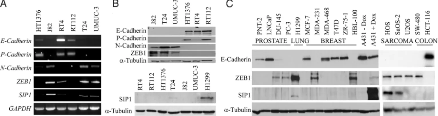
17 months (Range: 4 to 120 months). Aberrant or absent E-cadherin staining compared with normal E-E-cadherin immunoreac-tivity did not predict TCC-related death (P⫽ 0.258, Log rank test) (Fig. 1A). This is in agreement with the results of a previous comprehensive study evaluating prognostic significance of E-cadherin status in bladder cancer (16). Similarly, ZEB1 staining did not correlate with cancer-specific survival (P⫽ 0.217, Log rank test) (Fig. 1B). However, patients with SIP1-immunopositive tumors were more likely to die of TCC compared with those who had SIP1-negative bladder tumors (P⫽ 0.005, Log rank test) (Fig. 1C). Cumulative survival at 5 years for patients with SIP1 immunon-egative tumors was 42% compared with only 11% for those with SIP1 immunopositive tumors. Of 14 patients alive at 5 years follow-up, 12 were SIP1-immunonegative. Although we have shown SIP1 immunopositivity to be a predictor of cancer-specific survival, a correlation between SIP1 expression levels, and lack of local response in the bladder to radiotherapy was statistically not signif-icant. However, we observed a trend toward better response to this treatment in patients with SIP1-negative tumors (Fig. 1D). ZEB Family Members and Mesenchymal Phenotype in Cultured Cells. We analyzed transcription of ZEB proteins in five bladder cancer cell lines derived from muscle invasive TCC and in the RT4 cell line originating from a superficial bladder papilloma. Morphologically, these cell lines represented two distinct types, epithelial (RT4, RT112, and HT1376) and mesenchymal (T24, UMUC3, and J82). Accordingly, cells of the first group expressed epithelial cadherins, whereas transcription of N-cadherin was detected exclusively in mesenchymal cell lines (Fig. 2A). Transcription of ZEB1 and SIP1 genes (zfhx1a and zfhx1b) was high in mesenchymal but low or absent in epithelial cells. Given that SIP1 and ZEB1 abundance is regulated post-transcriptionally, we analyzed their expression at protein level. Interestingly, full length SIP1 protein was expressed only in T24 cells; and its expression level was low. In contrast, approximately 140 kDa ZEB1-immunoreactive bands were de-tected in all carcinoma cell lines possessing mesenchymal features (Fig. 2B). The perfect inverse correlation between ZEB1 and E-cadherin levels in bladder cancer cell lines prompted us to analyze
Fig. 1. Cancer-specific survival of patients with muscle invasive TCC treated with radical radiotherapy according to immuno-positivity of (A) E-cadherin; (B) ZEB1; (C) SIP1. Note that SIP1 positivity, but not E-cadherin status or ZEB1 levels predict the outcome of the disease. (D) Proportion of tumors with a lack of response to radiotherapy divided into groups according to SIP1 or E-cadherin immunopositivity. SIP(-), no SIP1-positive cells detected in the sample; SIP(⫹/⫺), samples mostly negative, but with a few areas with faintly stained cells; SIP1(⫹), samples with 20–50% cells having strong nuclear staining; SIP1(⫹⫹), ⬎50% cells with strong nuclear staining. E-cad(⫹), membranous localization of E-cadherin throughout the whole section; E-cad(-, -/⫹), E-cadherin staining is absent either completely or focally. Examples of staining patterns are available asSI Methods(Fig. S2). Dotted line shows a percentage of nonresponding tumors in the whole series (n⫽ 56). The data demonstrate a trend toward a lack of response to radiotherapy in patients with SIP1-positive tumors.
CELL
the effect of ZEB1 on E-cadherin promoter activity. ZEB, SNAI, Twist, and E47 proteins repress E-cadherin transcription by inter-acting with two proximal E-boxes within the E-cadherin promoter. We used transient transfection assays with luciferase reporters driven either by wild-type or a mutant E-cadherin core promoter containing mutated E-boxes 1 and 2. Data obtained showed that ZEB1 was primarily responsible for the silencing E-cadherin pro-moter in E-cadherin-negative bladder cancer cells (Fig. S4).
Having demonstrated lack of SIP1 expression in E-cadherin-negative bladder cancer cell lines, we asked whether this is a general feature of carcinoma cell lines derived from other tumor types. Except for H1299 lung carcinoma cells, none of the carcinoma cell lines analyzed expressed SIP1. In contrast, we detected high levels of SIP1 protein in two out of three sarcoma cell lines. ZEB1 expression perfectly correlated with the lack of E-cadherin in all cell lines analyzed (Fig. 2C).
To address the functional aspects of ZEB proteins in bladder cancer cells, we transiently expressed ZEB1 or SIP1 in epithelial RT112 cells followed by double immunofluorescence staining for ZEB proteins along with the EMT markers. Cells expressing ZEB1 or SIP1 showed a tendency for localization at the periphery of clusters formed by groups of nontransfected cells. Expression of either ZEB1 or SIP1 lead to the disappearance of epithelial cadherins and cadherin-associated-catenin from cell-cell borders. In addition, ZEB1 activated expression of N-cadherin (Fig. S5).
In breast cancer cells, ZEB1 inhibition reactivated expression of E-cadherin and induced other EMT features (8). However, in contrast to MDA-231 breast carcinoma cells, knockdown of ZEB1 was insufficient to reactivate epithelial markers in J82 or UMUC-3 cells (Fig. S6). Despite this, ZEB1 depletion impaired cell motility by 50–80% in J82, UMUC-3, or MDA-231 cells independently of reexpression of epithelial cadherins (Fig. S6). Previously, we de-scribed Rb-cyclin D1-dependent cell cycle arrest in A431 cells expressing SIP1 (17). To address whether both ZEB proteins affect G1/S phase transition, we used ScanRˆ microscopy. By determining DNA content in cells expressing ZEB proteins and in MOCK-transfected cells we found that SIP1, but not ZEB1, significantly attenuated G1/S transition in RT112 cells (Fig. S7).
SIP1 Protects Bladder Cancer Cells from DNA-Damage-Induced
Apo-ptosis. Given a trend toward a worse response to radiotherapy
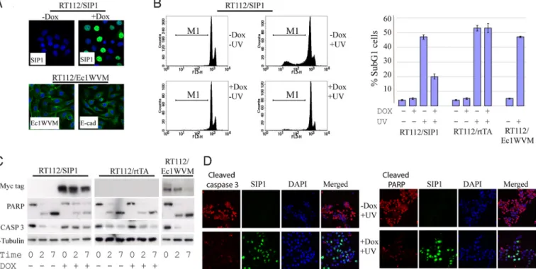
existed in patients with SIP1-positive bladder tumors (Fig. 1D), we wished to investigate whether SIP1 interferes with DNA-damage-induced apoptotic pathways. To this end, we generated a mixed population of RT112 cells with doxycycline- (DOX)-regulated expression of myc-tagged SIP1 in 60–70% of the cells (Fig. 3A). RT112/SIP1 cells were treated with UV, and 16 h later DNA
fragmentation was quantified by FACS analysis. SIP1 strongly decreased UV-induced DNA fragmentation: the proportion of cells in subG1dropped from 46% to 20% (Fig. 3B). As expected, DOX treatment of a parental rtTA-expressing clone had no effect on cell survival. As cell interactions, including intercellular adhesion mediated by cadherins, regulate cell viability (18), we speculated that inhibition of cell-cell adhesion and EMT-associated changes in cell morphology may have an impact on survival of UV-treated cells. Ec1WVM, a dominant-negative E-cadherin mutant (19) induced morphological transition in the squamous carcinoma cell line A431 via functional inactivation of E- and P-cadherins (20). Therefore, we expressed Ec1WVM in RT112 cells and analyzed its effect on UV-induced apoptosis. Although Ec1WVM induced cytoplasmic relocalization of E-cadherin and RT112/Ec1WVM cells acquired mesenchymal cell shape (Fig. 3A), their sensitivity to UV was not altered (Fig. 3B). To determine the extent of apoptosis and confirm our DNA fragmentation results, we analyzed biochem-ical hallmarks of apoptosis such as caspase activation and PARP cleavage. Consistently with the DNA fragmentation data, we observed that pro-caspase-3 and PARP cleavage was decreased in UV-treated SIP1-expressing cells, but not in parental cells or in cells expressing Ec1WVM (Fig. 3C).
SIP1 Protects Squamous Carcinoma Cells from DNA Damage-Induced
Apoptosis Independently of Cell Cycle Arrest.Arrest in the G1phase
of the cell cycle can cause resistance to different apoptotic stimuli. SIP1 induced accumulation of cells in G1 phase in RT112 cells (Fig. S7) and, to a larger extent, in A431 squamous carcinoma cells (17). Previously, we found that the underlying mechanism for this in A431 cells was a direct repression of cyclin D1 transcription by SIP1. Moreover, enforced expression of cyclin D1 uncoupled SIP1-induced cell cycle arrest from EMT (17). To study the link between the cell cycle control and anti-apoptotic functions of SIP1, and to generalize the findings obtained in RT112 cells, we exploited previously generated clones of A431 cells expressing SIP1; SIP1 and cyclin D1 in combination or Ec1WVM. As a negative control, we used A431 cells expressing SIP1ZFmut, a SIP1 mutant with an inactivated C-terminal Zn finger domain. This mutant was incom-petent in inducing EMT or cell cycle arrest (17). Consistent with the data obtained in RT112 cells, expression of SIP1 strongly protected A431 cells from DNA fragmentation (the proportion of cells in subG1 has fallen from 47% to 12% in the presence of SIP1; Fig. 4A). Interestingly, simultaneous induction of SIP1 and cyclin D1 inhib-ited UV-induced DNA fragmentation to the same level, suggesting that the anti-apoptotic activity of SIP1 is independent of cell cycle progression. In agreement with the results generated in RT112 cells, induction of Ec1WVM in A431/Ec1WVM cells had no effect
Fig. 2. ZEB family members are differentially expressed in cultured cancer cells. (A) Transcription of genes coding for cadherins and ZEB family members was analyzed in bladder cancer cell lines by semi quantitative RT-PCR. GAPDH was used as an internal control. (B) Western blot analysis of the expression of ZEB proteins and cadherins in bladder cancer cell lines. A H1299 lung adenocarcinoma lysate was used as a positive control. To control for equal loading blots were probed with anti-␣-tubulin antibody. (C) Protein expression of SIP1, ZEB1, and E-cadherin in a panel of human cancer cell lines. Note the correlation of the presence of ZEB1 and absence of E-cadherin. SIP1 expression was only detected in H1299, HOS, and SaOS-2 cell lines.
on UV-induced DNA fragmentation (Fig. 4A), indicating that anti-apototic function of SIP1 is independent of cell adhesion. Similarly, A431 clones with constitutive Ec1WVM expression were as sensitive to UV-induced apoptosis as epithelial A431 clones. Predictably, expression of SIP1ZFmut had no effect on DNA fragmentation (Fig. 4A). We observed that pro-caspase-3 and PARP cleavage was decreased in UV-treated SIP1-expressing cells, but not in cells expressing the SIP1ZFmut mutant or Ec1WVM. Of note, the extent of cleavage was similar in A431/SIP1 and A431/
SIP1/cyclD1–2 cells, that is, apoptotic hallmarks were induced by UV independently of the cell cycle (Fig. 4B).
SIP1 Interferes with Upstream Events in the Apoptotic Response.
After having demonstrated that SIP1 inhibits apoptotic hallmarks in UV-treated cells, we aimed to examine upstream components of the DNA damage response. Mitochondrial depolarization leading to cytochrome c release is a central event in the apoptotic cascade upstream of caspase-3 activation. We used TMRE staining and
Fig. 3. SIP1, but not a dominant negative E-cadherin mutant (Ec1WVM), protects RT112 cells from UV-induced apoptosis. (A) Ectopic expression of SIP1 and Ec1WVM in RT112 cells. RT112/SIP1 cells were maintained with or without DOX for 48 h. Cells were immunostained with an anti-SIP1 antibody. RT112/Ec1WVM cells were stained either with anti-myc antibody detecting mutant E-cadherin or with the anti-Ecadherin antibody recognising endogenous, but not mutant, E-cadherin (clone C20820; BD Biosciences). Cells were counterstained with DAPI; and merged images are presented. (B) SIP1, but not Ec1WVM reduced the extent of DNA fragmentation in UV-treated cells. RT112/SIP1, parental RT112/rtTA, and RT112/Ec1WVM were UV-treated (80 mJ/cm2) to induce apoptosis. RT112/SIP1 and RT112/rtTA cells were
maintained with or without DOX for 48 h before UV treatment. Cells were harvested, stained for DNA and analyzed by flow cytometry for subG1DNA content; the
percentage of subG1cells in each population is shown in the bar chart. The results show mean⫾ SD of triplicate experiments. In some experiments DOX-treated
RT112/SIP1 cells were stained for SIP1, the fraction of SIP1-positive cells was gated and compared with the untreated and ungated cells. Flow cytometry profiles show an example of decreased apoptosis in UV-treated cells gated for SIP1. (C and D) Analysis of PARP and pro-caspase 3 cleavage in RT112/SIP1, RT112/rtTA and RT112/Ec1WVM cells. Cells were treated as described in B; and expression of caspase 3 and PARP was analyzed by western blotting (C) or immunofluorescence (D). Blots were probed with an anti-␣-Tubulin antibody to control for equal loading. Note decreased cleavage of caspase 3 and PARP in cells expressing SIP1.
Fig. 4. SIP1 has a G1arrest-independent antiapoptotic activity in A431 cells. (A) Flow cytometry analyses of A431 cells with DOX-regulated expression of SIP1,
SIP1⫹cyclin D1 (clone A431/SIP1/cyclD1–2), SIP1 mutant (SIP1ZFmut), or Ec1WVM. Cells with DOX-regulated expression of indicated proteins were maintained with or without DOX for 48 h before UV treatment (60 mJ/cm2). 6 h after irradiation, the cells were harvested and analyzed by flow cytometry. The diagram shows the results
(mean⫾ SD) of four independent experiments. (B) UV-induced PARP and caspase 3 cleavage in different A431 clones. DOX-treated and untreated cells were exposed to UV and, at the indicated time points, cells were harvested and lysed. The expression of PARP, pro-caspase 3, SIP1, SIP1ZFmut, and Ec1WVM was analyzed by western blotting. Anti-␣-tubulin antibody was used to confirm equal loading.
CELL
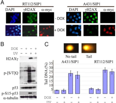
flow cytometry to analyze mitochondrial membrane potential in UV-treated RT112 and A431 cells with or without SIP1 expression. In the absence of DOX, all cell lines analyzed demonstrated an increase in the number of cells with depolarized mitochondria reaching 70–80% in 4 h after UV treatment. Mitochondrial depo-larization was strongly reduced by SIP1, but not by SIP1ZFmut or Ec1WVM (Fig. S8). One of the most upstream events preceding mitochondrial depolarization is the activation of ATM (ataxia-telangiectasia mutated) and ATR (ATM- and Rad3-related) pro-tein kinases, the sensors of DNA damage, which phosphorylate target proteins and coordinate diverse cellular responses including apoptosis. Rapid phosphorylation of a histone H2AX by ATM/ ATR kinases in response to both ionizing (21) and UV radiation (22) is critical for the recognition of DNA lesions. Immunofluo-rescent analyses of RT112 and A431 cells maintained for 2.5 h after UV treatment have shown that␥-H2AX foci formation was com-promised in SIP1-expressing cells (Fig. 5A). Western blotting confirmed SIP1-mediated reduction in phosphorylation of H2AX and another essential ATM/ATR target, p53, in A431 cells. Simi-larly, phosphorylation of various other ATM/ATR substrates at a common amino acid motif (serine/threonine followed by glutamine, the [S/T]Q-motif) was delayed or inhibited by SIP1 (Fig. 5B). We addressed whether expression of SIP1 has an effect on the extent of DNA damage induced by UV. For these purposes we used the alkali-denaturing comet assay, a method allowing to estimate levels of single-strand DNA breaks in response to DNA-damaging agents. SIP1-expressing and nonexpressing RT112 or A431 cells were treated with different UV doses, immediately lysed and subjected to the assay. The results show that UV-dependent DNA damage was not affected by SIP1 (Fig. 5C).
Taken together, our data show that SIP1 suppresses all steps in the DNA damage-induced apoptotic response, phosphorylation of
ATM/ATR substrates, mitochondrial depolarization, activation of caspase-3, PARP cleavage and DNA fragmentation. However, it does not affect the extent of DNA damage. The anti-apoptotic activity of SIP1 is largely independent of its effects on cell adhesion or cell cycle progression (Fig. 6).
Discussion
In agreement with the data previously obtained in breast cancer cell lines (5, 23), transcription of both ZEB family members correlated with mesenchymal characteristics of bladder carci-noma cells. However, at the protein level, various mesenchymal carcinoma cell lines of different origins expressed ZEB1 but not SIP1. SIP1 has been shown to induce G1phase cell cycle arrest in squamous carcinoma cells by repressing cyclin D1 transcrip-tion (17). Here we demonstrated that SIP1 significantly re-pressed G1/S transition in RT112 cells, whereas the effect of ZEB1 was insignificant. In hepatocellular carcinoma cells, SIP1 repressed hTERT, resulting in replicative senescence (24). In contrast, ZEB1 was required for proliferation of mouse embryo fibroblasts, and ZEB1⫺/⫺mouse embryo fibroblasts underwent premature replicative senescence (25). Differential expression of ZEB1 and SIP1 proteins in carcinoma cell lines may ref lect selection for SIP1-negative cells during cell culture propagation. ZEB1 plays an important role in carcinoma cells in vitro. Our data show that ZEB1 is responsible for silencing E-cadherin promoter in UMUC-3 cells in transient transfection assay (Fig. S4). Enforced expression of either ZEB protein resulted in down-regulation of epithelial cadherins, but ZEB1 depletion, although retarding cell motility, was not sufficient for E-cadherin restoration and mesenchymal-epithelial transition. These data suggest that transient activation of ZEB proteins triggers EMT, but its mainte-nance in growing tumors may require additional, possibly epige-netic mechanisms. This is in agreement with the notion that DNA methylation is involved in transcriptional silencing of E-cadherin gene in bladder cancer cell lines and in tumor tissues (26).
Despite the absence of SIP1 in cultured carcinoma cells, negative effects of SIP1 on cell cycle progression and its ability to induce replicative senescence, the idea that SIP1 may function as a tumor suppressor in bladder cancer is contradicted by the IHC data. We found that SIP1 is highly expressed in bladder tumors and is an independent predictor of poor cancer-specific survival. We observed a trend toward a better local response in the bladder to radiotherapy in SIP1-negative tumors. However, this trend could not be confirmed statistically, possibly because of the relatively small sample size available for this
Fig. 5. SIP1 inhibits phosphorylation of ATM/ATR substrates without affecting the degree of DNA damage. (A) RT112/SIP1 and A431/SIP1 cells were maintained with or without DOX for 48 h, irradiated with 80 or 60 mJ/cm2UV respectively,
incubated for 2.5 h, fixed and double-stained for SIP1 and␥-H2AX. (B) Western blot analysis of phosphorylated ATM/ATR substrates in SIP1-expressing or non-expressing A431 cells before and 2 h after UV treatment. (C) A431/SIP1 or RT112 cells were cultured with or without DOX for 48 h and treated with different UV doses as indicated. A denaturing comet assay was performed and DNA was stained and examined by fluorescence microscopy as described inSI Methods
section. The percentage of DNA in the tail of the comet (% tail DNA) was calculated for each cell by the Komet Analysis software. Results are means⫾ SD of the four gels. Images show examples of UV-treated and -untreated nuclei.
Fig. 6. Scheme depicting effects of SIP1 and an E-cadherin dominant-negative mutant on cell fate-regulating pathways. SIP1 and Ec1WVM activate cell motility, morphological EMT, and expression of vimentin. SIP1, but not Ec1WVM, affects cell fate by regulating cell proliferation, senescence, and DNA damage-induced apoptosis.
analysis. Because ZEB proteins activate tumor cell invasiveness (7, 10, 17) and survival (this study), we propose SIP1 as a biomarker of biological aggressiveness and micrometastases. Further studies will reveal whether SIP1 expression level is also a predictor of the response to different treatment modalities.
Loss of E-cadherin expression is a central event in EMT pro-grams. Its functional inhibition is sufficient to activate cell motility, invasiveness and other aspects of EMT (20). Different mechanisms such as somatic mutations or proteolytic cleavage by proteases secreted by stromal cells can cause loss of E-cadherin in cancer and trigger EMT-related programs (27, 28). The Ec1WVM mutant does not affect cell viability, indicating that the susceptibility to apoptosis can differ in cancer cells using different EMT pathways (Fig. 6). Consistent with the previous study (16), we found that complete or partial absence of E-cadherin staining correlated with tumor stage, but not with patients’ survival. Overall, our data suggest that the nature of an EMT pathway utilized by a given tumor defines the aggressiveness of the disease.
We describe a biological activity of SIP1 in tumor cells, protection from DNA damage-induced cell death. SIP1 suppresses mitochon-drial depolarization, cleavage of PARP, pro-caspase 3, and phos-phorylation of ATM/ATR substrates. In experiments with cells ectopically expressing cyclin D1 and a dominant-negative form of E-cadherin, Ec1WVM, we show that the anti-apoptotic activity of SIP1 is largely independent of G1arrest or functional inhibition of
epithelial junctional complex. Apoptosis resistance strongly con-tributes to the clonal expansion of cancer cells in primary tumors and is also important for malignant cells to disseminate and form metastases (29). Among other proapoptotic stimuli present in developing tumors, activated oncogenes may induce stalling and collapse of DNA replication forks, DNA breakage and activation of DNA damage response leading to apoptosis or senescence (30). We hypothesize that SIP1 may contribute to tumor progression by protecting cancer cells from apoptosis including DNA-damage-dependent apoptotic pathways induced by activated oncogenes.
Therefore, the anti-apoptotic function of SIP1 may underlie the frequent expression of SIP1 in bladder cancers and provide a mechanistic explanation of the association between SIP1 immuno-positivity and the disease aggressiveness. SIP1 is not a unique EMT-inducing transcription factor in possessing anti-apoptotic activity. In different systems, SNAIL and SLUG have been shown to act as survival factors interfering with p53 and Akt pathways (31–33). Moreover, down-regulation of PTEN is likely to be a part of anti-apoptotic signaling activated by SNAIL (33). RT112 and A431 cells used in this study contain mutant forms of p53; and SIP1 does not repress PTEN in these cells, suggesting that the above mechanisms do not contribute to a cell survival pathway regulated by SIP1. However, SIP1 inhibits␥H2AX foci formation and phos-phorylation of ATM/ATR substrates, suggesting that it interferes with upstream events in DNA damage pathways. Our data dem-onstrating that a single protein (SIP1) can control both cell motility and the response to DNA damage may shed light on the link between cancer metastasis and genetic instability.
Materials and Methods
For subG1DNA analysis, cells were trypsinized, pelleted, and fixed with 70%
ice-cold ethanol. After 2 h incubation at⫺20°C, cells were washed with PBS and incubated for 30 min with primary antibody in FACS solution (0.5% saponin, 3% BSA in PBS). Next, cells were washed three times with FACS solution. Alexa-488 conjugated secondary antibodies were applied in FACS solution for 30 min and cells were washed three times with FACS solution. To stain for DNA, cells were incubated with 0.260 U RNase (in PBS) for 20 min followed by 20 min incubation with 50M propidium iodide (dissolved in PBS). SubG1DNA content was analyzed using a flow cytometer (FACSCalibur, BD
Biosciences).
For additional materials and methods seeSI Methods.
ACKNOWLEDGMENTS. We thank Geert Berx, Amparo Cano, and Gerry Cohen for sharing reagents. This work was supported by Cancer Research United King-dom Grants C8851/A10844 (to E.T.) and C13560/A4661 (to G.D.D.J.), British Uro-logical Foundation (H.Q.), Foundation for Hope Against Cancer (M.K.), Associa-tion for InternaAssocia-tional Cancer Research Grant 07– 0083 (to E.T.), and Scientific and Technological Research Council of Turkey Grant 104S243 (to T.Y.).
1. Yang J, Weinberg RA (2008) Epithelial-mesenchymal transition: At the crossroads of development and tumor metastasis. Dev Cell 14:818 – 829.
2. Peinado H, Olmeda D, Cano A (2007) Snail, Zeb and bHLH factors in tumor progression: An alliance against the epithelial phenotype? Nat Rev Cancer 7:415– 428.
3. De Craene B, van Roy F, Berx G (2005) Unraveling the signaling cascades for the Snail family of transcription factors. Cell Signal 17:535–547.
4. Mani SA, et al. (2008) The epithelial-mesenchymal transition generates cells with proper-ties of stem cells. Cell 133:704 –715.
5. Gregory PA, Bracken CP, Bert AG, Goodall GJ (2008) MicroRNA as regulators of epithelial-mesenchymal transition. Cell Cycle 7:3112–3118.
6. Postigo AA, Depp JL, Taylor JJ, Kroll KL (2003) Regulation of Smad signaling through a differential recruitment of coactivators and corepressors by ZEB proteins. EMBO J 22:2453– 2462.
7. Vandewalle C, et al. (2005) SIP1/ZEB2 induces EMT by repressing genes of different epithelial cell-cell junctions. Nucleic Acids Res 33:6566 – 6578.
8. Aigner K, et al. (2007) The transcription factor ZEB1 (␦EF1) promotes tumor cell dedifferenti-ation by repressing master regulators of epithelial polarity. Oncogene 26:6979–6988. 9. Spoelstra NS, et al. (2006) The transcription factor ZEB1 is aberrantly expressed in
aggres-sive uterine cancers. Cancer Res 66:3893–3902.
10. Spaderna S, et al. (2006) A transient, EMT-linked loss of basement membranes indicates metastasis and poor survival in colorectal cancer. Gastroenterology 131:830 – 840. 11. Dohadwala M, et al. (2006) Cyclooxygenase-2-dependent regulation of E-cadherin:
Pros-taglandin E(2) induces transcriptional repressors ZEB1 and snail in non-small lung cancer. Cancer Res 66:5338 –5345.
12. Graham TR, et al. (2008) Insulin-like growth factor-I-dependent up-regulation of ZEB1 drives epithelial-to-mesenchymal transition in human prostate cancer cells. Cancer Res 68:2479 –2488.
13. Rosivatz E, et al. (2002) Diffrential expression of the epithelial-mesenchymal transition regulators snail, SIP1 and twist in gastric cancer. Am J Pathol 161:1881–1891. 14. Alves CC, et al. (2007) Slug is overexpressed in gastric carcinomas and may act
syner-gistically with SIP1 and Snail in the down-regulation of E-cadherin. J Pathol 211:507– 515.
15. Knowles MA (2008) Molecular pathogenesis of bladder cancer. Int J Clin Oncol 13:287–297.
16. Baumgart E, et al. (2007) Identification and prognostic significance of an epithelial-mesenchymal transition expression profile in human bladder tumors. Clin Cancer Res 13:1685–1694.
17. Mejlvang J, et al. (2007) Direct repression of cyclin D1 by SIP1 attenuates cell cycle progression in cells undergoing an epithelial mesenchymal transition. Mol Biol Cell 18:4615– 4624.
18. Grossman J (2002) Molecular mechanisms of ‘‘detachment-induced apoptosis-anoikis.’’ Apoptosis 7:247–260.
19. Chitaev N, Troyanovsky S (1998) Adhesive but not lateral E-cadherin complexes require calcium and catenins for their formation. J Cell Biol 142:837– 846.
20. Andersen H, et al. (2005) Immediate and delayed effects of E-cadherin inhibition on gene regulation and cell motility in human epidermoid carcinoma cells. Mol Cell Biol; 25:9138 – 9150.
21. Bonner WM, et al. (2008) GammaH2AX and cancer. Nat Rev Cancer 8:957–967. 22. Marti TM, et al. (2006) H2AX phosphorylation within the G1 phase after UV irradiation
depends on nucleotide excision repair and not DNA double-strand breaks. Proc Natl Acad Sci USA 103:9891–9896.
23. Eger A, et al. (2005)␦EF1 is a transcriptional repressor of E-cadherin and regulates epithelial plasticity in breast cancer cells. Oncogene 24:2375–2385.
24. Ozturk N, et al. (2006) Reprogramming of replicative senescence in hepatocellular carci-noma-derived cells. Proc Natl Acad Sci USA 103:2178 –2183.
25. Lin Y, et al. (2008) Zeb1 links epithelial-mesenchymal transition and cellular senescence. Development 135:579 –588.
26. Yates DR, et al. (2007) Promoter hypermethylation identifies progression risk in bladder cancer. Clin Cancer Res 13:2046 –2053.
27. Lochter A, et al. (1997) Matrix metalloproteinase stromelysin-1 triggers a cascade of molecular alterations that leads to stable epithelial-to-mesenchymal conversion and pre-malignant phenotype in mammary epithelial cells. J Cell Biol 139:1861–1872. 28. Huguenin M, et al. (2008) The metalloprotease meprin beta processes E-cadherin and
weakens intercellular adhesion. PLoS ONE 3:e2153.
29. Glinsky GV, Glinsky VV (1996) Apoptosis and metastasis: A superior resistance of metastatic cancer cells to programmed cell death. Cancer Lett 101:43–51.
30. Bartek J, Bartkova J, Lukas J (2007) DNA damage signaling guards against activated oncogenes and tumour progression. Oncogene 26:7773–7779.
31. Kajita M, McClinic KN, Wade P (2004) Aberrant expression of the transcription factors Snail and Slug alters the response to genotoxic stress. Mol Cell Biol 24:6559 – 6566. 32. Vega S, et al. (2004) Snail blocks the cell cycle and confers resistance to cell death. Genes
Dev 18:1131–1143.
33. Escriva M, et al. (2008) Repression of PTEN phosphatase by Snail transcription factor during gamma radiation-induced apoptosis. Mol Cell Biol 28:1528 –1540.
CELL