Giant Pelvic Schwannoma:
A Case Report
AABBSS TTRRAACCTT Schwannomas are benign peripheral nerve sheath tumors. Pelvic schwannomas which are usually seen in young adults are quite rare tumors and usually present as large masses causing vague symptoms. When asymptomatic huge pelvic masses are detected, it may not always be pos-sible to differentiate benign and malignant lesions without histopathological examination. These tu-mors do not have specific clinical, laboratory, and radiological findings. Therefore preoperative diagnosis is usually not adequate and complete resection of the mass might be essential. Herein this report, we aimed to describe the clinical, radiological, and pathological findings of a giant pelvic schwannoma detected in a 53-year-old man.
KKeeyy WWoorrddss:: Neurilemmoma; nerve sheath neoplasms Ö
ÖZZEETT Schwannomalar iyi huylu periferal sinir kılıfı tümörleridir. Sıklıkla genç erişkinlerde görü-len pelvik yerleşimli schwannomalar oldukça seyrek görügörü-len tümörlerdir ve genellikle belirsiz semptomlara neden olan geniş boyutlu kitleler olarak karşımıza çıkarlar. Herhangi bir semptoma neden olmayan büyük boyutlu pelvik kitleler saptandığında benign ve malign kitle lezyonları arasında ayırıcı tanı yapmak her zaman mümkün olmayabilir. Bu tümörlere ait spesifik klinik, la-boratuar ve radyolojik bulgu yoktur. Bu nedenle preoperatif tanı çoğu zaman yeterli değildir ve kitlenin tümüyle çıkarılması ve histopatolojik inceleme mutlak gerekli olabilir. Bu yazıda 53 yaşında bir erkek hastada saptanan dev bir pelvik schwannomanın klinik, radyolojik ve patolojik bulgu-larının tanımlanması amaçlanmıştır.
AAnnaahh ttaarr KKee llii mmee lleerr:: Nörilemmoma, sinir kılıfı tümörleri
TTuurrkkiiyyee KKlliinniikklleerrii JJ MMeedd SSccii 22001100;;3300((22))::775500--44 Ahmet Hakan HALİLOĞLU, MD,a
Hatim Yahya USLU, MD,b
Nuket ÜZÜM, MD,c
Nuray HALİLOĞLU, MD,d
Orhan GÖĞÜŞ, MD,a
Departments of aUrology bGeneral Surgery,
Ufuk University Faculty of Medicine, Ankara
cMikropat Pathology Laboratory, İstanbul
dDepartment of Radiology Ankara University Faculty of Medicine, Ankara Ge liş Ta ri hi/Re ce i ved: 13.06.2008 Ka bul Ta ri hi/Ac cep ted: 11.09.2008 Ya zış ma Ad re si/Cor res pon den ce: Ahmet Hakan HALİLOĞLU, MD, Ufuk University Faculty of Medicine, Department of Urology, Ankara, TÜRKİYE/TURKEY
chwannomas are infrequent peripheral nerve sheath tumors which ac-count for 5% of benign soft tissue tumors. More than 50% of the lesions are seen in the head and neck region and extremities.1Pelvic
nomas are quite rare tumors, compromising less than 1% of benign schwan-nomas.2,3When seen in the pelvic region they may present with symptoms
like abdominal pain, hypertension, and frequent urination. Nevertheless the symptoms are usually vague and nonspesific hence there is frequently a delay in the detection of the tumor. It should be kept in mind that although rare, schwannomas can be malignant and as accurate preoperative diagnosis is dif-ficult, and the mass has to be removed totally by disseccing from the neigh-bouring structures. We present here a histopathologically proven huge perivesical schwannoma with a brief review of the literature.
CASE REPORT
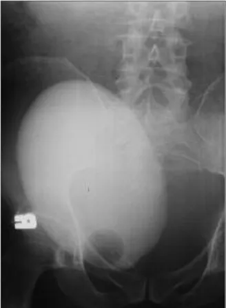
A 53-year old man was admitted to the urology de-partment with left flank pain and gross hematuria with a history of 3-months. There was no signifi-cant finding in his personal or family medical his-tory. A huge hard mass below the umblikus was detected on physical examination. Cystograpy re-vealed displacement of the bladder to the right side but there was no filling defect in the cavity and the bladder margins were smooth (Figure 1). There was no significant finding on neurological examination. Suprapubic ultrasonography (US) revealed a hy-poechoic solid mass located posterolaterally to the left wall of the bladder with dimensions of 18 x 13 x 8 cm. There was no obvious relation between the soft tissue mass and the prostate gland on transrec-tal sonography. On computed tomography (CT), the well demarcated hypodense mass with minimal contrast enhancement was neighbouring the small intestine mesenteric fat superiorly and running down to the ischioanal fossa inferiorly. No
calcifi-cations were detected on CT images. The mass was displacing the bladder anterolateraly and rectum laterally. Magnetic resonance imaging (MRI) was performed in order to evaluate the exact nature of the lesion and to determine its relation to the neigh-bouring structures. MR images revealed a het-erogenous mass with solid and cystic components located in the presacral region which was lying downwards to the left ischioanal fossa with dimen-sions of 24 x 14 x 11 cm. On contrast enhanced T1-weighted images the central part of the mass was enhancing predominantly and signal void areas rep-resenting vessels were present on T2-weighted im-ages (Figures 2, 3). However MRI also failed to show the exact origin of the mass but it revealed that there was no invasion to the surrounding tissues.
As the preoperative diagnosis was not accu-rate, surgical exploration was considered to be es-sential. Previously, a cystoscopy was performed in order not to miss an occult intravesical lesion which may cause hematuria. The high-positioned bladder was hardly enrolled during the cystoscopic examination performed by 21 F rigid cystoscop. No intravesical lesions were detected and the ureteral orifices were normal.
During the exploration, the mass seemed to occupy the whole left perivesival region and ex-tended towards to the bladder neck and vesicula seminalis but as it was not adhesive to the neigh-bouring structures and it could easily be removed with no significant bleeding at all. Since as the dis-tal part of the left ureter was running so close to the mass, it was damaged during the exploration and ureterouretereostomy was performed. There was no need for blood transfusion during or after the surgery and no complications were noted in the postoperative period.
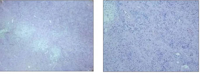
On the histopathological examination, macro-scopically, the total specimen was well-circum-scribed, encapsulated, with gelatinous appearance and gray in color, measured 30 cm by 23 cm and 20 cm. Microscopically, the tumor was exhibited a fas-cicular and from place to place whorled growth pat-tern with thin, loose and intact fibrous capsule. Hypercellular (Antony A component) and hypocel-lular (Antony B) components were seen in
low-FIGURE 1: On cystographic examination the bladder seems to be normal
power field. The cells were similar with elongated cell shape and wavy nuclei, in both areas. There were nuclear palisading areas in tumor. Prominent thick hyaline vessel walls and widespread myxoid areas were present within the tumor. Mitoses were
ex-tremely scanty (Figure 4). There were no cytological atypia or necrosis. The immunohistochemical study revealed a diffuse positive staining in both cyto-plasms and nuclei of the cells with S-100 (Figure 5). The patient was discharged on the 5th day of the surgery. Pelvic CT was performed in 6 months time and there were no pathological findings. He is still under follow-up.
DISCUSSION
Schwannomas originate from the Schwann cells of the neural sheath, and especially when multiple le-sions are present, they can be associated with neu-rofibromatosis type I.4,5The tumor may occur in any
nerve trunk except for the cranial nerves I and II which lack schwann cells. Pelvic schwannomas mostly arise from the lumbar and sacral nerves.6,7In
the present case, the presacral tumor was multilobu-lated and it was running down to the ischioanal fossa, but there was no pedicle of the tumor attaching it to the lumbar or sacral nerve roots suggesting a neuro-genic origin.
Schwannomas slow-growing tumors but as they do not cause symptoms unless they reach a large size enough to compress adjacent structures, and as retroperitoneum is a flexible location allow-ing the tumor grow-up easily, pelvic schwannomas may present as huge masses with cystic degenera-tion at the initial diagnosis.4,8
It has been estimated that pelvic schwannomas are freqently located in the lower retroperitoneum, and they are usually ovoid or spherical in shape with smooth, well-defined borders. As the size of the mass increases, the amount of cystic degenera-tion remains.4Although there is no imaging
char-acteristic finding unique to this tumor, presence the cystic degeneration in a semisolid tumor on US may suggest the diagnosis, as retroperitoneal tumors are usually not cystic. On CT scans, schwannomas ap-pear as heterogenous masses, and if present punc-tate or mottled calcifications can easily be detected by CT.9MRI is the most accurate imaging method
in the diagnosis of soft-tissue tumors with its high contrast resolution and multiplanar capability. Schwannomas are usually hypointense or isointense with muscle on T1- weighted images, and
hyperin-FIGURE 2: T1-weighted axial MR image (A) reveals a well-defined huge
pelvic mass displacing the bladder anterolateraly (short arrow) and rectum lat-erally (long arrow). The central part of the mass is mildly hyperintense when compared to the muscle. On fat saturated T2-weighted axial image (B) there are signal void areas representing vasculary elements (arrows). The mass is continuing down to the ischioanal fossa (C).
FIGURE 3: On contrast enhanced T1-weighted sagittal MR image the
cen-tral part of the presacral mass enhances predominantly whereas the upper parts seems to be cystic.
tense on T2-weighted images. Nevertheless Heterogenity due to cystic and hemorrhagic com-ponents can also be detected on MR images. Solid components would enhance after intravenous con-trast medium administration.4MRI is also capable
of showing the relationship of the tumor with adja-cent anatomic structures and delineating the vascu-lar nature of the tumor.10
CT-guided biopsy is usually not recommended since cellular pleomorphism may lead to misinter-pretion as malignancy. On the other hand, if the mass is malignant, tumor seeding and the other well known risks of percutaneous biopsy like hem-orrhage or infection may occur.7,9
The ideal choice of treatment is complete ex-cision with preservation of the associated nerve. As malignant schwannomas are aggressive neoplasms acting as high-grade sarcomas and the risk of ma-lignancy can not be excluded preoperatively or by frozen section analysis, neighbouring structures can be sacrificied in order to attain negative tumor margins. In the case we presented here, the left ureter was running so close to the mass that it was damaged during exploration. Oppositely, one can consider partial resection or simple enucleation re-lying on the common benign nature of the tumor and also considering the age, general health, and symptoms of the patient.9,10
On histopathological examination two pat-terns exist for schwannomas, Antoni A and B types representing high and low cellularity, respectively.
Antoni A areas are composed of spindle-shaped cells and Antoni B areas are composed of edema-tous and degenerated tissues with fewer cells. Dif-fuse staining of S-100 protein is helpfull to confirm the diagnosis of schwannoma.6,8The present tumor
consisted of both Antoni A and Antoni B cells and immunohistochemical study revealed a diffuse pos-itive staining of the cells with S-100 protein. There was no sign of malignant transformation.
In conclusion; there is no spesific imaging finding for benign pelvic schwannomas, and al-though rare, malignant transformation may occur. Therefore, complete surgical resection should be the choice of treatment depending on the patient’s general status, and the relationship of the tumor with the adjacent vital structures.
FIGURE 5: S-100 immunoreactivity in schwann cells (Streptavidin-biotin
peroxidase x 200)
a b
FIGURE 4: Antoni type A (cellular areas) and antoni type B (hypocellular areas) tissue. (HE x 40) (A). The myxoid matrix and hyaline vessel walls in tumor (HE
1. Gir gin C, Oz kan U, Se zer A, Tug yan N. Lar ge pel vic schwan no ma ca u sing bi la te ral hydro-nep hro sis. Int J Urol 2003;10(11):616-8. 2. Fe lix EL, Wo od DK, Das Gup ta TK. Tu mors of
the ret ro pe ri to ne um. Curr Probl Can cer 1981;6(1):1-47.
3. Guz BV, Wo od DP Jr, Mon ti e JE, Pon tes JE. Ret ro pe ri to ne al ne u ral she ath tu mors: Cle ve -land Cli nic ex pe ri en ce. J Urol 1989;142(6): 1434-7.
4. Hug hes MJ, Tho mas JM, Fis her C, Mos ko vic EC. Ima ging fe a tu res of ret ro pe ri to ne al and
pel vic schwan no mas. Clin Ra di ol 2005;60(8): 886-93.
5. Ha be ral A, Tur gut F, Tur gut M, Koç S, Kö se F, Öz fut tu A, et al. [A ca se of ma lig nant schwan-no ma of the pel vic sympat he tic ple xus in a girl with Ne u ro fib ro ma to sis type 1]. Tur ki ye Kli nik -le ri J Gyne col Obst 1994;4(3):203-5. 6. Tong RS, Col li er N, Ka ye AH. Chro nic sci a ti
-ca se con dary to ret ro pe ri to ne al pel vic schwan-no ma. J Clin Ne u ros ci 2003;10(1):108-11. 7. Da nesh mand S, Yo us sef za deh D, Cha mi e K,
Bos well W, Wu N, Ste in JP, et al. Be nign ret ro
pe ri to ne al schwan no ma: a ca se se ri es and re vi -ew of the li te ra tu re. Uro logy 2003;62(6):993-7. 8. Ue da M, Oka mo to Y, Ue ki M. A pel vic ret ro
-pe ri to ne al schwan no ma ari sing in the right pa-ra col pi um. Gyne col On col 1996;60(3):480-3. 9. Goh BKP, Tan YM, Chung YFA, Chow PKH,
Oo i LPJ, Wong WK. Ret ro pe ri to ne al schwan-no ma. Am J Surg 2006;192(1):14-8. 10. Oh ta I, Lin PH, Ra u CL, Wang KC. Eva lu a ti on
of pe ri nep hric, ret ro pe ri to ne al schwan no mas: ca se re port and re vi ew of the li te ra tu re. So uth Med J 2007;100(1):80-2.