A GIPC1-Palmitate Switch Modulates Dopamine Drd3 Receptor
Trafficking and Signaling
Margarita Arango-Lievano,a,b,cOzge Sensoy,dAmélie Borie,a,b,cMaithé Corbani,a,b,cGilles Guillon,a,b,cPierre Sokoloff,e* Harel Weinstein,f Freddy Jeanneteaua,b,c
Inserm, U1191, Institute of Functional Genomics, Montpellier, Francea
; CNRS, UMR-5203, Montpellier, Franceb
; Université de Montpellier, Montpellier, Francec ; Istanbul Medipol University, School of Engineering and Natural Sciences, Istanbul, Turkeyd
; Unit of Neurobiology and Molecular Pharmacology, INSERM U573, Centre Paul Broca, Paris, Francee
; Department of Physiology and Biophysics, Weill Medical College of Cornell University, New York, New York, USAf
Palmitoylation is involved in several neuropsychiatric and movement disorders for which a dysfunctional signaling of the
dopamine D3 receptor (Drd3) is hypothesized. Computational modeling of Drd3’s homologue, Drd2, has shed some light
on the putative role of palmitoylation as a reversible switch for dopaminergic receptor signaling. Drd3 is presumed to be
palmitoylated, based on sequence homology with Drd2, but the functional attributes afforded by Drd3 palmitoylation have
not been studied. Since these receptors are major targets of antipsychotic and anti-Parkinsonian drugs, a better
character-ization of Drd3 signaling and posttranslational modifications, like palmitoylation, may improve the prospects for drug
development. Using molecular dynamics simulations, we evaluated in silico how Drd3 palmitoylation could elicit
signifi-cant remodeling of the C-terminal cytoplasmic domain to expose docking sites for signaling proteins. We tested this model
in cellulo by using the interaction of Drd3 with the G-alpha interacting protein (GAIP) C terminus 1 (GIPC1) as a
tem-plate. From a series of biochemical studies, live imaging, and analyses of mutant proteins, we propose that Drd3
palmitoyl-ation acts as a molecular switch for Drd3-biased signaling via a GIPC1-dependent route, which is likely to affect the mode
of action of antipsychotic drugs.
T
he C terminus of G-protein-coupled receptors (GPCRs) has
been reported to take part in a large repertoire of
protein-protein interactions and represents a functional component of
GPCR signaling that is characterized by the malleability of the
interface it provides (
1
,
2
). In addition, the GPCR C termini can
switch between a soluble form in the cytoplasm and an acylated
form anchored to the membrane (
3
). Among the latter, the
palmi-toylated form is established by the covalent linkage of a palmitic
acid moiety through a thioester bound on one or more cysteine
residues often localized in proximity to the conserved
amphi-pathic helical motif 8 (helix-8) (
4
). The palmitate moiety is
thought to be captured in cholesterol-rich membrane
environ-ments in order to stabilize GPCRs (
5
,
6
). The helix-8 may adopt a
helical structure in the presence of membranes, thereby affecting
structural docking sites involved in GPCR dimerization and
sig-naling (
7–10
). Various suggestions have been offered in the
liter-ature regarding the possible regulation of cellular processes by a
palmitoylation-dependent conformational switch of the helix-8 in
GPCR C-terminal tails. These include G-protein coupling (
11
),
oligomerization (
12
,
13
), regulation of activation (
14
), receptor
turnover (
15
), and trafficking (
16
).
Dopamine receptors Drd1 and Drd2 are palmitoylated on one
or more cysteine residues within the C-terminal domains, and
mutations involving these cysteines result in functional
impair-ment of dopamine signaling (
3
,
17
,
18
). However, the molecular
mechanisms by which palmitoylation contributes to these effects
are not understood. Recent structural modeling of Drd2 helix-8 in
silico, based on the X-ray structures of Drd3 (
19
), suggested a
putative role for palmitoylation in the anchoring of the Drd2 C
terminus into the membrane environment, thus masking some of
its molecular motifs (
10
). In turn, depalmitoylation was
hypoth-esized by molecular modeling to expose the Drd2 C terminus to
the aqueous environment, thereby opening possibilities for
pro-tein-protein interactions with cytoplasmic partners (
10
). One
il-lustrative example is the interaction of GIPC1 with the Drd2 C
terminus through a region that overlaps with the palmitoylation
site (
Fig. 1A
) (
18
,
20
,
21
). Computational modeling of the
Drd2-GIPC1 interaction based on the X-ray structures of Drd3 and the
PDZ domain of GIPC2 predicted that Drd2 C-terminal
palmitoyl-ation could be inhibitory (
10
), but this has not been tested
exper-imentally. This is all the more intriguing, as the process could be
dynamic, catalyzed by palmitoyl transferases and inhibited by
thioesterases (
22
). Because palmitoylation activity was previously
mapped in several subcellular compartments, like the biosynthetic
pathway (
23
) and at the plasma membrane of cells in culture (
24
),
cycles of Drd2 palmitoylation-depalmitoylation between
subcel-lular compartments could bias the Drd2-GIPC1 interaction and
subsequent signaling.
The interest in signaling modulation by various structural
components of GPCRs is related to the growing importance of
biased signaling in new drug development. Drd2 and Drd3 are
Received 2 October 2015 Returned for modification 27 October 2015 Accepted 6 January 2016
Accepted manuscript posted online 19 January 2016
Citation Arango-Lievano M, Sensoy O, Borie A, Corbani M, Guillon G, Sokoloff P, Weinstein H, Jeanneteau F. 2016. A GIPC1-palmitate switch modulates dopamine Drd3 receptor trafficking and signaling. Mol Cell Biol 36:1019 –1031.
doi:10.1128/MCB.00916-15.
Address correspondence to Freddy Jeanneteau, [email protected]. * Present address: Pierre Sokoloff, PSAdvice, Belleserre, France.
The Unit of Neurobiology and Molecular Pharmacology, INSERM U573 research unit, no longer exists and has been integrated into INSERM UMR894, Center for Psychiatry and Neuroscience, Paris, France.
Copyright © 2016, American Society for Microbiology. All Rights Reserved.
on July 2, 2020 at Medipol University
http://mcb.asm.org/
prime targets of antipsychotic and anti-Parkinsonian drugs (
25
,
26
,
27–31
). The two dopamine receptor subtypes share common
signaling cascades, including inhibition of cyclic AMP (cAMP)
formation through G
i/G
Ocoupling and phospholipase C,
regula-tion of ion channel activities, and stimularegula-tion of mitogenesis
through the activation of mitogen-activated protein kinases and
Akt (
32–35
). The close homology between Drd2 and Drd3
sug-gests that C-terminal palmitoylation could be a common
mecha-nism for controlling signaling via the helix-8. To explore this
hy-pothesis, we undertook in silico and in cellulo analyses of the
impact of palmitoylation on Drd3 signaling via GIPC1, an
inter-acting protein (
21
).
GIPC1 has previously been identified as an interacting protein
for several transmembrane and membrane-associated proteins,
including but not limited to GAIP, a regulator of G protein
signal-ing (
36
),
1-adrenergic receptors (
37
), semaphorin M-SemF (
38
),
glucose transporter GLUT1 (
39
), tyrosine kinase receptors like the
neurotrophin receptors tropomyosin-related kinases (TrkA and
TrkB) (
40
), insulin-like growth factor 1 (IGF-1) receptor (
41
),
transforming growth factor
 (TGF-) receptor type III (
42
), and
lysophosphatidic acid receptor 1 (LPA1) (
43
). These studies
sug-gested a possible role for GIPC1 in the regulation of vesicular
trafficking (
36
,
40
,
43
), receptor surface expression (
39
,
42
), and G
protein signaling (
36
,
37
,
40
,
41
). In the case of Drd2 and Drd3,
dopamine signaling via the cAMP route is negatively regulated by
GIPC1 by way of a direct interaction with the PDZ (PSD95/Dig/
ZO-1) and the acyl carrier protein (ACP) domains (
21
). Previous
studies implicated other ACP domains in the transfer of acyl
co-enzyme A (acyl-CoA) moieties, including palmitoyl-CoA, to the
catalytic domain of acyl transferases and thioesterases (
44–46
).
The questions remain whether Drd3 is indeed palmitoylated and if
the presence of the ACP domain provides GIPC1 with regulatory
functions. The Drd3 C terminus consists of a conserved
amphi-pathic helical motif (Hx8) that contains the Lys-Ser-Cys motif for
GIPC1 binding and a putative C-terminal palmitoylation site (
Fig.
1A
). The two sites overlap, suggesting the possibility of
competi-tive interaction between GIPC1 binding and palmitoylation.
MATERIALS AND METHODS
Computational modeling. The computational modeling of the dynamic
behavior of Helix-8 of Drd3 in a membrane system was performed as
previously described for Drd2 (10). A series of coarse-grained MD simu-lations (CGMD) was carried out for a system composed of the transmem-brane 7 (TM7)– helix-8 segment from Drd3. The CGMD simulations were done with the Martini force field, following established protocols (47–49). Briefly, an integration step of 0.04 ps was used, and long-range electrostatic and van der Waals interactions were calculated with shift functions. For the latter, we used a twin-range cutoff scheme of 0.9 and 1.2 Å, with the neighboring list updated every 10 ps. The Berendsen algorithm was used to maintain constant temperature and pressure (50). Simula-tions were performed in a lipid membrane composed of 70% sphingomy-elin and 30% cholesterol to mimic lipid rafts that act as membrane mi-crodomains for the assembly of signaling molecules (51,52). All the systems were neutralized at 0.15 M NaCl (47,53). The complete simulated system is composed of the Drd3 entire TM7 (residues 372 to 388) together with parts of the extracellular loop 3 (residues 368 to 371) and the entire amphipathic helix-8 (residues 389 to 400) of human Drd3 (hDrd3). The backbone atoms of TM7 were restrained to their initial coordinates to maintain the orientation adopted by TM7 within the membrane in the complete structure of the receptor. The palmitoylated C terminus was obtained by patching the palmitate group to the terminal cysteine residue of helix-8 and was considered to be in the zwitterionic form. The effect of reversible palmitoylation on the depth of membrane penetration of the entire amphipathic helix-8 in this segment was evaluated. Simulations of (non)palmitoylated Hx8 were performed for 4s (effective time). To determine convergence, the backbone insertion depth of each residue of helix-8 was monitored between successive predetermined lengths of win-dows over the entire trajectory; 100-ns winwin-dows were used. A cutoff of 0.05 Å was used as the convergence criterion between subsequent win-dows. The depth of insertion of the helix-8 backbone was calculated by including the nitrogen, C␣ atom, and the carbonyl group. The average insertion was determined with respect to the phosphorous atoms of the membrane lipid headgroups present within a 2-Å cutoff distance from the backbone of helix-8.
Plasmids. The human Drd3 was tagged at the N terminus with the
enhanced green fluorescent protein (GFP) fused to the nicotinic receptor ␣7 subunit signal peptide (54) using the pCEP4 vector (Invitrogen Corp.). The GFP-Drd3⌬C receptor mutant was previously described (21), the GFP-Drd3CS mutant was generated by site-directed mutagenesis with QuikChange kit (Agilent Technologies), and GFP-Drd3/2AR was gen-erated by PCR using the following oligonucleotides (underlined se-quences specify restriction sites for cloning): sense, 5=-CCCAAGCTTGG AAGGCGGGGCAG-3=, and antisense 5=-GCTCTAGAGCTCGAGTCAT AGTAGCGATCTTCTGAGGCAGCAGCAGGACAGGATCTTGAGGA AGG-3=. C-terminal domains of Drd3 (animo acids 389 to 400) and
mu-FIG 1 Membrane insertion profile of the (non)palmitoylated Drd3 C terminus. (A) The binding site of GIPC1 to the C-terminal domain of Drd3 contains a
putative palmitoylation site. (B) Average backbone insertion values (means⫾ SEM of 8 replicates) for (non)palmitoylated Drd3 C termini were calculated as described in Materials and Methods. The data show the level of the phosphorous atom of lipids at the water-membrane interface. The model predicted that the Drd3 C terminus is inserted more deeply in the membrane environment when palmitoylated (paired t test; *, P⬍ 0.0001).
on July 2, 2020 at Medipol University
http://mcb.asm.org/
tants (Drd3⌬C and Drd3/2AR) were inserted in frame downstream of the B42 activation domain into a pEG202 vector and GIPC1 subcloned into pJG4.5 downstream of the LexA domain to perform the 2-hybrid binary complementation assay (OriGene Technologies, Rockville, MD) as previously described (21). GIPC1 and a mutant lacking the ACP domain (⌬ACP, lacking amino acids 223 to 316) were constructed by PCR and subcloned into the pcDNA3.1HisC vector (Invitrogen Corp., San Diego, CA). The resulting GIPC1 contained a dual 6⫻His/Xpress tag at the N terminus. All constructs were verified by automated nucleotide sequenc-ing (Licor, Lincoln, NE).
Reagents. Dopamine, ascorbic acid, hydroxylamine,
N-ethylmaleim-ide, palmitic acid, and 2-bromohexadecanoic acid (2BrP) were obtained from Sigma. Commercial antibodies were as follows: anti-extracellular signal-regulated kinases 1 and 2 (anti-ERK1/2; catalog number M3807; Sigma) and the phospho-ERK1/2[T202/Y204] (catalog num-ber 9101), phosphor-AKT[S473] (catalog numnum-ber 9271), and AKT (catalog number 9272) antibodies were from Cell Signaling Technol-ogies. The 6⫻His tag and streptavidin-horseradish peroxidase (HRP) were from ThermoScientific. GFP antibodies (catalog number Ab13970) were from Abcam. Homemade antibodies against GIPC1 and Drd3 were previously described (21,55).
Cell culture and transfections. HEK293 cells and CHO cells were
cultured in Dulbecco’s modified Eagle’s medium (DMEM; Life Technol-ogies, Rockville, MD) supplemented with 10% fetal calf serum and 100 g/ml penicillin-streptomycin in a humidified atmosphere of 5% CO2, 95% air. Striatal primary neuron cultures derived from time-pregnant Sprague-Dawley rat (Janvier Lab) embryos at day 18 (E18) were prepared on glass coverslips coated with poly-D-lysine (0.1g/ml) and grown for 3 weeks in neurobasal medium supplemented with B27 (Invitrogen) with a 1/10 proportion of cortical neurons. The protocol complied with the Eu-ropean Communities Council Directive (86/609/EEC) and was approved by the Ministère de la Recherche Française (agreement number CEEA-LR-00651.01). Transfections were performed with Lipofectamine 2000 (Life Technologies, Rockville, MD). Palmitic acid and 2-bromo-palmitate (Sigma) were prepared fresh as stock solutions in ethanol (EtOH; 3% final concentration) and diluted further with culture medium. The vehicle consisted of 3% EtOH.
[3H]palmitate protein labeling assay. HEK293 cells were starved
from serum for 1 h prior to the addition of 0.4 mCi/ml de-[3H]palmitate ([9,10-3H]palmitate; 30 to 60 Ci/mmol; NEN) for 50 min. Cells were rinsed several times in phosphate-buffered saline (PBS) before solubiliza-tion in a digitonin-cholate lysis buffer (50 mM Na/Na2PO4[pH 7.4], 300 mM NaCl, 4% digitonin, 1% cholate-Na, and protease inhibitors) for 45 min at 4°C. Lysates were diluted with 1 volume of double-distilled water and centrifuged at 50,000⫻ g for 40 min. Supernatant containing the solubilized Drd3 was collected, and the protein concentrations were mea-sured with Coomassie stain (Bio-Rad) and bovine serum albumin (BSA) as the standards. Normalized protein levels were further diluted 20 times with 50 mM Na/Na2PO4(pH 7.4) and cleared with normal rabbit IgG (Santa Cruz Biotechnology) prior to immunoprecipitation of the Drd3 receptors with anti-GFP antibodies (Abcam) overnight and a 2-hour in-cubation with a 50% slurry of protein A-Sepharose (Amersham Pharma-cia Biotech). Beads were rinsed 5 times with the diluted solubilization buffer and suspended in a nonreducing sample loading buffer. Proteins were separated by SDS-PAGE (4 to 12% acrylamide gradient; Life Tech-nologies). Proteins were fixed in the gels by using 10% acetic acid and 40% isopropanol in water and further incubated with 1 M hydroxylamine (pH 7) prior to fluorographic signal amplification (Amplify; Amersham Phar-macia Biotech). Finally, gels were dehydrated and exposed to MP films (Amersham Pharmacia Biotech) for 4 to 8 weeks. Autoradiographic films were digitalized, and signals were measured based on the optical density, using the public domain NIH Image program version 1.63 (developed at the U.S. National Institutes of Health and available on the Internet at
http://rsb.info.nih.gov/nih-image/).
Nonradioactive detection of palmitoylation by ABE. Palmitoylation
of the GFP-Drd3 and mutant CS expressed transiently in HEK293 cells was assayed using the acyl-biotin exchange (ABE) method as previously described (56,57). In brief, GFP-Drd3 constructs were immunoprecipi-tated with GFP antibodies in lysis buffer supplemented with 50 mM N-ethylmaleimide to block nonpalmitoylated cysteines. Further treatment with 1 M hydroxylamine cleaved the thioester bonds at palmitoylated cysteines. GFP-Drd3 contructs were treated with thiol-reactive biotin-BMCC (ThermoScientific), and palmitoylated cysteine was further de-tected with streptavidin-HRP (ThermoScientific) by Western blotting.
[125I]iodosulpride binding assays. Drd3 ligand binding
experi-ments were performed on purified membrane fractions using 0.1 nM [125I]iodosulpride (Amersham Pharmacia Biotech) as previously de-scribed (58). To detect Drd3 present at the cell surface, we used the Drd3 ligands dopamine and iodosulpride, which cannot diffuse through membranes. To this end, whole cells were scratched off culture dishes, harvested by centrifugation, suspended in DMEM–F-12 supplemented with ascorbic acid (50g/ml), and incubated in quadruplicate for 30 min at 30°C with 0.1 nM [125I]iodosulpride (Amersham Pharmacia) in the increasing concentrations of nonradioactive dopamine. Nonspecific binding was measured in the presence of eticlopride (1M). Binding was stopped by filtration through GF/C filters coated with polyethyleneimine (0.3%, vol/vol), and radioactivity was counted on the filter by gamma scintigraphy. Dopamine competition curves were analyzed by nonlinear regression with a two-site model by using Prism software (GraphPad, San Diego, CA). The protein concentration was estimated with the Coomassie protein assay reagent using a wide range of BSA concentrations as stan-dards.
125I-cAMP accumulation assay. Cells were preincubated with 10M
3-isobutyl-1 methylxanthine in␣-minimal essential medium for 25 min and treated with increasing concentrations of dopamine for 10 min in the presence of 0.5M forskolin. The reaction was stopped by addition of 50 l of ice-cold 0.1 M HCl. Cells were sonicated, and cAMP accumulation was assayed with a125I-cAMP radioimmunoassay kit (DuPont/NEN, Bos-ton, MA).
Binary two-hybrid protein interactions. Binding of the Drd3 C
ter-minus to GIPC1 was measured with the DupLex-A two-hybrid system kit (OriGene Technologies) in the Saccharomyces cerevisiae strain EGY48, which harbors the reporter genes for LEU1 and-galactosidase under the control of upstream LexA binding sites. Transformants were grown on selective medium and assayed for-galactosidase (-Gal) to verify and quantify interactions in a solid support assay and liquid culture assay, respectively, with 5-bromo-4-chloro-3-indolyl--D-galactopyranoside (X-Gal) and o-nitrophenyl-D-galactopyranoside (ONPG) as substrates, according to the yeast protocols handbook from Clontech (Palo Alto, CA). One unit of-Gal was defined as the amount that hydrolyzed 1 mol of ONPG per min per cell.
Endoplasmic reticulum exit assay. Nonpermissive temperature shifts
have been previously used to reduce protein trafficking in the secretory pathway and accumulate transmembrane cargo proteins in the pericen-triolar intermediate compartment (59). Transfected HEK293 cells were grown at a restrictive temperature of 20°C overnight to block GFP-Drd3 cargo in the pericentriolar endoplasmic reticulum (ER) to the Golgi com-partment. Back at a permissive temperature of 37°C, cells started to ex-press GFP-Drd3 at the plasma membrane. Cycloheximide (CHX) was added to cells for at least 1 h prior to temperature shift (from 20°C to 37°C) to prevent the synthesis of new GFP-Drd3 cargo and follow the ER exit of the existing pool of GFP-Drd3 cargo. Therefore, the thermal shift ER exit assay combined with CHX permitted the analysis of GFP-Drd3 targeting to the plasma membrane as a function of GIPC1 overexpression and palmitoylation in cell preparations fixed with ice-cold 2% parafor-maldehyde (PFA).
Immunolabeling. Cells were grown on collagen-coated coverslips and
fixed in 2% paraformaldehyde–PBS (pH 7.4) for 20 min at room temper-ature, washed twice in PBS-glycine buffer (0.1 M, pH 7.4), and permeated
on July 2, 2020 at Medipol University
http://mcb.asm.org/
for 20 min with 0.05% saponin while blocking in 10% fetal bovine serum– PBS. GIPC1 was labeled with an anti-Xpress antibody (1:3,000 dilution; Life Technologies), and Drd3 was labeled with a GFP antibody (1:3,000 dilution; Abcam) in incubation buffer (0.1% BSA–PBS supplemented with 10% fetal bovine serum). A-LexA-conjugated secondary antibodies were used for detection (1:1,000 dilution; Molecular Probes). GFP-Drd3 prominently localized to the plasma membrane after treatment with 20 g/ml CHX for at least 5 h to clear the intracellular biosynthetic pathway. Previously described home-made antibodies were used to stain the endog-enous GIPC1 and Drd3 on adult rat brain sections fixed with 4% parafor-maldehyde (21). Images were acquired with a laser scanning confocal image system (TCS SPII software; Leica, Deerfield, IL) coupled to a Leica DM R fluorescence microscope. When necessary, fluorescent images were transformed into grayscale pixels, and ImageJ plug-ins were applied to trace the regions of interest (ROI; the plasma membrane and the intracel-lular compartment).
GFP antibody feeding assay. GFP antibodies were applied in the
cul-ture medium of living cells at 4°C for 30 min to limit vesicular transport. This allowed for the labeling of the GFP-Drd3 constructs present at the cell surface under detergent-free conditions with Alexa Fluor 555-conju-gated secondary antibodies. ImageJ was utilized to measure the surface covered by Alexa Fluor 555 and GFP signals in the same ROIs of at least 10 cells per condition.
Fluorescence imaging. Time-lapse imaging of transfected GFP-Drd3
in HEK293 cells seeded on poly-D-lysine-coated Ibidi bottom dishes (Bio-valley) was performed with an inverted epifluorescence microscope (1X70; Olympus) coupled with a Coolsnap HQ camera (Roper Scientific, France). ImageJ plug-ins were used to align image stacks and track vesicle movement in time series images.
Statistical analysis. For simulation data, the standard error (SE)
from 8 replicates was calculated according to the following formula: SE⫽
再冑
⌺共s⫽1兲 m ⌺ 共i⫽1兲 n Y is2 共ny⫺1兲共ny兲冎
1 ⁄ 2, where s is the series number, i is the point number in series s, m is the number of series for point y in the chart, n is the number of points in each series, Yisis the data value for
series s and the ith point, and nyis the total number of data values in all
series. For in cellulo data, the averages⫾ standard errors of at least three independent experiments are presented. For [125I]iodosulpride binding,125I-cAMP accumulation, and binary 2-hybrid assays, each sample was analyzed in triplicate. Significance (P⬍ 0.05) was deter-mined using the two-tailed Student t test.
RESULTS
In silico simulations show that palmitoylation increases Drd3
C-terminal penetration in the membrane. We investigated by
using computational approaches and a Drd3 three-dimensional
model the relationship between palmitoylation and membrane
insertion of the Drd3 C terminus by CGMD simulations, as
de-scribed in detail in the Materials and Methods section. We found
that the helix-8 inserted deeper in a membrane composed of 70%
to 30% sphingomyelin-cholesterol (
Fig. 1B
). Palmitoylation
sta-tistically increased the overall penetration depth of each residue
of the helix-8 residues (
Fig. 1B
). Therefore, we concluded that the
C terminus tail of the Drd3 is more accessible to the aqueous
environment when depalmitoylated, raising the possibility that
Drd3 palmitoylation and GIPC1 binding are competitive
pro-cesses.
In cellulo, GIPC1 diminished Drd3 palmitoylation. We
mon-itored Drd3 palmitoylation by using a reconstituted system
con-sisting of HEK293 cells transfected transiently with the human
Drd3 construct fused with EGFP at the N terminus to preserve
C-terminal motifs and facilitate biochemical and functional
stud-ies. Metabolic labeling by incorporation of [
3H]palmitate revealed
Drd3 palmitoylation after immunoprecipitation with specific
GFP antibodies and fluorography (
Fig. 2A
). To examine the
impact of GIPC1, we cotransfected GIPC1 with GFP-Drd3 in
HEK293 cells and recapitulated the metabolic labeling with
[
3H]palmitate followed by fluorography on the GFP
immunopre-cipitates. We found that GIPC1 decreased GFP-Drd3
palmitoyl-ation (
Fig. 2A
and
B
), an effect that did not result from the reduced
expression levels of GFP-Drd3. Furthermore, the N-terminal GFP
tag did not interfere with either dopamine binding (mean
⫾ SEM
for Drd3 of 20.94
⫾ 1.12 nM, compared to results for GFP-Drd3,
25.61
⫾ 1.38 nM; n ⫽ 3 independent experiments) or Drd3
pal-mitoylation (
Fig. 2C
) monitored by [
3H]palmitate incorporation
followed by the immunoprecipitation of Drd3 with a specific
an-tiserum previously described (
55
).
Palmitoylation diminished Drd3 colocalization with GIPC1.
To block palmitoylation in cellulo, we used 2-bromopalmitate
(2BrP) as a competitive and irreversible general inhibitor of
palmitoyl transferases (
60
,
61
). We found that 2BrP inhibited the
palmitoylation of GFP-Drd3 when incubated before metabolic
labeling with [
3H]palmitate (
Fig. 3A
and
B
). We then monitored
the colocalization of GFP-Drd3 and GIPC1 as a function of
cellu-lar palmitoylation. In control cells treated with vehicle,
colocaliza-tion of GFP-Drd3 with GIPC1 was detected at the plasma
mem-brane and in vesicles (
Fig. 3C
) as previously described (
21
). In
comparison, cells treated with 2BrP demonstrated colocalization
FIG 2 Drd3 palmitoylation and effect of GIPC1. (A) Incorporation of [3H]palmitate in HEK293 cells transiently transfected with GFP-Drd3 and His6-GIPC1.
Results of fluorography of immunopurified GFP-Drd3 using anti-GFP antibodies are shown. (B) Means⫾ SEM collected from 4 independent experiments (unpaired t test; P⬍ 0.05). (C) Incorporation of [3H]palmitate in Drd3 stably expressed in CHO cells. Fluorography results are show for the radioactivity
incorporated into Drd3 and for Western blotting of Drd3 protein immunopurified from cell lysates with specific antibodies against Drd3.
on July 2, 2020 at Medipol University
http://mcb.asm.org/
of GFP-Drd3 with GIPC1 at the plasma membrane and vesicles in
the proximal region (
Fig. 3C
, inset d) but not in the pericentriolar
region (
Fig. 3C
, inset c). Finally, cells treated with palmitate
dem-onstrated little colocalization between GIPC1 and GFP-Drd3 (
Fig.
3C
, inset b). The results indicated that only a subset of
GFP-Drd3-containing vesicles colocalized with GIPC1 as a function of
palmi-toylation in HEK293 cells. Likewise, GFP-Drd3 colocalized,
par-tially, with GIPC1 in the adult rat brain, thus confirming the
physiological relevance of the punctate staining observed in
HEK293 cells (
Fig. 3D
).
Alterations of GFP-Drd3 cell surface presentation and
sig-naling by palmitoylation and GIPC1. Inhibition of
palmitoyl-ation by 2BrP resulted in the accumulpalmitoyl-ation of GFP-Drd3 in
cyto-plasmic vesicles of transfected primary medium spiny neurons
(MSNs) (
Fig. 4A
) as well as in transfected HEK293 cells (
Fig. 5A
).
This effect was reproduced by the overexpression of GIPC1 with
no additive effects of 2BrP in HEK293 cells (
Fig. 5B
).
Conse-quently, a deficiency of Drd3 palmitoylation could impinge on the
delivery of Drd3 to the cell surface. To test this hypothesis, we
monitored Drd3 anterograde targeting in transfected cells grown
from a nonpermissive to a permissive temperature. At 20°C,
GFP-Drd3 constructs accumulate in the ER and move to the plasma
membrane when returned to 37°C (
Fig. 5C
). The impact of
cellu-lar palmitoylation was tested with 2BrP treatment during the
per-FIG 3 Disruption of Drd3 palmitoylation and colocalization of Drd3 with GIPC1. (A) Pharmacological blockage of GFP-Drd3 palmitoylation with 100M 2BrP added overnight prior labeling of transfected HEK293 cells with [3H]palmitate. Fluorography results with immunopurified GFP-Drd3 with GFP antibodies are
shown. (B) Means⫾ SEM of data collected from 3 independent experiments (unpaired t test; P ⬍ 0.05). (C) HEK293 cells cotransfected with GFP-Drd3 and GIPC1 constructs treated with 20M palmitate or 20 M 2BrP overnight. GIPC1 and Drd3 partially colocalized at the plasma membrane and in vesicles of vehicle- and 2BrP-treated cells (insets a and d, respectively). GFP-Drd3 did not colocalize with GIPC1 in palmitate-treated cells (inset b) and in the perinuclear region of 2BrP-treated cells (inset c). (D) Immunolabeling of endogenous GIPC1 and Drd3 in the adult rat brain on a coronal section at⫹1.2 mm from bregma, as depicted on a Nissl-stained slice (top). Arrows point to colocalization found in the granular cells in the islands of Calleja (Islm) and in medium-sized spiny neurons in the nucleus accumbens (Nacc).
on July 2, 2020 at Medipol University
http://mcb.asm.org/
missive state, and the role of GIPC1 was assessed by transfection
from the nonpermissive state onwards. We found that 2BrP and
GIPC1 diminished the localization of GFP-Drd3 at the plasma
membrane with no additive effects (
Fig. 5D
). To assess the
func-tional consequence of palmitoylation on Drd3 signaling, we
mea-sured the inhibition by dopamine of forskolin (FSK)-stimulated
cAMP accumulation, since Drd3 is negatively coupled to this
pathway (
30
,
34
). Dopamine inhibited cAMP accumulation by
66%
⫾ 4% in the vector-transfected control cells, and this
re-sponse was impaired by addition of 2BrP (30%
⫾ 14%; P ⫽ 0.0001
compared to vehicle). The response to dopamine was less efficient
in the presence of GIPC1 (40%
⫾ 6.4%; P ⫽ 0.0068 compared to
vector), and 2BrP had no greater more effect (45%
⫾ 6.6%; P ⫽ 0.
608 compared to GIPC1 plus vehicle) (
Fig. 5F
).
Additionally, we monitored other Drd3 signaling pathways via
the kinases Akt (
Fig. 5G
) and Erk (
Fig. 5H
). Dopamine inhibited
Akt phosphorylation independently of GIPC1 and 2BrP
pretreat-ment. In contrast, the activation of the Erk pathway by dopamine
depended on pretreatment with 2-BrP with no obvious effects of
GIPC1. Together, the results indicated that only the cAMP
path-way is sensitive to both palmitoylation and GIPC1.
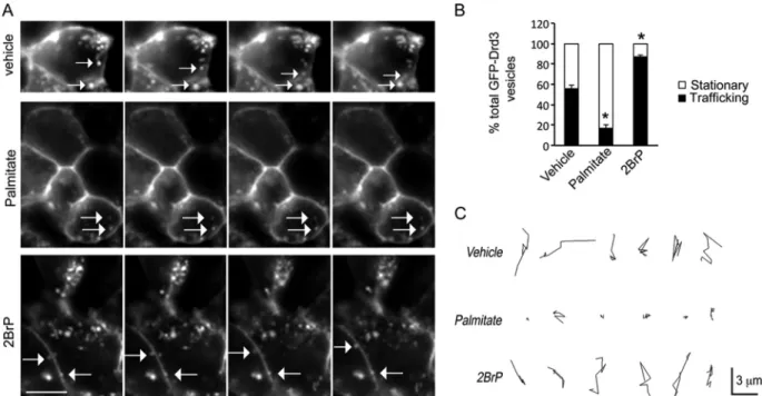
Palmitoylation reduced GFP-Drd3 cargo trafficking. To
in-vestigate the role of cellular palmitoylation on Drd3 cellular
traf-ficking, we performed time-lapse microscopy of GFP-Drd3
car-goes in HEK293 cells cotransfected with GIPC1 (
Fig. 6A
). Control
cells treated with vehicle exhibited equal numbers of stationary
and trafficking vesicles. Treatment with palmitate increased
sig-nificantly the proportion of stationary GFP-Drd3-containing
ves-icles. In contrast, inhibition of cellular palmitoylation with 2BrP
increased significantly the proportion of GFP-Drd3 cargoes
traf-ficking. Overall, the proportion of stationary GFP-Drd3 cargoes
increased and decreased, respectively, with palmitate and 2BrP
treatment (
Fig. 6B
). The size of a single GFP-Drd3 cargo vesicle
was
⬃0.3 m, and trafficking velocity was 0.24 ⫾ 0.02 m/s.
Interestingly, the velocity of GFP-Drd3 cargoes did not change
with treatments. However, the total distance traveled by cargoes
significantly diminished upon treatment with palmitate (2.13
⫾
0.58
m/40 s) compared to 2BrP-treated cells (9.1 ⫾ 0.47 m/40
s) or vehicle controls (9.63
⫾ 0.81 m/40 s). The data are
consis-tent with an increased proportion of stationary cargoes in cells
exposed to palmitate, whereas the reverse was observed upon
treatment with 2BrP (
Fig. 6C
).
Role of a GIPC1 mutant that cannot bind to Drd3. Previous in
silico structural modeling using Drd2 as the template predicted
that (i) the GIPC1 PDZ domain alone is not sufficient to bind to
the palmitoylated C-terminal PDZ binding motif and that (ii)
depalmitoylation of the C-terminal cysteine enables a distancing
of the PDZ binding motif away from the membrane environment
(
10
). Given that both the PDZ and ACP domains are required to
bind the Drd3 C terminus in a yeast two-hybrid assay (
21
), we
investigated the role of the ACP domain in GFP-Drd3 subcellular
distribution and signaling. To this end, we generated a mutant
deprived of the ACP domain only (GIPC1
⌬ACP) (
Fig. 7A
) and
confirmed by coimmunoprecipitation that GIPC1 required the
ACP to interact with GFP-Drd3 in HEK293 cells (
Fig. 7B
).
Func-tionally, transfection of GIPC1⌬ACP failed to reduce the cAMP
response to dopamine (
Fig. 7C
) and failed to reduce the
presen-tation of GFP-Drd3 to the plasma membrane (
Fig. 7D
)
com-pared to wild-type GIPC1. Additionally, 2BrP treatment
re-duced GFP-Drd3 cell surface presentation in cells transfected
with GIPC1
⌬ACP but not in the cells transfected with GIPC1
(
Fig. 7E
). Therefore, the ACP domain is required for GIPC1 to
recapitulate the effects of 2BrP.
Role of a palmitoylated Drd3 mutant that cannot bind to
GIPC1. According to the hypothesis, a deletion mutant lacking
the C-terminal cysteine (GFP-Drd3⌬C) should impair both Drd3
palmitoylation and GIPC1 binding (
21
). To dissociate Drd3
pal-mitoylation from the interaction of Drd3 with GIPC1, we
gener-ated a chimera of Drd3 and
2AR C-terminus tails (GFP-Drd3/
2AR) that preserved the Drd3 palmitoylation site, Leu-Ser-Cys,
as an internal motif followed by the C-terminal PDZ binding
mo-tif, Ser-Leu-Leu
COOHof the
2AR, because it cannot bind to
GIPC1 (
37
). Use of these mutants confirmed two predictions: (i)
Drd3⌬C and Drd3/2AR failed to bind to GIPC1 in a binary
two-hybrid assay in yeast (
Fig. 8A
), and (ii) GFP-Drd3
⌬C failed to be
palmitoylated, in contrast to GFP-Drd3 and GFP-Drd3/2AR
(
Fig. 8B
), indicating that the C-terminal cysteine residue is the
primary palmitoylation site for Drd3.
Further fluorescence microscopy of transfected cells indicated
that GFP-Drd3 and GFP-Drd3/2AR were distributed mostly at
the plasma membrane, compared to GFP-Drd3
⌬C, which
accu-mulated in cytoplasmic compartments (
Fig. 8C
). Whole-cell
[
125I]iodosulpride binding confirmed that GFP-Drd3
⌬C did not
efficiently reach the plasma membrane, compared to GFP-Drd3
FIG 4 Localization of GFP-Drd2 and GFP-Drd3 in primary medium spiny neurons. Representative distribution of GFP-Drd3 (A) and GFP-Drd2 (B) in rat
embryo-derived striatal neurons cotransfected after 3 weeks in culture with the vector or GIPC1 and after treatment with 50M 2-BrP overnight.
on July 2, 2020 at Medipol University
http://mcb.asm.org/
or GFP-Drd3/2AR (
Fig. 8D
). Yet, GFP-Drd3⌬C responded to
dopamine by activating Erk phosphorylation as a function of
2-BrP pretreatment (
Fig. 8E
). In contrast, GFP-Drd3⌬C failed to
inhibit Akt phosphorylation with or without pretreatment with
2BrP (
Fig. 8E
). To determine the role of Drd3 palmitoylation
in-dependently of GIPC1 binding, we treated GFP-Drd3/
2AR-transfected cells with 2BrP. We found that inhibition of cellular
palmitoylation was sufficient to reduce whole-cell [
125I]iodosul-pride binding to floor levels of the GFP-Drd3 control (
Fig. 8F
). In
contrast, cell surface [
125I]iodosulpride binding of the
GFP-Drd3
⌬C construct was low and independent of 2BrP treatment
(
Fig. 8F
).
Role of a nonpalmitoylated Drd3 mutant that can bind to
GIPC1. To determine the role of Drd3 palmitoylation
indepen-FIG 5 Disruption of palmitoylation with 2BrP impaired GFP-Drd3 functions. (A) Representative images of GFP-Drd3 subcellular distribution in HEK293 cellstreated with 100M palmitate or 100 M 2BrP overnight. Bar, 20 m. (B) Proportion of GFP-Drd3 fluorescence in the cytoplasm of HEK293 cells cotransfected with GIPC1 or with the control vector relative to the total fluorescence (unpaired t test: *, P⫽ 0.033 for vehicle versus 2-BrP; #, P ⫽ 0.042 for vector versus GIPC1). (C) Thermal shift to 20°C overnight resulted in the accumulation of transfected GFP-Drd3 in the ER that resolved when cells were returned to 37°C for several hours. The effect of palmitoylation was assessed in the presence of 100M 2BrP or 100 M palmitate. Bar, 15 m. (D) Means ⫾ SEM of data collected from 3 independent experiments, expressed as the percentage of cells targeting Drd3 to the plasma membrane (PM). Two-way analysis of variance for the interaction of time with palmitoylation on Drd3 presentation at the cell surface [F(4, 18)⫽ 3.385; *, P ⫽ 0.0312) is lost in cells overexpressing GIPC1 (F(4, 18)⫽
0.1515; P⫽ 0.95). (E) It is known that Drd3 activation inhibits cAMP formation and Akt phosphorylation and that it activates Erk1/2, but the role of GIPC1 and palmitoylation is unknown. (F) Effect of 15M 2-BrP overnight on the inhibition of cAMP accumulation by a 15-min stimulation with 0.1 M dopamine (DA) in the presence of 0.5M FSK in HEK293 cells cotransfected with GFP-Drd3 and GIPC1 or the control vector. Mean ⫾ SEM data were collected from 6 experiments (unpaired t test; P⬍ 0.05). (G) Effect of 50 M 2-BrP overnight on the inhibition of Akt phosphorylation by a 4-min stimulation with 0.1 M DA in HEK293 cells cotransfected with GFP-Drd3 or the control vector or GIPC1. (H) Effect of 50M 2-BrP overnight on the phosphorylation of Erk1/2 by a 4-min stimulation with 0.1M DA in HEK293 cells cotransfected with GFP-Drd3 and the control vector or GIPC1.
on July 2, 2020 at Medipol University
http://mcb.asm.org/
dently of GIPC1 binding, we generated a Drd3 mutant that
coim-munoprecipitated and colocalized with GIPC1 (
Fig. 9A
and
C
)
but failed to be palmitoylated in HEK293 cells (
Fig. 9B
). This
mutant, GFP-Drd3C/S, was not properly targeted to the plasma
membrane of transfected HEK293 cells and failed to respond to
pretreatment with 2-BrP (
Fig. 9D
and
E
), compared to the
wild-type GFP-Drd3 (WT). Functionally, GFP-Drd3C/S failed to
re-spond to dopamine in inhibiting the cAMP pathway, either with
or without pretreatment with 2BrP (
Fig. 9F
). Yet, GFP-Drd3C/S
fully responded to dopamine for inhibiting the phosphorylation
of Akt and for activating the Erk pathway (
Fig. 9G
). Together, the
results indicate that Drd3 palmitoylation is dispensable for Erk
and Akt signaling but essential for the cAMP signaling route.
DISCUSSION
In this study, we extended the in silico modeling of the
GIPC1-Drd2 interaction (
10
) to the cognate GIPC1-Drd3 interaction, as
the template, and then validated the model in in cellulo
experi-ments. Molecular dynamic (MD) simulations predicted that
C-terminal palmitoylation of Drd3 would enhance the penetration
of each residue of the C terminus, including the helix-8, into the
membrane environment, thus reducing accessibility to
interac-tion domains with intracellular proteins, notably, the PDZ
do-main of GIPC1, as proposed for the Drd2. In cellulo experiments
confirmed the prediction, thereby providing a framework for
un-derstanding how reversible Drd3 palmitoylation controls dopamine
signaling via the GIPC1 route. These findings could be significant for
Drd3 pleiotropic functions in physiology and treatment with
antipsy-chotic and anti-Parkinsonian drugs.
In two different cell lines, Drd3 was constitutively
palmitoyla-ted at multiple molecular mass isoforms between 40 and 55 kDa
(65 and 80 kDa for GFP-Drd3) which are likely to correspond
to intermediates of glycosylation, given that constitutive
pal-FIG 6 2BrP increased the motility of GFP-Drd3 cargo vesicles. (A) Effect of 20M palmitate or 20 M 2BrP on the trafficking of GFP-Drd3 cargo vesicles in HEK293 cells cotransfected with GIPC1. Images were taken every 5 s. Scale bar, 4m. (B) Relative proportion of GFP-Drd3 vesicles that are stationary or trafficking. Means ⫾ SEM for data collected from 20 cells per condition are shown (unpaired t test; P⬍ 0.0001). (C) Representative tracks used by individual vesicles.
FIG 7 Description of a GIPC1 mutant that cannot bind to Drd3. (A)
Sche-matic representation of GIPC1 and mutant lacking the ACP domain. (B) GIPC1⌬ACP did not bind to GFP-Drd3, as shown by coimmunoprecipitation from lysates of HEK293 cells cotransfected with GFP-Drd3 and the GIPC1 construct. (C) Inhibition of cAMP accumulation by a 15-min stimulation with 0.1M DA in HEK293 cells cotransfected with GFP-Drd3 and GIPC1 or the control vector in the presence of 0.5M FSK. Means ⫾ SEM of data from 3 independent experiments (unpaired t test; #, P⬍ 0.0001; **, P ⫽ 0.0034; *, P ⫽ 0.0221). (D) Representative HEK293 cells cotransfected with GFP-Drd3 and GIPC1⌬ACP after stimulation with 100 M palmitate or 100 M 2BrP over-night. Scale bar, 12m. (E) Effects of GIPC1⌬ACP on the binding of 0.1 nM [125I]iodosulpride to whole-cell preparations of HEK293 cells cotransfected
with GFP-Drd3 and GIPC1 constructs. Means⫾ SEM of data from 4 experi-ments (unpaired t test; *, P⫽ 0.0294; **, P ⫽ 0.0047).
on July 2, 2020 at Medipol University
http://mcb.asm.org/
mitoylation is an early posttranslational modification
occur-ring in the ER and Golgi apparatus, where glycosylation occurs
(
62
). Lack of Drd3 palmitoylation by nonselective
pharmaco-logical blockade with 2BrP or by targeted mutation of the
C-terminal cysteine resulted in the accumulation of Drd3 in
intracellular membrane compartments. Further experiments
using an anterograde transport assay, whole-cell Drd3
radioli-gand binding, antibody feeding assay, and live imaging of Drd3
cargo vesicles consistently supported the idea that
palmitoyl-ation is required to target and stabilize Drd3 at the cell surface.
The use of the chimeric Drd3/
2AR mutant and Drd3-C/S
mutant was instrumental to demonstrate that Drd3
palmitoyl-FIG 8 Description of a palmitoylated Drd3 mutant that cannot bind to GIPC1. (A) Binding of GIPC1 to Drd3 mutants monitored by-galactosidase activity using ONPG as the substrate in yeast. The Drd3⌬C lacks the C-terminal cysteine residue, whereas the Drd3/2AR chimera harbors the palmitoylation site of Drd3 and the C-terminal PDZ ligand of the2AR. Means ⫾ SEM of data collected from 3 independent experiments are shown (unpaired t test; *, P ⬍ 0.01). (B) Palmitoylation of GFP-Drd3 mutants in HEK293 cells. (C) Subcellular distribution of GFP-Drd3 mutants transfected in HEK293 cells. Scale bar, 20m. PM, plasma membrane. (D) Binding of [125I]iodosulpride at the cell surface of HEK293 cells transfected with the GFP-Drd3 constructs in competition with increasing
concentrations of dopamine. Means⫾ SEM of data collected from 3 independent experiments (unpaired t test; *, P ⫽ 0.0017 for WT versus Drd3/2AR; #, P ⬍ 0.0001 for WT versus Drd3⌬C). (E) Effect of Drd3⌬C on Erk and Akt phosphorylations upon a 4-min stimulation with 0.1 M DA in HEK293 cells pretreated with 50M 2-BrP or vehicle overnight. (F) Effect of 100 M palmitate and 100 M 2-BrP on [125I]iodosulpride binding at the cell surface of HEK293 cells
transfected with the indicated GFP-Drd3 constructs. Means⫾ SEM of data collected from 3 independent experiments (unpaired t test, vehicle versus 2-BrP; *, P⫽ 0.0313; **, P ⫽ 0.0054).
on July 2, 2020 at Medipol University
http://mcb.asm.org/
ation is essential for Drd3 presentation at the cell surface and
that GIPC1 binding favors a vesicular distribution for Drd3.
This is consistent with the physiological distribution of Drd3
and GIPC1 in the adult rat brain, as well as in embryo-derived
cultured primary MSNs. Interestingly, palmitoylation
de-creased the motility of Drd3 cargo vesicles, perhaps by
reduc-ing access of Drd3 cargoes to cytoskeletal motor proteins for
which GIPC1 is an established adaptor (
39
,
63
,
64
).
GIPC1 colocalized with Drd3 in vesicles and at the plasma
membrane of HEK293 cells. Inhibition of cellular
palmitoyl-ation with 2BrP rearranged this colocalizpalmitoyl-ation, particularly in
vesicles close to the cell surface and at the plasma membrane.
Given that transfected GIPC1 decreased the levels of Drd3
pal-mitoylation, we anticipate that GIPC1 binds to
depalmitoyla-ted Drd3, as predicdepalmitoyla-ted for Drd2 in the in silico model. This is
consistent with a competition at the Drd3 C terminus for
over-lapping consensus sites for palmitoylation and GIPC1 binding
(
Fig. 1A
). This possibility is corroborated experimentally by
the disruption of the interaction between GIPC1 and Drd3
with a nonpalmitoylated synthetic peptide mimetic of the Drd3
C terminus (
21
). In addition, molecular modeling predicts that
helix-8 is not structurally changed much by palmitoylation but
that it is more deeply inserted into the membrane
environ-ment. In contrast, a depalmitoylated C terminus is more
ex-posed to the aqueous environment and presumably protein
partners like GIPC1, thus reinforcing the opposition between
the palmitoylation and GIPC interaction at Drd3. This may
also be true for Drd2, as suggested by results of transfection
experiments in primary MSNs (
Fig. 4B
) (
10
). The presence of
an ACP domain in GIPC1 suggests a putative function in the
FIG 9 Description of a nonpalmitoylated Drd3 mutant that can bind to GIPC1. (A) Coimmunoprecipation of GIPC1 with GFP-Drd3 or GFP-Drd3C/S mutants
from HEK293 transfected cells using 6⫻His antibodies. (B) Mutant GFP-Drd3C/S was not palmitoylated in HEK293 transfected cells compared to GFP-Drd3. Palmitoylation was measured using acyl-biotin exchange (see Materials and Methods). HAM, hydroxylamine. (C) The GFP-Drd3C/S mutant colocalized with GIPC1 in transfected HEK293 cells. (D) GFP antibody feeding assay on transfected HEK293 cells to label GFP-Drd3 and mutant GFP-Drd3C/S at the cell surface (red) compared to total GFP expression (green). The nuclei were labeled with Hoechst 33342 (blue). (E) Percentage of Drd3 at the cell surface. Means⫾ SEM of data from at least 10 cells per condition in 2 independent experiments are shown (unpaired t test; *, P⫽ 0.0003; #, P ⬍ 0.0001). (F) Effect of Drd3C/S on the inhibition of cAMP accumulation by a 15-min stimulation with 0.1M DA in the presence of 0.5 M FSK in HEK293 cells pretreated with 15 M 2-BrP or vehicle overnight. Means⫾ SD (n ⫽ 3) are shown (unpaired t test; *, P ⫽ 0.007). (G) Effect of Drd3C/S on Erk and Akt phosphorylation upon a 4-min stimulation with 0.1M DA in HEK293 cells pretreated with 50 M 2-BrP or vehicle overnight.
on July 2, 2020 at Medipol University
http://mcb.asm.org/
acylation of target PDZ cargoes (
36
). We found that the GIPC1
ACP domain contributed to the interaction with Drd3 in yeast
and HEK293 cells. However, the exact role of the ACP domain
has not been extensively studied and is beyond the scope of this
study.
Palmitoylation is a reversible and dynamic process (
22
). It is
thought that palmitoylation of GPCRs can sometimes be stable
(e.g., 5-HT1a [
65
,
66
]) or, most commonly, reversible (e.g.,
5-HT4a and 5-HT7a [
67
,
68
]), resulting in different loop
con-formations as the palmitate groups penetrate into the lipid
bilayer, thereby changing GPCR dimerization and signaling
properties (
3
,
12
). Several studies reported distinct effects of
ligand-induced activation on palmitoylation. For example,
ac-tivation of the
2AR with isoproterenol did not change its
palmitoylation rate (
69
). In contrast, increased turnover of
pal-mitoylation was demonstrated at least for the CHRM2, AVPR2,
and 5-HT4a receptors (
67
,
70
,
71
). These studies suggested that
one possible role for palmitoylation is to change the turnover
of target transmembrane proteins and thus their functional
properties. An example of this phenomenon is the
mannose-6-phosphate receptor, for which the half-life is 40 h when
depal-mitoylated and 2 h when paldepal-mitoylated (
72
). Consistently,
overexpression of GIPC1 decreased Drd3 palmitoylation and
Drd3 lysosomal degradation (
21
). Future experiments will
de-termine if Drd3 undergoes cycles of palmitoylation and
depal-mitoylation during its lifetime. Such a dynamic process could
facilitate the trafficking and signaling of depalmitoylated Drd3
cargoes by a GIPC1 signaling route.
Amid the complexity of dopamine signaling pathways (
26
,
73
,
74
), the cAMP pathway inhibited by Drd3 was most
sensi-tive to GIPC1 and palmitoylation. Interestingly, the Erk pathway
was sensitive to 2BrP but not to Drd3 mutations, indicating that
cel-lular palmitoylation rather than Drd3 palmitoylation is involved in
this process, as previously suggested in cancer cells (
75
). On the
con-trary, dopamine signaling via the Akt pathway required GIPC1
regardless of 2BrP treatment, given that Drd3⌬C failed to inhibit
Akt phosphorylation by dopamine. This is corroborated by results
in a previous study, indicating that the GIPC1 expression level can
modulate Akt phosphorylation by bovine serum albumin (
43
).
In summary, GIPC1 competes with Drd3 palmitoylation,
thereby impacting the presentation of the Drd3 at the plasma
membrane, the access of extracellular ligands, and the
dopami-nergic signal transduction. Given the important role of Drd3 in
the mode of action of antipsychotic drugs for treating
schizo-phrenia (
29
), in the underlying mechanisms of
levodopa-in-duced dyskinesia (
31
), and as a genetic predisposition factor in
essential tremor (
30
), the GIPC1-palmitoylation switch of
Drd3 provides novel mechanistic insights for manipulating
do-paminergic signaling when it is impaired.
ACKNOWLEDGMENTS
The work was supported by the Fondation pour la Recherche Médicale, the Fondation de France, and the French Ministry of Research.
We thank M. Asari for figure editing.
M.A.-L., P.S., and F.J. conceived all experiments. O.S. and H.W. de-signed the computational experiments. M.A.-L. and F.J. collected bio-chemical data. M.A.-L. and M.C. produced time-lapse fluorescence im-aging data. G.G. and A.B. did binding and cAMP experiments. O.S. performed in silico simulation studies. M.A.-L. and F.J. wrote the manu-script, with contributions from O.S., H.W., and P.S.
We declare no competing interests.
FUNDING INFORMATION
Fondation pour la recherche medicale provided funding to Pierre Sokoloff and Freddy Jeanneteau. French ministry of research provided funding to Freddy Jeanneteau. Fondation de France provided funding to Margarita Arango-Lievano and Freddy Jeanneteau.
REFERENCES
1. Bockaert J, Marin P, Dumuis A, Fagni L. 2003. The ‘magic tail’ of G protein-coupled receptors: an anchorage for functional protein networks. FEBS Lett 546:65–72.http://dx.doi.org/10.1016/S0014-5793(03)00453-8. 2. Dunn HA, Ferguson SS. 2015. PDZ protein regulation of GPCR traffick-ing and signaltraffick-ing pathways. Mol Pharmacol 88:624 – 639.http://dx.doi .org/10.1124/mol.115.098509.
3. Qanbar R, Bouvier M. 2003. Role of palmitoylation/depalmitoylation reactions in G-protein-coupled receptor function. Pharmacol Ther 97:1– 33.http://dx.doi.org/10.1016/S0163-7258(02)00300-5.
4. Goddard AD, Watts A. 2012. Regulation of G protein-coupled receptors by palmitoylation and cholesterol. BMC Biol 10:27.http://dx.doi.org/10 .1186/1741-7007-10-27.
5. Oates J, Watts A. 2011. Uncovering the intimate relationship between lipids, cholesterol and GPCR activation. Curr Opin Struct Biol 21:802– 807.http://dx.doi.org/10.1016/j.sbi.2011.09.007.
6. Zheng H, Pearsall EA, Hurst DP, Zhang Y, Chu J, Zhou Y, Reggio PH,
Loh HH, Law PY. 2012. Palmitoylation and membrane cholesterol
sta-bilize mu-opioid receptor homodimerization and G protein coupling. BMC Cell Biol 13:6.http://dx.doi.org/10.1186/1471-2121-13-6. 7. Palczewski K, Kumasaka T, Hori T, Behnke CA, Motoshima H, Fox BA,
Le Trong I, Teller DC, Okada T, Stenkamp RE, Yamamoto M, Miyano M. 2000. Crystal structure of rhodopsin: a G protein-coupled receptor.
Science 289:739 –745.http://dx.doi.org/10.1126/science.289.5480.739. 8. Scheerer P, Park JH, Hildebrand PW, Kim YJ, Krauss N, Choe HW,
Hofmann KP, Ernst OP. 2008. Crystal structure of opsin in its
G-protein-interacting conformation. Nature 455:497–502.http://dx.doi.org/10.1038 /nature07330.
9. Krishna AG, Menon ST, Terry TJ, Sakmar TP. 2002. Evidence that helix 8 of rhodopsin acts as a membrane-dependent conformational switch. Biochemistry 41:8298 – 8309.http://dx.doi.org/10.1021/bi025534m. 10. Sensoy O, Weinstein H. 2015. A mechanistic role of Helix 8 in GPCRs:
computational modeling of the dopamine D2 receptor interaction with the GIPC1-PDZ-domain. Biochim Biophys Acta 1848:976 –983.http://dx .doi.org/10.1016/j.bbamem.2014.12.002.
11. Ernst OP, Meyer CK, Marin EP, Henklein P, Fu WY, Sakmar TP,
Hofmann KP. 2000. Mutation of the fourth cytoplasmic loop of
rhodop-sin affects binding of transducin and peptides derived from the carboxyl-terminal sequences of transducin alpha and gamma subunits. J Biol Chem
275:1937–1943.http://dx.doi.org/10.1074/jbc.275.3.1937.
12. Cherezov V, Rosenbaum DM, Hanson MA, Rasmussen SG, Thian FS,
Kobilka TS, Choi HJ, Kuhn P, Weis WI, Kobilka BK, Stevens RC. 2007.
High-resolution crystal structure of an engineered human2-adrenergic G protein-coupled receptor. Science 318:1258 –1265.http://dx.doi.org/10 .1126/science.1150577.
13. Shan J, Khelashvili G, Mondal S, Mehler EL, Weinstein H. 2012. Ligand-dependent conformations and dynamics of the serotonin 5-HT(2A) receptor determine its activation and membrane-driven oli-gomerization properties. PLoS Comput Biol 8:e1002473. http://dx.doi .org/10.1371/journal.pcbi.1002473.
14. Delos Santos NM, Gardner LA, White SW, Bahouth SW. 2006. Char-acterization of the residues in helix 8 of the human1-adrenergic receptor that are involved in coupling the receptor to G proteins. J Biol Chem
281:12896 –12907.http://dx.doi.org/10.1074/jbc.M508500200. 15. Tetsuka M, Saito Y, Imai K, Doi H, Maruyama K. 2004. The basic
residues in the membrane-proximal C-terminal tail of the rat melanin-concentrating hormone receptor 1 are required for receptor function. Endocrinology 145:3712–3723.http://dx.doi.org/10.1210/en.2003-1638. 16. Kuwasako K, Hay DL, Nagata S, Murakami M, Kitamura K, Kato J.
2013. Functions of third extracellular loop and helix 8 of family B GPCRs complexed with RAMPs and characteristics of their receptor trafficking. Curr Protein Peptide Sci 14:416 – 428.http://dx.doi.org/10.2174/1389203 7113149990058.
17. Jin H, Zastawny R, George SR, O’Dowd BF. 1997. Elimination of palmitoylation sites in the human dopamine D1 receptor does not affect
on July 2, 2020 at Medipol University
http://mcb.asm.org/
21. Jeanneteau F, Diaz J, Sokoloff P, Griffon N. 2004. Interactions of GIPC with dopamine D2, D3 but not D4 receptors define a novel mode of reg-ulation of G protein-coupled receptors. Mol Biol Cell 15:696 –705. 22. Fukata Y, Fukata M. 2010. Protein palmitoylation in neuronal
develop-ment and synaptic plasticity. Nat Rev Neurosci 11:161–175.http://dx.doi .org/10.1038/nrn2788.
23. Blanpain C, Wittamer V, Vanderwinden JM, Boom A, Renneboog B,
Lee B, Le Poul E, El Asmar L, Govaerts C, Vassart G, Doms RW, Parmentier M. 2001. Palmitoylation of CCR5 is critical for receptor
traf-ficking and efficient activation of intracellular signaling pathways. J Biol Chem 276:23795–23804.http://dx.doi.org/10.1074/jbc.M100583200. 24. Dunphy JT, Greentree WK, Manahan CL, Linder ME. 1996. G-protein
palmitoyltransferase activity is enriched in plasma membranes. J Biol Chem 271:7154 –7159.http://dx.doi.org/10.1074/jbc.271.12.7154. 25. Berthet A, Bezard E. 2009. Dopamine receptors and L-dopa-induced
dyskinesia. Parkinsonism Relat Disord 15(Suppl 4):S8 –12.http://dx.doi .org/10.1016/S1353-8020(09)70827-2.
26. Beaulieu JM, Gainetdinov RR. 2011. The physiology, signaling, and pharmacology of dopamine receptors. Pharmacol Rev 63:182–217.http: //dx.doi.org/10.1124/pr.110.002642.
27. Deng H, Le W, Jankovic J. 2007. Genetics of essential tremor. Brain
130:1456 –1464.http://dx.doi.org/10.1093/brain/awm018.
28. Joyce JN, Millan MJ. 2007. Dopamine D3 receptor agonists for protection and repair in Parkinson’s disease. Curr Opin Pharmacol 7:100 –105.http: //dx.doi.org/10.1016/j.coph.2006.11.004.
29. Sokoloff P, Leriche L, Diaz J, Louvel J, Pumain R. 2013. Direct and indirect interactions of the dopamine D(3) receptor with glutamate path-ways: implications for the treatment of schizophrenia. Naunyn-Schmiedebergs Arch Pharmacol 386:107–124.http://dx.doi.org/10.1007 /s00210-012-0797-0.
30. Jeanneteau F, Funalot B, Jankovic J, Deng H, Lagarde JP, Lucotte G,
Sokoloff P. 2006. A functional variant of the dopamine D3 receptor is
associated with risk and age-at-onset of essential tremor. Proc Natl Acad Sci U S A 103:10753–10758.http://dx.doi.org/10.1073/pnas.0508189103. 31. Bezard E, Ferry S, Mach U, Stark H, Leriche L, Boraud T, Gross C,
Sokoloff P. 2003. Attenuation of levodopa-induced dyskinesia by
normal-izing dopamine D3 receptor function. Nat Med 9:762–767.http://dx.doi .org/10.1038/nm875.
32. Missale C, Nash SR, Robinson SW, Jaber M, Caron MG. 1998. Dopa-mine receptors: from structure to function. Physiol Rev 78:189 –225. 33. Sokoloff P, Diaz J, Le Foll B, Guillin O, Leriche L, Bezard E, Gross C.
2006. The dopamine D3 receptor: a therapeutic target for the treatment of neuropsychiatric disorders. CNS Neurol Disord Drug Targets 5:25– 43.
http://dx.doi.org/10.2174/187152706784111551.
34. Griffon N, Pilon C, Sautel F, Schwartz JC, Sokoloff P. 1997. Two intracellular signaling pathways for the dopamine D3 receptor: opposite and synergistic interactions with cyclic AMP. J Neurochem 68:1–9. 35. Rasolonjanahary R, Gerard C, Dufour MN, Homburger V, Enjalbert A,
Guillon G. 2002. Evidence for a direct negative coupling between
dop-amine-D2 receptors and PLC by heterotrimeric Gi1/2 proteins in rat an-terior pituitary cell membranes. Endocrinology 143:747–754.http://dx .doi.org/10.1210/endo.143.3.8697.
36. De Vries L, Lou X, Zhao G, Zheng B, Farquhar MG. 1998. GIPC, a PDZ domain containing protein, interacts specifically with the C terminus of RGS-GAIP. Proc Natl Acad Sci U S A 95:12340 –12345.http://dx.doi.org /10.1073/pnas.95.21.12340.
37. Hu LA, Chen W, Martin NP, Whalen EJ, Premont RT, Lefkowitz RJ. 2003. GIPC interacts with theadrenergic receptor and regulates 1-adrenergic receptor-mediated ERK activation. J Biol Chem 278:26295– 26301.http://dx.doi.org/10.1074/jbc.M212352200.
protein signaling downstream of insulin-like growth factor 1 receptor. J Biol Chem 277:6719 – 6725.http://dx.doi.org/10.1074/jbc.M108033200. 42. Blobe GC, Liu X, Fang SJ, How T, Lodish HF. 2001. A novel mechanism
for regulating transforming growth factor beta (TGF-beta) signaling. Functional modulation of type III TGF-beta receptor expression through interaction with the PDZ domain protein, GIPC. J Biol Chem 276:39608 – 39617.http://dx.doi.org/10.1074/jbc.M106831200.
43. Varsano T, Taupin V, Guo L, Baterina OY, Jr, Farquhar MG. 2012. The PDZ protein GIPC regulates trafficking of the LPA1 receptor from APPL signaling endosomes and attenuates the cell’s response to LPA. PLoS One
7:e49227.http://dx.doi.org/10.1371/journal.pone.0049227.
44. Beld J, Finzel K, Burkart MD. 2014. Versatility of acyl-acyl carrier protein synthetases. Chem Biol 21:1293–1299.http://dx.doi.org/10.1016/j.chem biol.2014.08.015.
45. Heath RJ, Rock CO. 1996. Regulation of fatty acid elongation and initi-ation by acyl-acyl carrier protein in Escherichia coli. J Biol Chem 271: 1833–1836.http://dx.doi.org/10.1074/jbc.271.4.1833.
46. Othman A, Lazarus C, Fraser T, Stobart K. 2000. Cloning of a palmitoyl-acyl carrier protein thioesterase from oil palm. Biochem Soc Trans 28: 619 – 622.http://dx.doi.org/10.1042/bst0280619.
47. Phillips JC, Braun R, Wang W, Gumbart J, Tajkhorshid E, Villa E,
Chipot C, Skeel RD, Kale L, Schulten K. 2005. Scalable molecular
dynamics with NAMD. J Comput Chem 26:1781–1802.http://dx.doi.org /10.1002/jcc.20289.
48. Marrink SJ, Risselada HJ, Yefimov S, Tieleman DP, de Vries AH. 2007. The MARTINI force field: coarse grained model for biomolecu-lar simulations. J Phys Chem B 111:7812–7824.http://dx.doi.org/10 .1021/jp071097f.
49. Cerutti DS, Duke RE, Darden TA, Lybrand TP. 2009. Staggered Mesh Ewald: an extension of the smooth particle-mesh Ewald method adding great versatility. J Chem Theory Comput 5:2322.http://dx.doi.org/10 .1021/ct9001015.
50. Berendsen HJC, Postma JPM, van Gunsteren WF, DiNola A, Haak JR. 1984. Molecular dynamics with coupling to an external bath. J Chem Phys
81:3684 –3690.http://dx.doi.org/10.1063/1.448118.
51. Hyvönen MT, Kovanen PT. 2003. Molecular dynamics simulation of sphingomyelin bilayer. J Phys Chem B 107: 9102–9108.http://dx.doi.org /10.1021/jp035319v.
52. Lim JB, Rogaski B, Klauda JB. 2012. Update of the cholesterol force field parameters in CHARMM. J Phys Chem B 116:203–210.http://dx.doi.org /10.1021/jp207925m.
53. Heinig M, Frishman D. 2004. STRIDE: a web server for secondary struc-ture assignment from known atomic coordinates of proteins. Nucleic Ac-ids Res 32:W500 –W502.http://dx.doi.org/10.1093/nar/gkh429. 54. Weill C, Ilien B, Goeldner M, Galzi JL. 1999. Fluorescent muscarinic
EGFP-hM1 chimeric receptors: design, ligand binding and functional properties. J Receptor Signal Transduct Res 19:423– 436.http://dx.doi.org /10.3109/10799899909036662.
55. Diaz J, Pilon C, Le Foll B, Gros C, Triller A, Schwartz JC, Sokoloff P. 2000. Dopamine D3 receptors expressed by all mesencephalic dopamine neurons. J Neurosci 20:8677– 8684.
56. Brigidi GS, Bamji SX. 28 February 2013. Detection of protein palmi-toylation in cultured hippocampal neurons by immunoprecipitation and acyl-biotin exchange (ABE). J Visual Exp http://dx.doi.org/10 .3971/50031.
57. Wan J, Roth AF, Bailey AO, Davis NG. 2007. Palmitoylated proteins: purification and identification. Nat Protoc 2:1573–1584.http://dx.doi.org /10.1038/nprot.2007.225.
58. Sokoloff P, Levesque D, Martres MP, Lannfelt L, Diaz G, Pilon C,
Schwartz JC. 1992. The dopamine D3 receptor as a key target for
on July 2, 2020 at Medipol University
http://mcb.asm.org/
psychotics. Clin Neuropharmacol 15(Suppl 1):456A– 457A.http://dx.doi .org/10.1097/00002826-199201001-00238.
59. Nuoffer C, Davidson HW, Matteson J, Meinkoth J, Balch WE. 1994. A GDP-bound of rab1 inhibits protein export from the endoplasmic retic-ulum and transport between Golgi compartments. J Cell Biol 125:225– 237.http://dx.doi.org/10.1083/jcb.125.2.225.
60. Davda D, El Azzouny MA, Tom CT, Hernandez JL, Majmudar JD,
Kennedy RT, Martin BR. 2013. Profiling targets of the irreversible
pal-mitoylation inhibitor 2-bromopalmitate. ACS Chem Biol 8:1912–1917.
http://dx.doi.org/10.1021/cb400380s.
61. Jennings BC, Nadolski MJ, Ling Y, Baker MB, Harrison ML, Deschenes
RJ, Linder ME. 2009. 2-Bromopalmitate and
2-(2-hydroxy-5-nitro-benzylidene)-benzo[b]thiophen-3-one inhibit DHHC-mediated palmi-toylation in vitro. J Lipid Res 50:233–242.
62. Grunewald S, Haase W, Reilander H, Michel H. 1996. Glycosylation, palmitoylation, and localization of the human D2S receptor in baculovi-rus-infected insect cells. Biochemistry 35:15149 –15161.http://dx.doi.org /10.1021/bi9607564.
63. Valdembri D, Caswell PT, Anderson KI, Schwarz JP, Konig I, Astanina
E, Caccavari F, Norman JC, Humphries MJ, Bussolino F, Serini G.
2009. Neuropilin-1/GIPC1 signaling regulates␣51 integrin traffic and function in endothelial cells. PLoS Biol 7:e25.http://dx.doi.org/10.1371 /journal.pbio.1000025.
64. Yano H, Ninan I, Zhang H, Milner TA, Arancio O, Chao MV. 2006. BDNF-mediated neurotransmission relies upon a myosin VI motor com-plex. Nat Neurosci 9:1009 –1018.http://dx.doi.org/10.1038/nn1730. 65. Papoucheva E, Dumuis A, Sebben M, Richter DW, Ponimaskin EG.
2004. The 5-hydroxytryptamine(1A) receptor is stably palmitoylated, and acylation is critical for communication of receptor with Gi protein. J Biol Chem 279:3280 –3291.http://dx.doi.org/10.1074/jbc.M308177200. 66. Gorinski N, Ponimaskin E. 2013. Palmitoylation of serotonin receptors.
Biochem Soc Trans 41:89 –94.http://dx.doi.org/10.1042/BST20120235. 67. Ponimaskin EG, Schmidt MF, Heine M, Bickmeyer U, Richter DW.
2001. 5-Hydroxytryptamine 4(a) receptor expressed in Sf9 cells is palmi-toylated in an agonist-dependent manner. Biochem J 353:627– 634.http: //dx.doi.org/10.1042/bj3530627.
68. Kvachnina E, Dumuis A, Wlodarczyk J, Renner U, Cochet M, Richter
DW, Ponimaskin E. 2009. Constitutive Gs-mediated, but not
G12-mediated, activity of the 5-hydroxytryptamine 5-HT7(a) receptor is mod-ulated by the palmitoylation of its C-terminal domain. Biochim Biophys Acta 1793:1646 –1655.http://dx.doi.org/10.1016/j.bbamcr.2009.08.008. 69. Trester-Zedlitz M, Burlingame A, Kobilka B, von Zastrow M. 2005.
Mass spectrometric analysis of agonist effects on posttranslational modi-fications of the beta-2 adrenoceptor in mammalian cells. Biochemistry
44:6133– 6143.http://dx.doi.org/10.1021/bi0475469.
70. Charest PG, Bouvier M. 2003. Palmitoylation of the V2 vasopressin receptor carboxyl tail enhances beta-arrestin recruitment leading to effi-cient receptor endocytosis and ERK1/2 activation. J Biol Chem 278: 41541– 41551.http://dx.doi.org/10.1074/jbc.M306589200.
71. Hayashi MK, Haga T. 1997. Palmitoylation of muscarinic acetylcholine receptor m2 subtypes: reduction in their ability to activate G proteins by mutation of a putative palmitoylation site, cysteine 457, in the carboxyl-terminal tail. Arch Biochem Biophys 340:376 –382.http://dx.doi.org/10 .1006/abbi.1997.9906.
72. Schweizer A, Kornfeld S, Rohrer J. 1996. Cysteine 34 of the cytoplasmic tail of the cation-dependent mannose 6-phosphate receptor is reversibly palmitoylated and required for normal trafficking and lysosomal enzyme sorting. J Cell Biol 132:577–584.http://dx.doi.org/10.1083/jcb.132.4.577. 73. Beaulieu JM, Sotnikova TD, Yao WD, Kockeritz L, Woodgett JR,
Gainetdinov RR, Caron MG. 2004. Lithium antagonizes
dopamine-dependent behaviors mediated by an AKT/glycogen synthase kinase 3 sig-naling cascade. Proc Natl Acad Sci U S A 101:5099 –5104.http://dx.doi.org /10.1073/pnas.0307921101.
74. Beaulieu JM, Tirotta E, Sotnikova TD, Masri B, Salahpour A,
Gainet-dinov RR, Borrelli E, Caron MG. 2007. Regulation of Akt signaling by D2
and D3 dopamine receptors in vivo. J Neurosci 27:881– 885.http://dx.doi .org/10.1523/JNEUROSCI.5074-06.2007.
75. Heakal Y, Woll MP, Fox T, Seaton K, Levenson R, Kester M. 2011. Neurotensin receptor-1 inducible palmitoylation is required for efficient receptor-mediated mitogenic-signaling within structured membrane mi-crodomains. Cancer Biol Ther 12:427– 435.http://dx.doi.org/10.4161/cbt .12.5.15984.