Comparative anatomical studies of some Teucrium sect.
Teucrium species: Teucrium alyssifolium Stapf, Teucrium
brevifolium Schreb. and Teucrium pestalozzae Boiss.
(Lamiaceae)
Gülay Ecevit-Genç1, Betül Büyükkılıç-Altınbaşak1,2,
Taner Özcan3, Tuncay Dirmenci3
1 Istanbul University, Faculty of Pharmacy, Department of Pharmaceutical Botany Istanbul, Turkey 2 Bezmialem Vakıf University, Faculty of Pharmacy, Department of Pharmaceutical Botany Istanbul, Turkey 3 Balıkesir Uni-versity, Faculty of Necatibey Education, Department of Biology Education Balıkesir, Turkey
Corresponding author: Tuncay Dirmenci ([email protected])
Academic editor: E. Fischer | Received 16 February 2018 | Accepted 21 March 2018 | Published 3 April 2018 Citation: Ecevit-Genç G, Büyükkılıç-Altınbaşak B, Özcan T, Dirmenci T (2018) Comparative anatomical studies of some Teucrium sect. Teucrium species: Teucrium alyssifolium Stapf, Teucrium brevifolium Schreb. and Teucrium pestalozzae Boiss. (Lamiaceae). PhytoKeys 96: 63–77. https://doi.org/10.3897/phytokeys.96.24498
Abstract
Teucrium alyssifolium Stapf (endemic), Teucrium pestalozzae Boiss. (endemic) and Teucrium brevifolium
Schreb. are three closely related taxa in Teucrium sect. Teucrium. The obtained data from the anatomical studies revealed that these three taxa represent the general anatomical characteristics of the Lamiaceae fam-ily. Leaves, anatomical features such as thick cuticle, abundant trichomes, rich palisade parenchyma layer in the mesophyll provide evidence that these three species are xeromorphic structures. Leaf and stem anatomy showed that the taxa have generally similar anatomical features. However, cuticle layers, epidermis cells size, indumentum density, mesophyll types, palisade parenchyma occupied in the mesophyll, presence of sphero-crystals in leaves and parenchyma, collenchyma and sclerenchyma layers in stems show differences amongst the taxa. Anatomical characters of leaf and stem of these taxa are examined for the first time in this study.
Keywords
Lamiaceae, Teucrium, leaf anatomy, stem anatomy
http://phytokeys.pensoft.net
Copyright Gülay Ecevit-Genç et al. This is an open access article distributed under the terms of the Creative Commons Attribution License (CC BY 4.0), which permits unrestricted use, distribution, and reproduction in any medium, provided the original author and source are credited.
Introduction
The genus Teucrium L. has approximately 300 species all over the world. Teucrium’s cosmo-politan distribution is mainly concentrated in Europe, North Africa and in the temperate parts of Asia (Ecevit-Genç et al. 2015, 2017). Teucrium is a large and polymorphic genus which is represented by 49 taxa (36 species) in Turkey. There are 18 endemic taxa (Go-vaerts 1999, Duman 2000, Dönmez 2006, Parolly and Eren 2007, Dönmez et al. 2010, Dinç et al. 2011a, 2011b, Dirmenci 2012, Özcan et al. 2015a, Vural et al. 2015, Dinç and Doğu 2016). The classification of Teucrium is based on sections. The main characters in the separation of sections are the calyx-shape and flower arrangement (Davis 1982; Na-varro et al. 2004; Dinç et al. 2008; Özcan et al. 2015a; Vural et al. 2015). Especially, leaf anatomy is important for the classification of the genus (Dinç et al. 2009). Also, absence or presence of trichomes and their types on the nutlets and vegetative parts are very important for classifying the species (Dinç et al. 2011a, Ecevit-Genç et al. 2015, 2017).
Teucrium species have traditionally been used in Turkey for abdominal pain,
stom-ach-ache, common cold, high fever, antipyretic, rheumatic pain and as an antidiabetic (Sezik et al. 2001, Aksoy-Sagirli et al. 2015).
T. alyssifolium is a narrowly distributed endemic species. It is classified as a
‘Con-servation Dependent (LR/cd)’ category of IUCN and it is a source of polyphenols and flavonoids and has confirmed antioxidant activities (Semiz et al. 2016). T. pestalozzae is an endemic species and its essential oil is characterised with β-caryophyllene (27.6%) and germacrene D (13.8%) as major constituents (Baser et al. 1997). Spathulenol and δ-cadinene are the main compounds of T. brevifolium essential oil and it has shown anti-tumour activities, a selective cytotoxicity on large lung carcinoma (IC50 value of 80.7 μg/ml) (Menichini et al. 2009).
The chromosome numbers are reported as 2n = 10, 14, 16, 18, 22, 26, 28, 30, 32, 36, 39, 48, 52, 56, 58, 60, 62, 64, 78, 80, 86, 90, 96 and 104 in the genus Teucrium (http://www.tropicos.org/Project/IPCN). The chromosome number of T. brevifolium examined in this study was determined as 2n = 30. Another member of the sect.
Teu-crium, T. sandrasicum, was studied and it was determined that the chromosome
num-ber is the same as T. brevifolium (Özcan et al. 2015b).
Pollen morphology supplies useful data at the taxonomic level in Teucrium (Na-varro et al. 2004, Marzouk et al. 2017). Oybak and İnceoğlu (1988) studied pollen morphology of some Turkish Teucrium members. They found out that the species be-longing to different sections had different pollen type and pollen shape, while pollen grain size and apocolpia size were the main characters used for distinguishing the spe-cies. Especially, T. alyssifolium could be easily separated from the other species of the sect. Teucrium according to pollen data.
There are several studies on Teucrium anatomy (Lakusic et al. 2006, 2010, Dinç et al. 2008, 2009, 2011a, 2011b, Dehshiri and Azadbakht 2012, Dinç and Doğu 2012, Doğu et al. 2013, Özcan 2013, Özcan and Eminagaoglu 2014, Ruiters et al. 2016). However, the anatomy of T. pestalozzae, T. brevifolium and T. alyssifolium has not been investigated. In our previous studies, we investigated the nutlet and leaf
micromorphol-ogy of some species belonging to the sect. Teucrium in Turkey (Ecevit-Genç et al. 2015, 2017). In the present study, we report on the anatomical features of the leaves and stems of T. alyssifolium, T. brevifolium and T. pestalozzae. The aim of this paper is to under-stand the anatomy of these three Teucrium species. Also, a better underunder-standing of sys-tematics helps the distinction of morphologically closely related taxa from each other.
Materials and methods
T. pestalozzae samples were collected from Antalya, T. brevifolium and T. alyssifolium
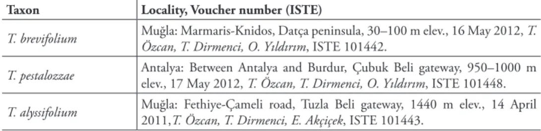
sam-ples were collected from Muğla provinces in Turkey (Figures 1, 3, 5). Voucher specimens are stored in the Herbarium of the Faculty of Pharmacy, Istanbul University (ISTE). Data about habitats of each investigated species are given in Table 1. Permanent micro-scopic preparations were made of plant materials fixed in 70% alcohol during the field studies. Cross-sections of the plant leaves and stems were taken manually and stained with Sartur solution (Çelebioğlu and Baytop 1949). Several slides were made and pho-tographed for each species with an Olympus BH-2 and Canon A 640 digital camera.
Results
The anatomy of the collected specimens were assessed by examination of leaf and stem cross sections (Figures 2, 4, 6). This is the first study about the anatomical features of the leaves and stems of T. alyssifolium, T. brevifolium and T. pestalozzae.
T. alyssifolium Stapf
Leaf anatomy
The epidermis at the both surfaces of the leaves is single layered. The epidermis con-sists of single-layer, ovoid or rectangular cells which are covered by thick cuticula. The upper epidermis cells are larger than the lower ones. Both leaf surfaces are covered by glandular and non-glandular trichomes. Also, the upper epidermis is covered with Table 1. Collection data of Teucrium taxa studied.
Taxon Locality, Voucher number (ISTE)
T. brevifolium Muğla: Marmaris-Knidos, Datça peninsula, 30–100 m elev., 16 May 2012, T. Özcan, T. Dirmenci, O. Yıldırım, ISTE 101442. T. pestalozzae Antalya: Between Antalya and Burdur, Çubuk Beli gateway, 950–1000 m elev., 17 May 2012, T. Özcan, T. Dirmenci, O. Yıldırım, ISTE 101448. T. alyssifolium Muğla: Fethiye-Çameli road, Tuzla Beli gateway, 1440 m elev., 14 April 2011,T. Özcan, T. Dirmenci, E. Akçiçek, ISTE 101443.
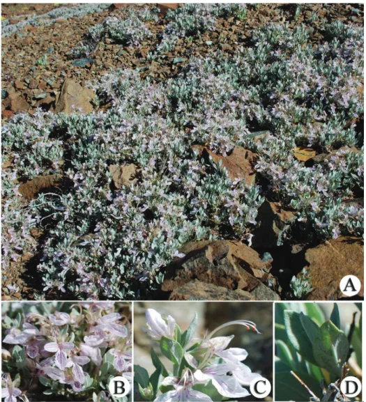
Figure 1. T. alyssifolium. A habitus B inflorescence C flower D leaf.
lower-density trichomes than the lower epidermis. The spherocrystals occur in the upper epidermis cells of the leaf in T. allysifolium. Leaves are isolateral. The mesophyll is differentiated into 1 layered palisade and 2–3-layered spongy parenchyma. The pali-sade parenchyma cells are under the upper and lower epidermis.
Their shapes are cylindrical in transverse section. The palisade parenchyma occu-pies about 60–65% of the mesophyll. The spongy parenchyma cells, ovoid or circular, are located between the palisade tissues. Both parenchyma tissues contain starch grains. The midrib has 3–4 layered collenchyma and 1–2 layered parenchyma below the lower epidermis. The vascular bundle is located in the central part of the midvein. Vascular bundles are collateral. The xylem layer is just below the collenchyma. 1–2 layered parenchyma and 5–6 layered collenchyma are located under the phloem (Figure 2A).
Stem anatomy
The stem is quadrangular shaped. The epidermis consists of single-layer, ovoid or rec-tangular cells which are covered by thick cuticula. There are glandular and non-glan-dular trichomes on the epidermis. Collenchyma with a single layer of cells between the corners but 4–5 layers of collenchyma at the corner of the stem. The cortex, consisting of 3–4 layered ovoidal parenchymatous cells, is located under the collenchyma. The endodermis is conspicuous as a single layer. The vascular tissue is surrounded by 1–2 Figure 2. T. alyssifolium, cross-section of the leaf (A), stem (B, C); cl: collenchyma; en: endodermis; gt: glandular trichomes; le: lower epidermis; p: parenchyma; ph: phloem; pp: palisade parenchyma;
sc: sclerenchyma; scr: sphaerocrystal, sg: starch grains; sp: spongy parenchyma; t: trichome; ue: upper epidermis; xy: xylem; Scale bars: 50 mμ.
layers of sclerenchyma fibres. The cambium is indistinguishable. Phloem and xylem members are conspicuous. The pith is present at the middle of the stem and it is com-pletely filled by orbicular parenchymatic cells (Figure 2B, C).
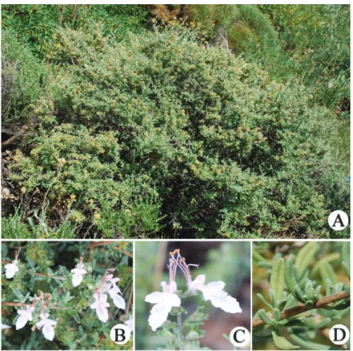
Teucrium brevifolium Schreb.
Leaf anatomy
The epidermis in both surfaces of the leaves is single layered. It is consists of single-lay-er, ovoid or rectangular cells which are covered by cuticula. Both surfaces are covered by a thick cuticula layer, with dense indumentum built of glandular and non-glandular trichomes. The upper epidermal cells are as large as the lower ones. Spherocrystals are observed in both epidermis cells. Leaves are dorsiventral. Palisade parenchyma has two layers and palisade parenchyma cells shapes are cylindrical in transverse section. The palisade parenchyma occupies about 60% of the mesophyll. Spongy parenchyma con-sists of four or five layers and their cells are ovoid or circular.
Figure 4. T. brevifolium, cross-section of the leaf (A), stem (B, C); cl: collenchyma; cu: cuticle; gt:
glandu-lar trichomes; le: lower epidermis; ph: phloem; pp: palisade parenchyma; sc: sclerenchyma; scr: sphaerocrys-tal, sg: starch grains; sp: spongy parenchyma; t: trichome; ue: upper epidermis; xy: xylem; Scale bars: 50 mμ.
Starch accumulated in both spongy and palisade parenchyma. Midrib has 5–6 layered collenchyma and 1–2 layered parenchyma below the lower epidermis. The col-lateral vascular bundle is located in the central part of the midvein. The xylem layer is found under the collenchyma. 1–2 layered parenchyma and 2–3 layered collenchyma are located under the phloem (Figure 4A).
Stem anatomy
The stem is rectangular shaped. The epidermis consists of single-layer, ovoid or rec-tangular cells which are covered by thick cuticula. It is covered by glandular and non-glandular trichomes. Underneath the epidermis, 6–7 layers of collenchyma are located at the corners, 3–4 layered collenchyma is located between the corners. Beneath the collenchyma, 5–6 layered rectangle shaped parenchymatous cells are located. Starch grains are also present in the parenchymatous cells. Endodermis and cambium are inconspicuous. 2–3 sclerenchymatic cell clusters are situated at the corners above the phloem. The pith is present in the middle of the stem and is completely filled by or-bicular parenchymatic cells (Figure 4B, C).
Teucrium pestalozzae Boiss.
Leaf anatomy
The epidermis in both surfaces of the leaves is single layered. It is consists of single-layer, ovoid or rectangular cells which are covered by cuticula. The upper epidermis cells are larger than the lower ones. The upper cuticle layer is slightly thicker than the lower ones.
Both surfaces are covered by glandular and non-glandular trichomes. Also, tri-chomes are abundant on the lower epidermis of leaves and sparse on the upper epider-mis of leaves. Leaves are dorsiventral. The spherocrystals occur in the upper epiderepider-mis of the mesophyll. Mesophyll is differentiated into 2-layered palisade and 5–6-layered spongy parenchyma. Palisade parenchyma cells are cylindrical shaped in transverse sec-tion. The palisade parenchyma occupies about 50–55% of the mesophyll.
The spongy parenchyma cells are ovoid or circular. Both parenchyma tissues dense-ly contain starch grains. Midrib has 5–6 layered collenchyma and 1 layer of parenchy-ma below the lower epidermis. The collateral vascular bundle is located in the central part of the midvein. The xylem layer is found under the collenchyma. 1–2 layered parenchyma and 4–5 layered collenchyma are located under the phloem (Figure 6A). Stem anatomy
The stem is rectangular shaped. The epidermis consists of single-layer, rectangular cells which are covered by cuticula. There are glandular and non-glandular trichomes on the epidermis. Underneath the epidermis, there is collenchyma with 1–2 layers between the corners but 7–8 layers of collenchyma at the corner of the stem. The cortex, con-sisting of 5–6 layers of ovoid shaped parenchymatous cells, is located under the collen-chyma. 1–2 layers of sclerenchyma fibres are located above the phloem. The cambium is indistinguishable. Phloem and xylem members are conspicuous. The pith is present in the middle of the stem and is completely composed of orbicular parenchymatic cells (Figure 6B, C).
Discussion
Sect. Teucrium is one of the eight Teucrium sections distributing in Turkey. The members of this section are perennial and shrubs or subshrubs. Leaves are entire to deeply dissected (in T. orientale subspecies, T. parviflorum, T. pruinosum) and revolute at the lower surface.
Flowers borne in racemes or spreading panicles or axillary in upper leaves. Pe-duncles/pedicels are 1–3-flowered. Calyx not gibbous, obconical-campanulate, teeth ± equal and triangular (Ekim 1982).
Figure 6. T. pestalozzae, cross-section of the leaf (A), stem (B, C); cl: collenchyma; cu: cuticle; e:
epider-mis; gt: glandular trichomes; le: lower epiderepider-mis; p: parenchyma; ph: phloem; pp: palisade parenchyma;
sc: sclerenchyma; scr: sphaerocrystal, sg: starch grains; sp: spongy parenchyma; t: trichome; ue: upper epidermis; xy: xylem; Scale bars: 50 mμ.
Three species showing the characteristic features of the section Teucrium, investigat-ed in the present study, have some significant distinguishing characters. Especially the size of parts of the flowers are very distinctive. T. alyssifolium easily differs with its leaf and bract shape, flower, filament and calyx size from T. brevifolium and T. pestalozzae in
their natural habitats (Table 2). T. alyssifolium is dwarf, suffruticose and T. brevifolium and T. pestalozzae are shrublet plants. Stamens of T. alyssifolium are longer than its lips, stamens subequal to lip in T. pestalozzae and slightly shorter than lips in T. brevifolium.
T. alyssifolium has the shortest and T. brevifolium has the longest stems.
In this study, morphologically related three taxa belonging to the sect. Teucrium have been investigated. Moreover anatomical features of these three species have been report-ed for the first time. Our results showreport-ed the general anatomical characteristics of three
Teucrium species as well those reported by Metcalfe and Chalk (1950) and Dinç et al.
(2008, 2009, 2011a, 2011b), Lakusic et al. (2010), Doğu et al. (2013), Özcan (2013). The results of the present study revealed that there were differences amongst the leaf anatomy of these three taxa (Table 2). The cuticle layer is on both sides and is of equal thickness to the epidermis for T. alyssifolium and T. brevifolium leaves. How-ever, the upper cuticle layer of T. pestalozzae leaves is slightly thicker than the lower ones. The upper epidermis cells are larger than the lower ones as T. alyssifolium and
T. pestalozzae, but both epidermis cells are the same size as in T. brevifolium. T. alys-sifolium and T. brevifolium have a more dense indumentum than T. pestalozzae. Also,
the indumentum of T. brevifolium has the same density on both sides, but the surface of the lower leaves of the other two species is denser than the upper ones. Mesophyll is dorsiventral in T. brevifolium and T. pestalozzae but isolateral in T. alyssifolium. The mesophilic organisation is an important distinguishing character for T. alyssifolium. Mesophyll types may be a good distinctive character in different species but sometimes it can be the same in some closer species (Erdoğan et al. 2012).
The palisade parenchyma shows a slight difference in mesophyll amongst the stud-ied taxa. However, these differences can be based on different ecological conditions. Collenchyma layers are different in midrib amongst these studied taxa. The spherocrys-tals occur in the upper epidermis of the leaf in T. alyssifolium and T. pestalozzae and both epidermis of the leaf in T. brevifolium. According to Metcalfe and Chalk (1950), druse and simple crystals are generally seen in dicotyledon plants. Absence or presence of the crystals and their density are used to distinguish the genera and their species (Salimpour et al. 2009, Güvenç and Kendir 2012). However, spherocrystals and raphi-des are less common crystal types for dicotyledons. Spherocrystals and raphiraphi-des have a diagnostic value for dicotyledons (Dinç et al. 2008, 2009, 2013, Ruiters et al. 2016). According to Dinç et al. (2008, 2009) and Ruiters et al. (2016), spherocrystals are an interspecific classification of sect Teucrium. Our results supported their observations.
Some characteristics of the leaf anatomy which indicates of xeromorphy have been reported before in previous studies (Metcalfe and Chalk 1983, Lakusic et al. 2010, Dinç et al. 2008, 2009). According to the results of our study, the three of taxa have leaves with xeromorphic features such as cuticula layer thickness, dense trichomes and a high proportion of the palisade parenchyma in the mesophyll.
In conclusion, this study shows that leaf and stem anatomy have a diagnostic value in the distinction of these three closely related Teucrium species in sect.
Teu-crium. Anatomical characters contribute to the separation of three species with the
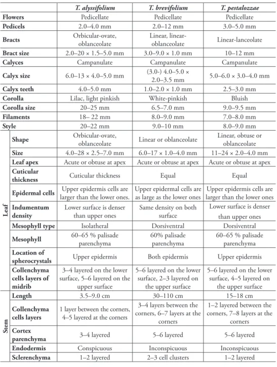
Table 2. Morphological and anatomical comparison of studied taxa.
T. alyssifolium T. brevifolium T. pestalozzae
Flowers Pedicellate Pedicellate Pedicellate
Pedicels 2.0–4.0 mm 2.0–12 mm 3.0–5.0 mm
Bracts Orbicular-ovate, oblanceolate Linear, linear-oblanceolate Linear-lanceolate
Bract size 2.0–20 × 1.5–5.0 mm 3.0–9.0 × 1.0 mm 10–12 mm
Calyces Campanulate Campanulate Campanulate
Calyx size 6.0–13 × 4.0–5.0 mm (3.0-) 4.0–5.0 × 2.0–3.5 mm 5.0–6.0 × 3.0–4.0 mm
Calyx teeth 4.0–5.0 mm 1.0–2.0 × 1.0 mm 2.5–3.0 mm
Corolla Lilac, light pinkish White-pinkish Bluish
Corolla size 20–25 mm 6.5–7.0 mm 9.0–9.5 mm
Filaments 18– 22 mm 8.0–9.0 mm 7.0–8.0 mm
Style 20–22 mm 9.0–10 mm 8.0–9.0 mm
Leaf
Shape Orbicular-ovate, oblanceolate Linear or oblanceolate Linear, obtuse or oblanceolate
Size 4.0–28 × 2.5–7.0 mm 6.0–17 × 1.0–4.0 mm 11–24 × 2.0–4.0 mm
Leaf apex Acute or obtuse at apex Acute or obtuse at apex Acute or obtuse at apex
Cuticular
thickness Cuticular thickness Equal Equal
Epidermal cells Upper epidermis cells are larger than the lower ones. Upper epidermal cells are as large as the lower ones larger than the lower onesUpper epidermis cells are Indumentum
density Lower surface is denser than upper ones Same density on both surface
Lower surface is denser than upper ones
Mesophyll type Isolatheral Dorsiventral Dorsiventral
Mesophyll 60–65 % palisade parenchyma 60% palisade parenchyma 60–65 % palisade parenchyma Location of
spherocrystals Upper epidermis Both epidermis Upper epidermis Collenchyma
cells layers of midrib
3–4 layered on the lower surface, 5–6 layered on the
upper surface
5–6 layered on the lower surface, 2–3 layered on
the upper surface
5–6 layered on the lower surface, 4–5 layered on
the upper surface
Stem
Length 3.5–9.0 cm 30–110 cm 15–18 cm
Collenchyma
cells layers 1 layer between the corners, 4–5 layered at the corners
3–4 layers between the corners, 6–7 layers at the
corners
1–2 layered between the corners, 7–8 layers at the
corners
Cortex
parenchyma 3–4 layered 5–6 layered 5–6 layered
Endodermis Conspicuous Inconspicuous Inconspicuous
Sclerenchyma 1–2 layered 2–3 cell clusters 1–2 layered
The stem is rectangle shaped in all species. In general, the stems of the family Lamiaceae species are rectangular (Metcalfe and Chalk 1950, Dinç et al. 2008a, 2009, Kahraman et al. 2009, Çalı 2014) or in some genera not (Khalik 2016). However, the stems of the sect.
Polium species in Turkey are not conspicuously rectangular. Parenchyma, collenchyma
and sclerenchyma layers have some differences amongst the stem of studied taxa. En-dodermis is conspicuous only in T. alyssifolium. Three studied taxa display general characteristics of Lamiaceae anatomy.
Acknowledgements
This work was supported by the Research Fund of İstanbul University (Project number 31081) and Research Fund of Balıkesir University (Project number 2012/8). We are grateful to Jessica Romero as a native speaker for her valuable contributions and lan-guage control of this manuscript.
References
Aksoy-Sagirli P, Ozsoy N, Ecevit-Genc G, Melikoglu G (2015) In vitro antioxidant activ-ity, cyclooxygenase-2, thioredoxin reductase inhibition and DNA protection properties of Teucrium sandrasicum L. Industrial Crops and Products 74: 545–550. https://doi. org/10.1016/j.indcrop.2015.05.025
Baser KHC, Demirçakmak B, Duman H (1997) Composition of the essential oils of three
Teucrium species from Turkey. Journal of Essential Oil Research 9: 545–549. https://doi.or
g/10.1080/10412905.1997.9700774
Çalı İÖ (2014) An anatomical study of medicinal species Ajuga orientalis L. (Lamiaceae) from Turkey. Journal of Medicinal Plants Research 8(6): 331–338.
Çelebioğlu S, Baytop T (1949) Bitkisel tozların tetkiki için yeni bir reaktif. Farmakolog 19: 301. Dehshiri MM, Azadbakht M (2012) Anatomy of Iranian species Teucrium polium (Lami-aceae) Journal of Biology and Today’s World 1(2): 48–52. https://doi.org/10.15412/J. JBTW.01010204
Dinç M, Dogu S, Bilgili B, Duran A (2009) Comparative anatomical and micromorphological studies on Teucrium creticum and Teucrium orientale var. orientale (Teucrium sect.
Teucri-um, Lamiaceae). Nordic Journal of Botany 27: 251–256.
https://doi.org/10.1111/j.1756-1051.2008.00323.x
Dinç M, Doğu S (2012) Anatomical and micromorphological studies on Teucrium sect.
Isotrio-don (Lamiaceae) in Turkey with a taxonomic note. Biologia 67(4): 663–672. https://doi.
org/10.2478/s11756-012-0049-2
Dinç M, Doğu S, Doğru KA, Kaya B (2011a) Anatomical and nutlet differentiation between
Teucrium montanum and T. polium from Turkey. Biologia 66(3): 448–453. https://doi.
org/10.2478/s11756-011-0035-0
Dinç M, Duran A, Pinar M, Ozturk M (2008) Anatomy, palynology and nutlet micromor-phology of Turkish endemic Teucrium sandrasicum (Lamiaceae). Biologia 63(5): 637–641. https://doi.org/10.2478/s11756-008-0137-5
Dinç M, Doğu S, Bağcı Y, (2011b) Taxonomic reinstatement of Teucrium andrusi from T.
paederotoides based on morphological and anatomical evidences. Nordic Journal of Botany
29: 148–158. https://doi.org/10.1111/j.1756-1051.2011.00894.x
Dinç M, Doğu S (2016) Teucrium pruinosum var. aksarayense var. nov. (Lamiaceae) from Central Anatolia, Turkey. Modern Phytomorphology (9): 13–17.
Dirmenci T (2012) Teucrium L. In: Güner A, Aslan S, Ekim T, Vural M, Babaç MT (Eds) Türkiye Bitkileri Listesi (Damarlı Bitkiler). Nezahat Gökyiğit Botanik Bahçesi ve Flora Araştırmaları Derneği Yayını, İstanbul, 595–598.
Doğu S, Dinç M, Kaya A, Demirci B (2013) Taxonomic status of the subspecies of Teucrium
lamiifolium in Turkey: Reevaluation based on macro-and micro-morphology, anatomy and
chemistry. Nord. J. Bot. 31: 198–207. https://doi.org/10.1111/j.1756-1051.2012.01452.x Dönmez AA (2006) Teucrium chasmophyticum Rech. f. (Lamiaceae): A new record for the flora
of Turkey. Turkish Journal of Botany 30: 317–320.
Dönmez AA, Mutlu B, Özçelik AD (2010) Teucrium melissoides Boiss. & Hausskn. ex Boiss. (Lamiaceae): A new record for Flora of Turkey. Hacettepe Journal of Biology and Chem-istry 38: 291–294.
Duman H (2000) Teucrium L. In: Güner A, Özhatay N, Ekim T, Başer KHC (Eds) Flora of Turkey and East Aegean Islands (Suppl. II). Edinburgh University Press, 197–198. Ekim T (1982). Teucrium L. In: Davis PH (Ed.) Flora of Turkey and the East Aegean Islands,
Vol. 7. Edinburgh University Press, Edinburgh, 53–75.
Ecevit-Genc G, Ozcan T, Dirmenci T (2017) Nutlet and leaf micromorphology in some Turkish species of Teucrium L. (Lamiaceae). Phytotaxa 312(1): 71–82. https://doi.org/10.11646/ phytotaxa.312.1.5
Ecevit-Genc G, Özcan T, Dirmenci T (2015) Micromorphological characters on nutlet and leaf indumentum of Teucrium sect. Teucrium (Lamiaceae) in Turkey. Turkish Journal of Botany 39: 439–448. https://doi.org/10.3906/bot-1406-18
Erdoğan E, Akçiçek E, Selvi S, Tümen G (2012) Comparative anatomical studies on the two
Stachys species (sect. Eriostomum, subsect. Germanicae) growing in Turkey. African Journal
of Pharmacy and Pharmacology 6(19): 1417–1427. https://doi.org/10.5897/AJPP12.267 Govaerts R (1999) World Checklist Seed Plants 3. Continental Publishing, Deurne, 1532. Güvenç A, Kendir G (2012) The leaf anatomy of some Erica taxa native to Turkey. Turkish
Journal of Botany 36(3): 253–262.
Kahraman A, Celep F, Doğan M (2009) Comparative morphology, anatomy and palynology of two Salvia L. species (Lamiaceae) and their taxonomic implications. Bangladesh Journal of Plant Taxonomy 16(1): 73–82. https://doi.org/10.3329/bjpt.v16i1.2749
Khalik KA (2016) A new species of Plectranthus (Lamiaceae) from Saudi Arabia. Turkish Jour-nal of Botany 40: 506–513. https://doi.org/10.3906/bot-1601-8
Lakusic B, Lakusic D, Jancic R, Stevanovic B (2006) Morpho-anatomical differentiation of the Balkan populations of the species Teucrium flavum L. (Lamiaceae). Flora 201: 108–119. https://doi.org/10.1016/j.flora.2005.05.001
Lakusic B, Stevanovic B, Jancic R, Lakusic D (2010) Habitat-related adaptations in morphol-ogy and anatomy of Teucrium (Lamiaceae) species from Balkan peninsula (Serbia and Montenegro). Flora 205: 633–646. https://doi.org/10.1016/j.flora.2010.04.018
Marzouk RI, Salama M, El-Darier M, Askar ABM (2017) Pollen morphology of Teucrium L. (Lamiaceae, Ajugoideae) in Libya. Bangladesh Journal of Plant Taxonomy 24(2): 219– 226. https://doi.org/10.3329/bjpt.v24i2.35118
Menichini F, Conforti F, Rigano D, Formisano C, Piozzi F, Senatore F (2009) Phytochemical composition, anti-inflammatory and antitumour activities of four Teucrium essential oils from Greece. Food Chemistry 115(2): 15 679–686.
Metcalfe CR, Chalk L (1950) Anatomy of the Dicotyledons I. Oxford University Press, London, 1041–1053.
Navarro T, El Oualidi J, Trigo MM (2004) Pollen morphology of Teucrium (Labiatae) and its taxonomic value. Belgian Journal of Botany 137(1): 70–84.
Oybak E, İnceoğlu, Ö (1988) Pollen morphology of some Teucrium L. (Labiatae) species. Communications Faculty of Sciences University of Ankara Series C: Biology 6: 133–146. https://doi.org/10.1501/Commuc_0000000133
Özcan M, Eminagaoglu O (2014) Stem and leaf anatomy of three taxa in Lamiaceae. Pakistan Journal of Botany 43(3): 345–352.
Özcan T (2013) Presence of Teucrium microphyllum in Turkey: Morpho-anatomical, karyological and ecological studies. Biodicon 6: 79–87.
Özcan T, Dirmenci T, Coskun F, Akcicek E, Güner Ö (2015a) A new species of Teucrium sect.
Scordium (Lamiaceae) from SE of Turkey. Turkish Journal of Botany 39: 310–317. https://
doi.org/10.3906/bot-1402-93
Özcan T, Dirmenci T, Martin E, Altinordu F (2015b) Cytotaxonomical study in five taxa of the genus Teucrium L. (Lamiaceae). Caryologia 68(1): 1–8. https://doi.org/10.1080 /00087114.2014.996037
Parolly G, Eren Ö (2007) Contributions to the flora of Turkey. 2. Willdenowia 37: 245–246. https://doi.org/10.3372/wi.37.37114
Ruiters AK, Tilney PM, Van Vuuren SF, Viljoen AM, Kamatou GPP, VanWyk BE (2016) The anatomy, ethnobotany, antimicrobial activity and essential oil composition of southern African species of Teucrium (Lamiaceae). South African Journal of Botany 102: 175–185. https://doi.org/10.1016/j.sajb.2015.06.008
Salimpour F, Mazooji A, Onsori S (2009) Stem and leaf anatomy of ten Geranium L. species in Iran. African Journal of Plant Science 3(11): 238–244.
Semiz G, Çelik G, Gönen E, Semiz A (2016) Essential oil composition, antioxidant activity and phenolic content of endemic Teucrium alyssifolium Staph. (Lamiaceae). Natural Product Research 30(19): 2225–2229. https://doi.org/10.1080/14786419.2016.1149703
Sezik E, Yeşilada E, Honda G, Takaishi Y, Takeda Y, Tanaka T (2001) Traditional medicine in Turkey X. Folk medicine in Central Anatolia. Journal of Ethnopharmacology 75: 95–115. https://doi.org/10.1016/S0378-8741(00)00399-8
Vural M, Duman H, Dirmenci T, Özcan T (2015) A new species of Teucrium Sect. Stachyobotrys (Lamiaceae) from South of Turkey. Turkish Journal of Botany 39: 318–324. https://doi. org/10.3906/bot-1403-50