Detection of Coxiella burnetii prevalence in bovine, ovine and caprine
herds
Uğur PARIN, Osman KAYA
Adnan Menderes University, Faculty of Veterinary Medicine, Department of Microbiology, Aydın, Turkey.
Summary: In this study, a total of 600 blood samples of 200 cattle, 200 sheep and 200 goats from 4 different farms in Aydin Province were investigated serologically and molecularly for the presence of C. burnetii. According to ELISA, the number of positive samples gathered from cattle, sheep and goats were 40 (20.0%), 58 (29.0 %) and 42 (21.0 %), respectively. According to IFA method, the number of positive samples gathered from cattle, sheep and goats were 44 (22.0%), 58 (29.0 %) and 46 (23.0 %), respectively. According to PCR analysis, the number of PCR positive samples of cattle were 96 (48.0%), sheep were 72 (36.0%) and goats were 46 (23.0%). In conclusion, PCR analysis was found as the most sensitive method for detecting C. burnetii from blood samples taken from animals which seem to be healthy.
Keywords: blood, cattle, C. burnetii, ELISA, goat, IFA, PCR, sheep.
Sığır, koyun ve keçi sürülerinde Coxiella burnetii prevalansının saptanması
Özet: Bu çalışmada Aydın ili ve çevresinde bulunan 4’er farklı çiftlikte bulunan 200 adet sığır, 200 adet koyun ve 200 adet keçinin her birinden kan alınmak üzere 600 adet örnekleme yapılarak C. burnetii etkeninin varlığı yönünden serolojik ve moleküler olarak incelenmiştir. ELISA yöntemine göre, sığırlardan koyunlardan ve keçilerden elde edilen serum örneklerinde pozitif örnek sayısı sırasıyla 40 (% 20.0), 58 (% 29.0) ve 42 (% 21.0) olarak belirlenmiştir. IFA yöntemine göre, sığırlardan koyunlardan ve keçilerden elde edilen serum örneklerinde pozitif örnek sayısı sırasıyla 44 (% 22.0), 58 (% 29.0) ve 46 adet (% 23.0) olarak belirlenmiştir. PCR yöntemine göre, sığırlardan elde edilen kan örneklerinde PCR pozitif örnek sayısı 96 (% 48.0), koyunlardan 72 (% 36.0), keçilerden ise 46 (% 23.0) olarak belirlenmiştir. Sonuç olarak sağlıklı görünen hayvanlardan alınan kanlardan C. burnetii varlığının araştırılması için en duyarlı metodun PCR olduğu ortaya konulmuştur.
Anahtar sözcükler: C. burnetii, ELISA, IFA, kan, keçi, koyun, PCR, sığır.
Introduction
Coxiella burnetii is an obligate, intracellular, gram
negative, pleomorphic and cocobacillary bacterial agent which is found in mammalian hosts and arthropods. This microorganism causes Q fever disease both in human and animals and this disease shows a worlwide spread. The disease also causes fever and influenza-like symptoms in affected individuals. The chronic form of the disease also causes endocarditis and hepatitis. The disease acquires en extra emerging situation, due to the host range of the disease including cattle, goats and sheep as well as wild animals. The determination of the disease sources is difficult, because the reservoir spectrum is wide and the agent Coxiella burnetii has a special resistancy against environmental conditions. Furthermore, the agent has been evaluated as a potential biological weapon since 1942 because of its aerosol spread, low infective dose and high morbidity rate (16).
The infection is generally seen in veterinarians, abattoir workers, laboratory staff and farmers whom are directly contacted with meat and milk products of the
infected animals. It is also reported that Q fever outbreak might be seen in the regions of individuals who have close contiguity with infected animals (11).
Cattle, sheep and goats are primary reservoirs of Q fever disease. The agent of Q fever, Coxiella burnetii is localized in the uterus and mammary glands of the infected animals. Beside this, C. burnetii may also infect avians and arthropods as well as mammalians. Previous outbreaks indicate that C. burnetii may infect wide reservoir species. It has been reported 18 outbreaks in 12 different country from 1999 to 2004. Six of the outbreaks emerged from sheeps, three from goats, two from being exposed to sheep and goat feces fertilizer, one of the outbreak emerged from wild animals and one from domestic carnivores. The sources of two outbreaks has been remained unknown (15).
Enzyme Linked Immunosorbent Assay and
Immunofluorescence Test are commonly used for detection of specific antibodies against C. burnetii (19). IFAT is a reference test for diagnosing Q fever disease and also the application of the method is simple. There
are some advantages of IFA test which are necessity of low quantity serum samples, and detectability of IgG, IgM and IgA antibodies formed against Phase I and Phase II agents. However the researchers have reported that IFA test is not applicable for excess amount of samples. This test is adaptable and practical for individual diagnoses, not for epidemiological researches (20).
Application of molecular methods for diagnosing Coxiellosis has been initiated lately due to its need of high biosafety level, rapid diagnosis and urgent application of antibiotic therapy. Isocytrate dehydrogenase genes, superoxide dysmutase genes and transposon-like repetitive regions are determined by PCR based techniques. PCR method is a practical method for identification of C. burnetii from biological samples. High level of specifity and sensitivity were acquired by PCR method applied with the primers consisting of repetitive transposon-like elements. However low level of DNA sequences are determined using these applications (9).
In our research, it was aimed to detect C. burnetii in bovine, ovine and caprine herds by serological and molecular methods.
Materials and Methods
In this study, blood samples were collected from four different cattle and sheep and goat farms in the region in the Aydın province. A total of 600 blood samples (200 cattle, 200 sheep and 200 goats) were collected from farms. The samples were brought to laboratory maintaining the cold chain. Full blood samples were kept in a deepfreeze (-20 °C). For seperation of sera, other blood samples were centrifuged at 5.000 rpm for 10 minutes. Adnan Menderes University Local Ethical Committee of Animal Experiments stated no ethical penalty for this research with the decision of document number of 2010/099-December 29th 2010.
Serological Tests: ELISA and IFA test were applied
to all of the collected samples. A commercial test kit (Vircell® C. burnetii ELISA IgG, G1001) was used for ELISA and applied according to manifacturer’s recommendation. Peroxidase linked anti-goat IgG conjugate were used in our study. A commercial test kit was used for IFA test (Vircell® C. burnetii IFA slide, SCOBU) and applied according to manifacturer’s recommendation.
DNA Extraction and Primers: A commercial
genomic DNA extraction kit (Fermentas® Genomic DNA Purification Kit, K0512) was used for DNA extraction. The extraction was performed by manifacturer’s recommendation. The oligonucleotide primer sequences which were used in the study were Trans I (5'-TGG TAT TCT TGC CGA TGA C-3') and Trans II (5'-GAT CGT AAC TGC TTA ATA AAC CG-3') which have been previously used by Lorenz et al. (1998).
Standard Strain: Purified and lyophilized C. burnetii Nine Mile Phase I strain was used as standard
strain which was found in laboratory stocks of our department.
DNA Amplification: PCR was used for amplification
of extracted DNA samples. The solutions and components used for PCR were prepared as consisting of 23.35 µl Milli-Q water, 5 µl taq reaction buffer 10X (500 mM KCl, 100 mM Tris HCl, pH 9.0), 1.25 µl Dntp mix, 5 µl MgCl2 (25 mM), 5 µl Trans-I and Trans-II (10
pm/ml from each primer), 0.4 µl Hot Start II Taq DNA polymerase (5 U/ml) and 5 µl extracted DNA, a total volume of 50 µl of PCR mixture (10).
The mastermixes prepared for PCR were applied to touchdown PCR process as 5 cycles 30 sec denaturation at 94 ºC, annealing at 66 ± 1ºC for 1 min (the temperature was decreased in each following step), 1 min extension at 72 ºC, and 40 cycles 30 sec denaturation at 94 ºC, primer annealing for 30 sec at 61 ºC, DNA extension for 1 min at 72 ºC (10).
Screening of PCR Products: The products obtained
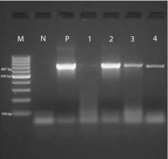
at the end of PCR were visualized by 2 % agarose gel electrophoresis and stained with ethidium bromide. Then, the products were screened in UV screening systems. 100 bp molecular weight marker (100 bp ladder, MBI Fermentas®, SM0241) were used as standard measure. Amplicons were observed at fragment space of 687 bp for C. burnetii (10).
Figure 1.Detection of C. burnetii in blood samples of cattle, sheep and goat by Trans-PCR M: 100 bp marker; N: Negative control (Mili-Q water); P: C. burnetii Nine Mile positive control; 1-2: Positive samples originating from cattle; 3: Positive sample originating from sheep; 4: Positive sample originating from goats.
Şekil 1. Sığır, koyun ve keçi kan örneklerinde Trans-PCR ile C.
burnetii varlığının tespiti - M: 100 bp isaretleyici; N: Negatif
kontrol (Mili-Q su); P: C. burnetii Nine Mile pozitif kontrol; 1-2: Sığır kökenli C. burnetii pozitif örnekler; 3: Koyun kökenli
C. burnetii pozitif örnek; 4: Keçi kökenli C. burnetii pozitif
Results
ELISA results of the samples: According to ELISA,
the number of positives from sera samples of cattle were 40 (20%), 58 (29%) from sheep sera, and 42 (21%) from goat sera.
IFA results of the samples: According to IFA
method, the number of positives from sera samples of cattle were 44 (22%), 58 (29%) from sheep sera and 46 (23%) from goat sera.
PCR results of the samples: According to PCR
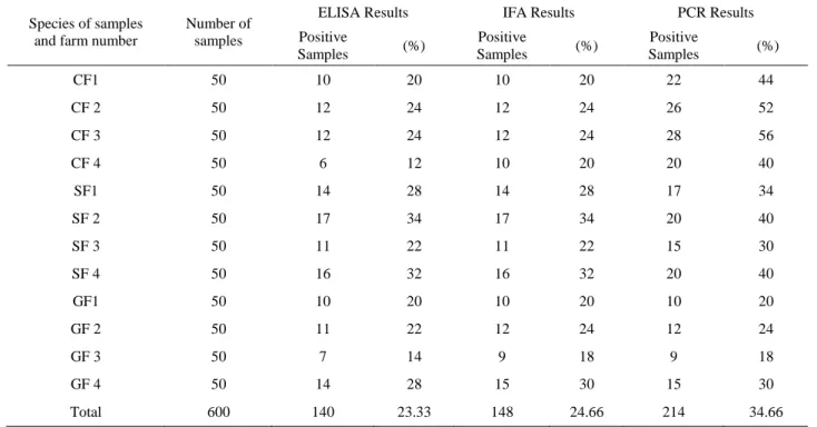
analysis, the number of positives from blood samples of cattle were 96 (48%), 72 (36%) from sheep blood and 46 (23%) from goat blood. As a result of screening PCR products, it is detected that C. burnetii positive products yielded 687 bp fragment size (Figure 1). The results of ELISA, IFA and PCR analysis applied for detection of the presence of C. burnetii is shown in Table 1.
Discussion and Conclusion
Q fever is seen as an endemic disease worldwide. This disease is identified as an important cause of the febrile diseases and the transmission of the agent generally occurs by inhalation (3).
Diagnosing Q fever according to the clinical symptoms is difficult as the disease generally occurs as subclinical infection. However, ELISA and IFAT are
very sensitive for diagnosing Q fever. These tests should also be applied using haemolytic and anti-complementary serum samples. The seroepidemiological researches have indicated that IFA and ELISA have the nearly same sentivity rates and there is no superiority over each other (3). In our study, 140 (23.33 %) samples were detected as positive by ELISA and 148 (26.66 %) samples were detected as positive by IFAT out of 600 samples. The results show that there is no significant difference between these two tests. The distribution of the samples taken from different farms showed that the positivity rates between cattle, sheep and goats did not differ significantly.
Q fever seroprevalence shows different ratios among different species, however prevalence rate goes between 9 and 21 % and the morbidity rate shows parallelism between different species (18). The first case of Q fever in Turkey was reported in 1946 by isolation of the causative agent from dairy milk (2). Different results were reported in Turkey dedicated to Q fever diseases from different regions from the seroprevalence studies made in recent years. C. burnetii IgG positivity was found in the ratio of 10 % in sheep and 5.8 % in cattle from a study performed in Elazığ province of Turkey (2). The seroprevalence of C. burnetii IgG was detected in the ratio of 3 % in Aydin province (8), the prevalence
Table 1. The results of ELISA, IFA and PCR in the detection of the presence of C. burnetii. Tablo 1. C. burnetii varlığının araştırılmasında kullanılan ELISA, IFA ve PCR sonuçları.
Species of samples and farm number
Number of samples
ELISA Results IFA Results PCR Results
Positive Samples (%) Positive Samples (%) Positive Samples (%) CF1 50 10 20 10 20 22 44 CF 2 50 12 24 12 24 26 52 CF 3 50 12 24 12 24 28 56 CF 4 50 6 12 10 20 20 40 SF1 50 14 28 14 28 17 34 SF 2 50 17 34 17 34 20 40 SF 3 50 11 22 11 22 15 30 SF 4 50 16 32 16 32 20 40 GF1 50 10 20 10 20 10 20 GF 2 50 11 22 12 24 12 24 GF 3 50 7 14 9 18 9 18 GF 4 50 14 28 15 30 15 30 Total 600 140 23.33 148 24.66 214 34.66
CF1: Cattle Farm 1, CF 2: Cattle Farm 2, CF 3: Cattle Farm 3, CF 4: Cattle Farm 4 SF1: Sheep Farm 1, SF 2: Sheep Farm 2, SF 3: Sheep Farm 3, SF 4: Sheep Farm 4 GF1: Goat Farm 1, GF 2: Goat Farm 2, GF 3: Goat Farm 3, GF 4: Goat Farm 4 CF1: Sığır Çiftliği 1, CF 2: Sığır Çiftliği 2, CF 3 Sığır Çiftliği 3, CF 4: Sığır Çiftliği 4 SF1: Koyun Çiftliği 1, SF 2: Koyun Çiftliği 2, SF 3: Koyun Çiftliği 3, SF 4: Koyun Çiftliği 4 GF1: Keçi Çiftliği 1, GF 2: Keçi Çiftliği 2, GF 3: Keçi Çiftliği 3, GF 4: Keçi Çiftliği 4
rate was also detected in the ratio of 6 % in Erzurum province of Turkey (17). The results of this study show the existance of C. burnetii infection in Aydin province of Turkey. The previous researches have reported that the antibodies detected by ELISA were found as positive in the ratio of 35.5 % from cattle with infertility problems, and out of 50 human serum sample, 23 (46 %) of them were detected as positive in İstanbul and Thrace region of Turkey (14). In our study, C. burnetii IgG antibodies were detected by ELISA from cattle, sheep and goat populations found in Aydin region. The seroprevalence rate was detected as 23.33 % by ELISA and this result was attractive.
The studies have previously been performed to detect the prevalence of Q fever disease in Turkey. These studies have noticed the risk of disease for human and animals. The seroprevalence rate in cattle was reported in the ratio of 5.8-21.7 % by Gökçen (1989). The seroprevalence rates of disease for cattle, sheep and human were detected in the ratio of 5.8 %, 10.5 % and 8 % respectively from Elazığ and its closer districts (2). The seropositivity rates were found in the ratio of 38.6 % from sheeps with abortion and 11 % from healthy sheeps (7). The same researcher has detected 20 % positivity rate in Elazığ, 20 %positivity rate in Malatya, 28 % positivity rate in Bingöl, and 27 % positivity rate in Muş provinces. The milk samples collected from sheep in Elazığ region had shown 3.5 % positivity rate in consequence of immunomagnetic seperation-PCR method (13). The serum samples collected from Antalya, Diyarbakır and Samsun provinces have shown 13.2, 6 and 1.8 % seropositivity ratio, respectively (1). The seroprevalence rate was shown up in Aydin region of Turkey for cattle, sheep and goat populations with our study.
The previous researches have reported that 7 (7.6 %) human samples were detected as Phase II positive from 20 samples by IFA test. C. burnetii IgG antibodies were found positive in 32 (34.8 %) of the cases and found susceptible in 9 (9.7 %) of the cases. IFA test was applied to 41 people and Phase II IgG antibodies were positive in 39 (42.3 %) individuals. The IFA test and C.
burnetii Phase I and Phase II IgG test were both positive
in 1 (2.4 %) person (4). The ELISA, IFAT and PCR results of our study also indicates the risk of exposion to Q fever disease for veterinarians and the working staff of the farms.
The PCR have been useful, rapid and sensitive technique for detection of C. burnetii agents recently. The detection rate of the agent has previously been found as 1-500 bacteria/ml in serum and 1 bacteria/mg in feces by conventional PCR (12). The mucZ gene was detected in the result of restriction fragment polymorphism studies (6). The result have been more efficient by application of multilocus tandem repetitive analysis method. The
existance of the agent should also be detected by multispacer sequence typing without the necessity of isolating the agent (17). The Trans-PCR method which we applied in our study exhibits that PCR method gives higher positivity rate than ELISA and IFAT.
The existence of C. burnetii was detected by molecular applications in Aydin region and transposon-like repetitive region genes of C. burnetii were targeted. In a previous study 6 (4.3 %) samples were detected PCR positive out of 138 samples (9). In our study, C. burnetii gene was detected in 34.66 % of the samples taken from 600 animals.
In this study, the positivity rates of the tests show that there is no significant difference between ELISA and IFA test. However, PCR results show that Trans-PCR method which were used for detection of transposon-like repetitive region genes of C. burnetii has given the highest positivity when compared with ELISA and IFAT. Furthermore, it was seen that C. burnetii agents exist in Aydın region when evaluating the distribution of the seropositivity between different farms and different animal species. The Q fever disease risk is present in all farms that samples were taken from, and in this case, it is recommended that the further studies on diagnosing Q fever and management procedures should be increased for ruminant husbandry regions in Aydın province of Turkey.
Acknowledgements
This work was the review of the doctorate thesis of the first author.This research was supported financially by Adnan Menderes University Scientific Reseaerch Projects Unit with the code of VTF-11017.
References
1. Berberoğlu U, Gozalan A, Kılıç S, Kurtoğlu D, Esen B (2004):A seroprevalence study of in Antalya, Diyarbakir
and Samsun provinces. Mikrobiyol Bült,38, 385-391.
2. Çetinkaya B, Kalender H, Ertas HB, Muz A, Arslan N, Ongor H, Gurcay M (2000):Seroprevalence of coxiellosis
in cattle, sheep and people in the east of Turkey. Vet Rec,
29, 146, 131-6.
3. Domingo P, Munoz C, Franquet T (1999):Acute Q fever
in adult patients: report on 63 sporadic cases in an urban area. Clin Infect Dis, 29, 874–879.
4. Eyigör M, Kırkan Ş, Gültekin B, Yaman S, Tekbıyık S, Aydın N (2006): Q Ateşi İçin Risk Gruplarında Coxiella
burnetii’ye Karşı Oluşan Antikorların ELISA ve IFA Testleri İle Saptanması. İnfeksiyon Dergisi (Turkish
Journal of Infection),20, 31-36.
5. Gökçen SS (1989):Ege Bölgesi sığırları arasında Q-fever
vakalarının yaygınlık derecesinin mikroaglütinasyon tekniği ile araştırılması. Etlik Mikrobiyol. Derg., 6, 79-85.
6. Jager C, Willems H, Thiele D, Baljer G (1998):Molecular characterization of Coxiella burnetii
7. Kalender H (2001): Elazığ ve komşu illerdeki koyunlarda
Coxiella burnetii enfeksiyonunun yaygınlığı. Turk J Vet
Anim Sci, 25, 51-55.
8. Kılıç S, Pasa S, Babur C, Ozlem MB (2005):
Investigation of Coxiella burnetii antibodies in sheep in Aydin region Turkey. Revue Med Vet, 156, 336-340.
9. Kırkan Ş, Kaya O, Tekbıyık S, Parın U (2008):
Detection of Coxiella burnetii in cattle by using PCR. Turk
J Vet Anim Sci, 32, 215-220.
10. Lorenz H, Cornelie J, Willems H, Baljer G (1998):PCR
Detection of Coxiella burnetii from Different Clinical Specimens, Especially Bovine Milk, on the Basis of DNA Preparation with a Silica Matrix. Appl Environ
Microbiol,64, 4234-4237.
11. Marrie TJ (1990):Acute Q fever. 125-160. In: eRe Press Inc Boca Raton Fla.
12. Nicollet P and Valognes A (2007):Current review of Q
fever diagnosis in animals. B. Acad. Vet. France, 160,
289-295.
13. Öngör H, Cetinkaya B, Karahan M, Açik MN, Bulut H, Muz A (2004):Detection of Coxiella burnetii by
immunomagnetic separation-PCR in the milk of sheep in Turkey. Vet Rec,154, 570-572.
14. Özgür NY, Hasöksüz M, Yılmaz H, İkiz S, Ilgaz A (1997):Detection of antibodies to Coxiella burnetii in
cattle with infertility problems and human sera by ELISA and its seroprevalence. Pendik Vet Mikrobiyol Derg, 28,
207-218.
15. Punda-Polic V, Leko-Grbic J, Radulovic S (2005):
Prevalence of antibodies to rickettsiae in the north-western part of Bosnia and Herzegovina. Eur J Epidemiol,11, 697-9.
16. Samuel JE, Kiss K, Varghees S (2003):Molecular
pathogenesis of Coxiella burnetii in a genomics era. Ann
NY Acad Sci,990, 653–663.
17. Seyitoğlu F, Özkurt Z, Okumuş B (2006): The
seroprevalance of Coxiellosis in Farmers and cattle in Erzurum District in Turkey. Turk J Vet Anim Sci,30, 71-75.
18. Uhaa IJ, Fishbein DB, Olson JG, Rives CC, Waag DM, Williams JC (1994): Evaluation of specificity of indirect
enzyme-linked immunosorbent assay for diagnosis of human Q fever. J Clin Microbiol,32, 1560-1565.
19. Varga V (1997): An explosive outbreak of Q-fever in
Jedl’ove Kostol’any, Slovakia. Cent Eur J Public Health,4,
180–182.
20. Waag D, Chulay J, Marrie T (1995): Validation of an
enzyme immunoassay for serodiagnosis of acute Q fever.
Eur J Clin Microbiol Infect Dis,14, 421–427.
Geliş tarihi: 10.03.2014/ Kabul tarihi: 17.10.2014
Address for correspondence:
Assist. Prof. Dr. Uğur Parın Adnan Menderes University, Faculty of Veterinary Medicine, Department of Microbiology, Işıklı, 09016, Aydın-TURKEY. e-mail: uparin@adu.edu.tr

