Summary
In this study, determination of resistance to methicilline and vancomycin in staphylococcus strains isolated from cow milk samples with subclinical and clinical mastitis was aimed. One hundred staphylococcus strains were isolated from total 306 quarter milk samples collected from the dairy cattle farms in the city of Burdur. Phenotypic resistance to methicillin and vancomycin was determined by Agar-Disc Diffusion Technique (ADDT) and Minimal Inhibitory Concentration (MIC) values for methicillin and vancomycin were determined by Agar Dilution Technique (ADT), according to Clinical and Laboratory Standards Institute (CLSI) protocols. Polymerase Chain Reaction (PCR) was performed for determination of mecA and vanA genes in the strains. The number of strains with both phenotypically and genotypically resistant and susceptible to methicillin were found as 20 and 25, respectively. Ten staphylococcus strains with reduced susceptibility to vancomycin were determined. None of the strains had vanA gene. In conclusion, it is stated that methicillin resistance in staphylococcus strains responsible from mastitis in cattle in Burdur province is high. In terms of vancomycin resistance there is no resistant staphylococcus strains, but presence of strains with reduced susceptibility to vancomycin should be taken into account.
Keywords: Mastitis, Methicillin, Staphylococcus spp., Vancomycin
Mastitisli Sığır Sütlerinden İzole Edilen Stafilokok Suşlarında
Metisilin ve Vankomisin Dirençliliğinin Belirlenmesi
Özet
Bu çalışmada, klinik ve subklinik mastitisli inek süt örneklerinden izole edilen stafilokok suşlarında metisilin ve vankomisin dirençliliğinin belirlenmesi amaçlanmıştır. Burdur ilindeki süt ineği çiftliklerinden toplanan 306 meme lobundan toplanan süt örneklerinden 100 stafilokok suşu izole edildi. Metisilin ve vankomisine fenotipik dirençlilik Agar-Disk Difüzyon Tekniği (ADDT) ile ve metisilin ve vankomisin için Minimal İnhibitör Konsantrasyonu (MİK) değerleri Agar Dilüsyon Tekniği (ADT) ile Klinik ve Laboratuvar Standartları Enstitüsü (CLSI) protokollerine göre belirlendi. Suşlarda mecA ve vanA genlerinin belirlenmesi için Polimeraz Zincir Reaksiyonu (PZR) gerçekleştirildi. Hem fenotipik hem de genotipik olarak metisiline dirençli ve duyarlı olan suşların sayısı sırasıyla 20 ve 25 olarak bulundu. On adet stafilokok suşu vankomisine duyarlılığı azalmış olarak belirlendi. Suşların hiç biri vanA genine sahip değil idi. Sonuç olarak, Burdur ilinde sığır mastitislerinden sorumlu stafilokok suşlarında metisilin dirençliliği yüksektir. Vankomisin dirençliliği açısından ise, dirençli suş bulunmamakta fakat vankomisine duyarlılığı azalmış suşların varlığı dikkate alınmalıdır.
Anahtar sözcükler: Mastitis, Metisilin, Staphylococcus spp., Vankomisin
Detection of Methicillin and Vancomycin Resistance in
Staphylococcus Strains Isolated from Bovine
Milk Samples with Mastitis
[1]Faruk PEHLIVANOGLU *
Hakan YARDIMCI **
[1] * **
Summarized from the First Author’s PhD Thesis
Department of Microbiology, Faculty of Veterinary Medicine, Mehmet Akif Ersoy University, Istiklal Campus, TR-15030 Burdur - TURKEY
Department of Microbiology, Faculty of Veterinary Medicine, Ankara University, TR-06110 Dışkapı, Ankara - TURKEY
Makale Kodu (Article Code): KVFD-2012-6642
Methicillin resistance in Staphylococcus spp. is described as resistance to beta-lactam antibiotics such as cloxacillin, dicloxacillin, methicillin, nafcillin, oxacillin which can not be hydrolized by beta lactamase enzymes produced by
staphylococcus strains 1. Methicillin resistant staphylococcus strains posses mec element in their genome that consists of mecA gene encoding PBP2a, mecI and mecRI regulatory elements controlling mecA gene transcription and mec
INTRODUCTION
İletişim (Correspondence) +90 248 2132063
associated DNA 2. PBP2a determines methicillin resistance because of its reduced affinity for beta-lactams and higher rates of release of the bound beta-lactams to itself 2,3. PBP2a can undertake the transpeptidation reactions when normal PBPs activities are blocked by the drug and allow the strain to form cell wall and continue to grow at lethal concentration of beta lactamase resistant antibiotics 2,3. On the other hand, overproduction of beta lactamases by the isolates and modification of normal PBPs may also be responsible from the methicillin resistance in staphylococcus strains 2,3.
Methicillin resistant strains are inherently cross-resistant to virtually all β-lactam antibiotics, regardless of in vitro antibiogram test results, which are the most effective and widely used class of antimicrobials for several bacterial infections 4. Additionally, methicillin resistant clinical strains are quite often multi-drug resistant (such as aminoglycosides, fluoroquinolones, macrolides, tetracycline) and this reduces significantly the therapeutic options for the treatment of staphylococcal infections 3,5.
Vancomycin is the drug of choice for treatment of multi-resistant methicillin multi-resistant staphylococcus infections. CLSI classifies vancomycin resistance in staphylococci as intermediate and full resistance 4. In intermediate resistant strains, excess amount of peptidoglycan production and thickening of cell wall are common characterics, and since thickening of cell wall causes trapping of vancomycin molecules in the cell wall peptidoglycan, vancomycin molecules can not reach to target close to cell membrane where synthesis of cell wall occurs 6-9. Intermediate resistance can not be transferable and form after vancomycin exposure. The mechanism for full resistance is mediated by vanA gene encoding D-Alanine-D-Lactate depsipeptide, which exist instead of D-Alanine D-Alanine dipeptide of NAM/ NAG of a vancomycin susceptible strain. Since D-Ala-D-Lac has a much lower affinity to vancomycin molecules than D-Ala-D-Ala, vancomycin can not bind and interrupt the peptidoglycan sysnthesis of cell wall and this result in full resistance 10. It is known that vanA gene placed on Tn1516 in staphylococcus strains and it was transfered from Enterecoccus faecalis by conjugation of Tn1516 or a conjugative plasmid carrying Tn151611,12.
Isolation of methicilline resistant staphylococcus strains responsible from bovine mastitis has been reported by many researchers worldwide 13-19. In terms of vancomycin resistance, full resistance has not been reported yet in staphylococcus strains from bovine mastitis cases 19-21, but recently, in a study conducted in Aydin province of Turkey, vancomycin intermediate S. aureus (VISA) strains were detected from bovine mastitis cases by ADT but it was stated that these isolates did not carry vanA gene 5.
This study is conducted to determine the resistance to methicillin and vancomycin by both phenotypical and geno- typical methods in Staphylococcus spp. strains isolated from bovine mastitic milk samples in the city of Burdur in Turkey.
MATERIAL and METHODS
Sampling
In the study, total 306 milk samples were collected from 107 lactating cows belonging to 57 dairy cattle managements in Burdur province located in southwest of Turkey. Out of 306 milk samples, 121 samples were collected from clinically infected udders and remaining 185 samples were collected from subclinically infected udders, with positive California Mastitis Test result. Firstly, teats were disinfected by 70% alchohol and at least 5 ml milk samples were collected into sterile tubes.
Isolation and Identification
Fifty microliters of each samples were streaked on 7% sheep blood agar plates within two hours of collection. The plates were incubated in air at 37°C for 24 h. Identification of staphylococcus strains were performed by the following tests: Gram staining, oxidation and fermentation test with glucose, oxidase test, catalase test, clumping factor and tube coagulase tests, mannitol fermentation test, Voges- Proskauer test (asetoin test), beta-galactosidase test (ONPG), carbonhydrate fermentation tests (glucose, fructose, lactose, maltose, mannitol, mannose, N-acetyl glucosamine, raffinose, sucrose, xylose, trehalose), urease test, and polymixin and novobiocin susceptibility tests 22,23.
Antimicrobial Susceptibility Testing
- Agar-Disc Diffusion Technique (ADDT)
CLSI protocols were followed 24. Briefly, the test was performed on Mueller Hinton Agar (MHA) supplemented with 2% NaCl for methicillin and on MHA for vancomycin resistance determination. The bacterial inoculum was prepared by suspending isolated staphylococcus strain colonies from an 24 h agar plate in tryptic soy broth to a turbidity matching a 0.5 McFarland Standard and the inoculum suspension was used within 15 min of preparation. One µg oxacillin commercial disc (Bioanalysis, Turkey) for methicillin resistance and 30 µg vancomycin (Bioanalysis, Turkey) for vancomycin resistance determination were placed manually on agar plates and the plates were incubated in an inverted position in air under 35°C for 24 h. The inhibition zones around discs were examined and measured. Transmitted light was used for light growth or microcolonies. The results were evaluated according to CLSI critical zone diameters (Oxacillin-S aureus: R≤10 mm, I:11-12 mm, S≥13 mm; CoNS: R≤17mm, S≥18 mm) 25, (Vancomycin-Staphylococcus spp.: R<15mm, S≥15 mm) 4.
- Agar Dilution Technique (ADT)
The Minimal Inhibitory Concentration (MIC) of vancomycin was determined by agar dilution technique as recommended by CLSI 26. The test was performed for methicillin resistance testing on MHA supplemented with 2%
NaCl and for vancomycin resistance testing on MHA. Firstly, the mentioned agar plates with different oxacillin (Sigma, USA) and vancomycin (Lek Pharmaceuticals, Slovenia) concentrations ranging from 0.25 µg/ml to 256 µg/ml were prepared. The inoculum was prepared by suspending isolated staphylococcus strain colonies from an 24 h agar plate in tryptic soy broth to a turbidity matching a 0.5 McFarland Standard and used within 15 min of preparation. The plates were inoculated with the inoculum suspension of a strain starting with the lowest concentration. Growth control plate without antibiotics were also inoculated with the strains. Then, the plates were incubated in air at 35°C for 24 h. The MIC was read as the first antibiotic concentration that inhibits the growth of the strain completely. The results were evaluated according to CLSI critical MIC values (Oxacillin-S. aureus: S ≤2 μg/ml, R ≥4 μg/ ml; CoNS: S≤0.25 μg/ml, R ≥0.5 μg/ml) 27, (Vancomycin-S.
aureus: S≤ 2 μg/ml, I: 4-8 μg/ml, R ≥16 μg/ml; CoNS: S ≤ 4
μg/ml, I:8-16 μg/ml, R ≥32 μg/ml) 4.
In methicillin testing, S. aureus ATCC 43300 (Ondokuz Mayıs University, Faculty of Veterinary Medicine) was used as positive control and S. aureus ATCC 25923 (Mehmet Akif Ersoy University, Faculty of Veterinary Medicine) was used as negative control. In vancomycin testing, Enterococcus
faecalis VanA gene positive avian isolate (Ondokuz Mayıs
University, Faculty of Veterinary Medicine) and S. aureus ATCC 43300 were used as positive and negative control strain, respectively.
Polymerase Chain Reaction (PCR)
- DNA Extraction
Single colonies of each isolates and control strains were inoculated into 1 ml nutritient broth (Oxoid, England) and incubated in air at 37°C for 24 h. The cultures were centrifujed at 8.000 rpm for 5 minutes. Pellets were resuspended in 180 μl of 20 mM Tris/Cl (Amresco, USA), 2 mM EDTA (Bioshop, Canada) and 1% Triton X-100 (Sigma, USA) (pH 8.0) solution supplemented with 0.2 mg/ml lysostaphin (Sigma, USA) and the tubes were incubated at 37°C for 30 min. Then, 25 μl proteinase K (Macherey-Nagel, Germany) was added into each tubes and vortexed and incubated at 56°C for 2 h. DNA was purified from the strains by using a DNA extraction kit (Macherey-Nagel, Germany) in accordance with the manufacturer’s instructions. Extracted DNA samples were kept at -20°C until use in PCR.
- PCR Reaction
PCR was achieved with 2 µL extracted template DNA, 2 µL primers and 10 µL 2 x Master mix (4 mM MgCl2, 0.4 mM each dNTPs, 0.05 U/µl recombinant Tag DNA Polymerase and reaction buffer) (Fermentas, Lithuania) in a final volume of 25 µL. A 314 bp fragment of the mecA gene was amplified using two oligonucleotide primers, forward 5’-CCT AGT AAA GCT CCG GAA-3’ and reverse 5’- CTA GTC CAT TCG GTC CA-3’ 28. A 1030 bp fragment of vanA gene was amplified
using forward 5’-CAT GAA TAG AAT AAA AGT TGC AAT A-3’ and reverse 5’- CCC CTT TAA CGC TAA TAC GAT CAA-3’ primers 29. Appropriate positive and negative controls were included in each PCR run. For mecA spesific PCR, the cycling profile comprised an initial denaturation step at 94°C for 10 min, followed by 30 cycles of denaturation at 94°C for 1 min, annealing at 54°C for 1 min, and extension at 72°C for 1 min and a single extension step of 72°C for 7 min. For vanA spesific PCR, the thermal cycling program was as follow: initial denaturation at 95°C for 10 min; followed by 30 cycles of denaturation (30 sec at 94°C), annealing (30 seconds at 58°C) and extension (30 sec at 72°C); and a single extension (10 min at 72°C).
- Electrophoresis
MecA and vanA spesific PCR products were
electro-phoresed in a 1.5% and 1.8% agarose gel 28,29, respectively, at 170 V for 60 min (Thermo Electron Corporation, USA), stained with ethidium bromide (0.5 μg/ml) and visualized and photographed under UV transilluminator (Genius, Bio-Imaging System, Syngene, England).
RESULTS
In this study, total 100 Staphylococcus spp. strains, 82 coagulase positive staphylococcus (CoPS) and 18 coagulase negative staphylococcus (CoNS) strains, were isolated from 127 out of 306 (41.5%) mastitic milk samples. Fifty five strains were isolated from 185 milk samples collected from sub-clinically infected udders and remaining 45 strains were from 121 milk samples collected from clinically infected udders. Among the strains isolated from clinical cases, 38 (84.4%) strains were CoPS and 7 (15.6%) strains were CoNS strains. The strains isolated from subclinical cases consist of 44 (80%) CoPS and 11 (20%) CoNS strains. In total, 63 cows out of 107 (58.9%) cows with mastitis in this study were diagnosed with staphylococcal mastitis.
Species distribution of Staphylococcus spp. strains were as follow: 65 S. aureus, 17 S. intermedius, 4 S. simulans, 3 S.
chromogenes, 3 S. lugdunensis, 3 S. xylosus, 2 S. hemolyticus,
1 S. hominis subsp. hominis, 1 S. cohnii subsp. urealyticum and 1 unidentified CoNS strain.
ADDT
Out of 100 strains, total 31 strains were found resistant and 64 strains were found susceptible to methicillin. Only 5 strains were determined as intermediate resistant to methicillin (Table 1). Out of 65 S. aureus strains tested, 15 strains were found resistant, 5 strains were found intermediate and 45 strains were found susceptible to methicillin. All of S. intermedius strains (n:17) were susceptible to methicillin (Table 2). In CoNS strains, 2 strains were susceptible and 16 strains were resistant to methicillin (Table 1). In terms of vancomycin resistance, all strains were found susceptible to vancomycin.
ADT
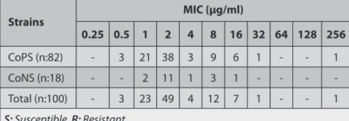
Total 38 strains were determined resistant and 62 strains were determined susceptible to methicillin (Table 1). Among 65 S. aureus strains, 20 strains were resistant and 45 strains were susceptible (Table 2). All S. intermedius strains were found susceptible to methicillin as found in agar disc diffusion technique. On the other hand, all CoNS strains were determined as resistant to methicillin with a MIC value equal to and greater than 0.5 µg/ml. MIC values of the strains for methicillin were presented on Table 3. In terms of vancomycin resistance, 90 out of 100 staphylococcus strains were found susceptible. The remaining 10 strains (7
S. aureus and 3 CoNS strains) were found to have reduced
susceptibility to vancomycin with MIC value 4-8 μg/ml. MIC values of the strains for vancomycin were presented on Table 4.
PCR
Total 57 strains were found carrying mecA gene and none of the strains were found having vanA gene. Total 20 strains were determined resistant to methicillin both pheno- typically and genotypically, and 25 strains were susceptible to methicilin both phenotypically and genotypically (Table 5).
DISCUSSION
After isolation of first methicillin resistant S. aureus (MRSA) strain from bovine mastitis cases in Belgium by Devriese et al.13, presence of MRSA in bovine mastitis cases has been reported by several studies in different countries. MRSA strains were detected by Kaszanyitzky et al.14 in Hungary, by Moon et al.15 in Korea, by Virgin et al.16 in USA, by Stastkova et al.17 in Check Republic, by Spohr et al.18in Germany, and by Kumar et al.19in India. Recently, there have been some studies conducting in Turkey showing that both phenotypic and genotypic resistant strains to methicillin exist in bovine mastitis cases. In Turkey, methicillin resistance rate in S. aureus strains isolated from bovine mastitis cases was reported as 60% in Aydin province 30 and 17.5% in Burdur province 31 by ADDT. In CoNS strains, methicillin resistance rate was reported as 73% in Aydin province 30, and 22.8% in Burdur province 31 by ADDT. In another study conducted in Aydin province, methicillin resistance in staphylococcus strains from bovine milk samples was found as 10.4% 32. Sareyyupoglu et al.33reported methiciline resistance by ADDT in S. aureus strains from mastitic bovine milk samples collected from Polatlı, Çubuk and Haymana districts of Ankara Province as 31.8%, 58.6% and 0%, and by mecA spesific PCR as 31.8%, 31% and 3.1%, respectively. The difference in resistance rates found in the studies can be attributed to strain differences among the regions, use of different sampling methods in the studies, difference in the number of isolate tested, use of different antimicrobial susceptibility testing protocols and difference in evaluation of the test results in the studies. Additionally, difference in Table 1. Staphylococcus strains and results of ADDT and ADT with oxacillin
Tablo 1. Stafilokok suşları ve oksasilin ile ADDT ve ADT sonuçları
Strains ADDT ADT
R I S R I S
CoPS (n:82) 15 5 62 20 n/a 62
CoNS (n:18) 16 n/a 2 18 n/a 0
Total (n:100) 31 5 64 38 n/a 62
R: Resistant, S: Susceptible, I: Intermediate, n/a: non applicable
Table 2. CoPS strains and results of ADDT and ADT with oxacillin Tablo 2. Koagülaz Pozitif Stafilokok suşları ve oksasilin ile ADDT ve ADT
sonuçları
CoPS Strains ADDT ADT
R I S R I S
S. aureus (n:65) 15 5 45 20 n/a 45
S. intermedius (n:17) 0 0 17 0 n/a 17
Total (n:82) 15 5 62 20 n/a 62
R: Resistant, S: Susceptible, I: Intermediate, n/a: non applicable
Table 3. Distribution of MIC values of the strains for oxacillin Tablo 3. Oksasilin için suşların MİK değerlerinin dağılımı
Strains MIC (µg/ml) 0.25 0.5 1 2 4 8 16 32 64 128 256 CoPS (n:82) - 3 21 38 3 9 6 1 - - 1 CoNS (n:18) - - 2 11 1 3 1 - - - -Total (n:100) - 3 23 49 4 12 7 1 - - 1 S: Susceptible, R: Resistant
Table 5. Comparison of ADT with oxacillin and PCR (mecA) results Tablo 5. Oksasilin ile ADT ve PZR (mecA) sonuçlarının karşılaştırılması
Test Staphylococcus spp. PCR Total
mecA + mecA -ADT CoPS (n:82) S. aureus (n:65) S R 25 12 20 8 45 20 S. intermedius (n:17) S R 12 0 5 0 17 0 CoNS (n:18) S R 0 8 0 10 0 18 Total (n:100) 57 43 100 R: Resistant, S: Susceptible
Table 4. Distribution of MIC values of the strains for vancomycin Tablo 4. Vankomisin için suşların MİK değerlerinin dağılımı
Strains MIC (µg/ml)
0.25 0.5 1 2 4 8 16 32 64 128 256
CoPS (n:82) - 2 48 25 7 - - -
-CoNS (n:18) - - 9 6 - 3 - - - -
-Total (n:100) - 2 57 31 7 3 - - - -
herd sizes and age of the cows sampled in the studies and history of antibiotic use for treatment in the cattle included in the studies were not generally indicated in the studies and all these factors can be the reason for quite differences in the results of the studies. In this study, methicillin resistance were found 23.1% and 88.9% for S. aureus and CoNS, respectively by ADDT. In this study, phenotypic methicillin resistance in CoNS were found higher than S. aureus strains and this supported the results of and Kırkan et al.30 and Turutoglu et al.31. The results of this study indicates that methicillin resistance in staphylococcus strains is high in Burdur province and this may cause the treatment failures for mastitis cases in the cows in the field.
In this study, two phenotypic antimicrobial susceptibility techniques were used and methicillin resistance for S.
aureus were found (15/65) 23.1% by ADDT and (20/65)
30.8% by ADT. The only difference between the results of two techniques was that 5 intermediately resistant S. aureus strains with 11-12 mm zone diameters had MIC value equal to and greater than 4 μg/ml in ADT and these were determined as resistant to methicilline in ADT. On the other hand, methicillin resistance in CoNS were found (16/18) 88.9% by ADDT and 100% by ADT. The only difference between two techniques was 2 CoNS strains found sensitive by ADDT with greater than 18 mm zone diameters had MIC values higher than 0.5 μg/ml and were determined as resistant in ADT. Since ADT is more sensitive than ADDT especially for detecting heterogenously resistant strains, the results of two techniques can be different. Thus, ADT is accepted as standard method by CLSI 26. Additionally, ADDT results can be affected easily by external factors and thus all resistant and borderline heterogen resistant strains can not be detected by ADDT34.
In the study, total 20 strains were determined resistant to methicillin both phenotypically and genotypically, and 25 strains were susceptible to methicilin both phenotypically and genotypically. Total 57 out of 100 staphylococcus strains were found carrying mecA gene. Twenty five S. aureus strains were phenotypically sensitive but mecA gene positive. The situation in these S. aureus strains can be attributed to suppression of mecA gene and heterogenous character of methicillin resistance 3. It is known that in phenotypically sensitive but mecA gene positive strains, expression of mecA gene are controlled by primarily regulator genes (mecI-
MecRI) and other chromosomal genes. Since mecI-MecR1
genes are intact in these strains, and beta lactam antibiotics can not activate mecR1 gene strongly, mecI supress the expression of mecA partially or completely. Thus, resistance to methicillin in these strains can not be determined by phenotypical tests 3,35. Development of resistance in such strains occurs by inactivation of mecI and continuous production of PBP2a due to mutations and deletions in operator and/or promoter regions of mecI or mecA after selective pressure of beta-lactam antibiotics 3. Additionally, inactivation of fem factors may result in alteration in
methicillin resistance level since they play role in biosynthesis of peptidoglycan structure of the bacteria 3. On the other hand, it is known that phenotypical tests are occasionally inadequate in detecting heterogenously resistant and borderline resistant strains due to existence of low number (1:106) of resistant bacteria in a culture of such bacteria 34,36. It has been stated that resistance in these subpopulation of a culture originate from additional chromosomal mutations other than mec elements 3. When such clinical strains are reported as sentitive to methicillin, it can result with treatment failure because of existence of resistant subpopulations 37,38. Also, several external factors (inoculum, incubation period and temperature, pH and NaCl concentration of medium, etc.) can influence the expression of resistance in vitro and result of the phenotypical tests 34,39. Similarly, Sareyyupoglu et al.33did not detect resistance by phenotypic test in the strains isolated from mastitic bovine milk samples in Haymana district of Ankara province but 3.1% of the strains were found positive for mecA gene by PCR. In this study, phenotypic resistance in S. aureus strains was found 30.8% (20/65) and genotypic resistance was found 56.9% (37/65).
On the other hand, in this study, out of 20 S. aureus strains found resistant by phenotypical tests, 12 strains (60%) were positive and 8 strains (40%) were negative for mecA gene. Out of 18 CoNS strains found resistant by phenotypical tests, 8 strains (44.4%) were positive and 10 strains (55.6%) were negative for mecA gene (Table 5). Similarly, the rates of having mecA gene in phenotypically methicillin resistant
S. aureus strains from bovine mastitic milk samples have
been reported by several researchers. It was reported by Türütoğlu et al.40as 16.7% in Burdur province, by Moon et al.15as 61.9% in Korea, by Kumar et al.19as 77% in India. In phenotypically resistant but mecA gene negative strains, several mechanisms, such as overexpression of beta-lactamase, overproduction of PBPs and point mutations in PBPs are held responsible from the resistance to methicilline 2. In the study, these strains (8 S. aureus and 10 CoNS) were tested by ADDT with amoxycillin clavulanic acid combination and found susceptible. This showed that phenotypic resistance in these strains are probably due to overproduction of beta lactamase by the strains. However, it is stated that other mechanisms for phenotypic resistance should not be disregarded completely in such strains 37.
Vancomycin is known as a last choice for treatment of methicillin resistant staphylococcus infections in human medicine. Even though vancomycin resistant S. aureus and vancomycin resistant CoNSstrains have been reported in human, a vancomycin resistant staphylococcus strain has not been reported yet from animal specimens 5,19-21. Also in this study, 100 staphylococcus strains were tested by ADDT and all strains were found susceptible to vancomycin. But in ADT, 7 S. aureus (MİK:4 μg/ml) and 3 CoNS (8 μg/ml) strains were detected. This showed that staphylococcus strains intermediately resistant to vancomycin may be isolated from bovine mastitis cases. Similarly, in Aydin province, Türkyılmaz
et al.5tested 16 MRSA strains isolated from bovine mastitis cases for vancomycin susceptibility by ADT and they found 7 VISA strains with 4 μg/ml MIC value. In this study, detection of intermediate resistant strains by only ADT, which is more sensitive than ADDT and accepted as standard technique by CLSI, can be due to heterogenous character of vancomycin resistance and existence of small subpopulations with different MIC values in a culture. In this study, as stated by CLSI, because of inadequacy of ADDT in vancomycin susceptibility testing of staphylococcus strains, ADT was also used 26.
VanA gene specific PCR results showed that both
sensitive (n:90) and intermediate resistant strains (n:10) for vancomycin did not carry vanA gene. Turkyilmaz et al.5also performed vanA specific PCR for intermediate resistant strains isolated from bovine mastitis cases and did not determine vanA gene. Since vanA gene is not present, mechanisms for 10 intermediate resistant strains can be explained with changes in biosynthesis of cell wall of the strains. In such strains, increase in peptidoglican synthesis, reduction in cross bridges in peptidoglican and increase in D-Ala-D-Ala residues have been stated by researchers 6-9.
Staphylococcus strains from bovine mastitic milk can pass to human during milking and processing of milk and can cause infections in human. In this study, it is shown that there is a possibility for transmission of MRSA and methicillin resistant coagulase negative staphylococcus (MRCoNS) strains from cow mastitic cases to human. Since all strains were sensitive to vancomycin that is a last treatment choice for MRSA and MRCoNS infections, possibility of untreatable vancomycin resistant staphylococcal infections in human can be disregarded. However, presence of staphylococcus strains with intermediately resistant to vancomycin is important. In case of being heterogen VISA, the sub-populations with 4-8 μg/ml MIC values in these strains can play key role as a precursor for development of homojen VISA strains after selection pressure of vancomycin use.
A
cknowledgementsThe authors thank to Assistant Professor Alper ÇİFTÇİ (Ondokuz Mayıs University, Faculty of Veterinary Medicine) for providing the standard strains in vancomycin and methicillin susceptibility testing, and to Prof. Dr. Hülya TÜRÜTOĞLU and Assistant Professor Dilek ÖZTÜRK (Mehmet Akif Ersoy University, Faculty of Veterinary Medicine) for providing the methicillin susceptible reference strain S.
aureus ATCC 25923. The authors are also grateful to Associate
Professor Barış SAREYYÜPOĞLU for his helpful comments.
REFERENCES
1. Unal S: MRSA problemi. ANKEM Derg, 23, 1-12, 2009.
2. Chambers HF: Methicillin resistance in Staphylococci: Molecular and biochemical basis and clinical implications. Clin Microbiol Rev, 10, 781-791, 1997.
3. Stapleton PD, Taylor PW: Methicillin resistance in Staphylococcus aureus:
Mechanisms and modulation. Sci Prog, 85, 57-72, 2002.
4. CLSI (Clinical and Laboratory Standards Institute): Performance standards for antimicrobial susceptibility testing. 16th informational supplement, Approved standard M100-S16, CLSI, Wayne, PA, 2006. 5. Turkyilmaz S, Tekbiyik S, Oryasin E, Bozdogan B: Molecular epidemiology and antimicrobial resistance mechanisms of methicillin-resistant Staphylococcus aureus isolated from bovine milk. Zoon Publ Health, 57, 197-203, 2010.
6. Hiramatsu K, Hanaki H, Ino T, Yabuta K, Oguri T, Tenover FC: Methicillin- resistant Staphylococcus aureus clinical strain with reduced vancomycin susceptibility. J Antimicrob Chemother, 40, 135-136, 1997.
7. Hanaki H, Kuwahara-arai K, Boyle-vavra S, Daum RS, Labischinski H, Hiramatsu K: Activated cell-wall synthesis is associated with vancomycin resistance in methicillin-resistant Staphylococcus aureus clinical strains Mu3 and Mu50. J Antimicrob Chemother, 42, 199-209, 1998.
8. Geisela R, Schmitza FJ, Thomasa BL, Bernsa G, Zetschec O, Ulrichc B, Fluitb AC, Labischinskyd H, Wittee W: Emergence of heterogeneous intermediate vancomycin resistance in Staphylococcus aureus isolates in the Düsseldorf area. J Antimicrob Chemother, 43, 846-848, 1999.
9. Hiramatsu K: Vancomycin-resistant S. aureus: A new model of antibiotic resistance. Lancet Infect Dis, 1, 147-155, 2001.
10. Tenover FC: Mechanisms of antibacterial resistance in bacteria. Am J Infect Control, 34, 3-10, 2006.
11. Tenover FC, Weigel LM, Appelbaum PC, McDougal LK, Chaitram J, McAllister S, Clark N, Killgore G, O’Hara CM, Jevitt L, Patel JB, Bozdogan B: Vancomycin-Resistant Staphylococcus aureus isolate from a patient in Pennsylvania. Antimicrob Agents Chemother, 48, 275-280, 2004. 12. Hageman JC, Patel JB, Carey RC, Tenover FC, McDonald LC: Investigation and control of vancomycin-intermediate and resistant Staphylococcus aureus: A Guide for Health Departments and Infection Control Personnel. Atlanta, Georgia, USA, 2006. http://www.cdc.gov/ ncidod/dhqp/pdf/ar/visa_vrsa_guide.pdf, Accessed: 22.09.2010. 13. Devriese LA, Vandamme LR, Fameree L: Methicillin (cloxacillin)-resistant Staphylococcus aureus strains isolated from bovine mastitis cases. Zbl Vet B, 19, 598-605, 1972.
14. Kaszanyitzky EJ, Egyed Z, Janosi SZ, Keseru J, Gal Z, Szabo I, Veres Z, Somogyi P: Staphylococci isolated from animals and food with phenotypically reduced susceptibility to β-lactamase-resistant β-lactam antibiotics. Acta Vet Hung, 52, 7-17, 2004.
15. Moon JS, Lee AR, Kang HM, Lee ES, Kim MN, Paik YH, Park YH, Joo YS, Koo HC: Phenotypic and genetic antibiogram of methicillin-resistant staphylococci isolated from bovine mastitis in Korea. J Dairy Sci, 90, 1176-1185, 2007.
16. Virgin JE, Van Slyke TM, Lombard JE, Zadoks RN: Short communication: Methicillin-resistant Staphylococcus aureus detection in US bulk tank milk. J Dairy Sci, 92, 4988-4991, 2009.
17. Stastkova Z, Karpiskova S, Karpiskova R: Findings of methicillin-resistant strains of Staphylococcus aureus in livestock. Czech J Food Sci, 27, 36-41, 2009.
18. Spohr M, Rau J, Friedrich A, Klittich G, Fetsch A, Guerra B, Hammerl JA, Tenhagen BA: Methicillin-resistant Staphylococcus aureus (MRSA) in three dairy herds in Southwest Germany. Zoon Publ Health, 58, 252-261, 2011. 19. Kumar R, Yadav BR, Singh RS: Genetic determinants of antibiotic resistance in Staphylococcus aureus isolates from milk of mastitic crossbred cattle. Curr Microbiol, 60, 379-386, 2010.
20. Pereira MS, Siqueira-Junior JP: Antimicrobial drug resistance in Staphylococcus aureus isolated from cattle in Brazil. Lett Appl Microbiol, 20, 391-395, 1995.
21. Huber H, Giezendanner N, Stephan R, Zweifel C: Genotypes, antibiotic resistance profiles and microarray-based characterization of methicillin-resistant Staphylococcus aureus strains isolated from livestock and veterinarians in Switzerland. Zoon Publ Health, 58, 343-349, 2011. 22. Koneman EW, Allen SD, Dowell VR, Janda WM, Sommers HM, Winn WC: Preliminary identification of aerobic and facultatively anaerobic Gram positive cocci. In, Color Atlas and Textbook of Diagnostic Microbiology.
3rd ed., Lippincott, Philadelphia, USA, 1988.
23. Winn W Jr, Allen S, Janda W, Koneman E, Procop G, Schreckenberger P, Woods G: Koneman’s Color Atlas and Textbook of Diagnostic Microbiology. 6th ed., pp. 623-671 Lippincott Williams and Wilkins, Baltimore, MD, USA, 2006. 24. CLSI (Clinical and Laboratory Standards Institute): Performance Standards for Antimicrobial Disk Susceptibility Test. Approved Standard M2-A9, 9th ed., CLSI, Wayne, PA, 2006.
25. CLSI (Clinical and Laboratory Standards Institute): Performance Standards for Antimicrobial Susceptibility Testing. 17th Informational Supplement. Approved Standard M100-S17, CLSI, Wayne, PA, 2007. 26. CLSI (Clinical and Laboratory Standards Institute): Methods for dilution antimicrobial susceptibility testing for bacteria that grow aerobically. Approved standard M7-A7, 7th ed., CLSI, Wayne, PA, 2006. 27. NCCLS (National Committee for Clinical Laboratory Standards): Methods for Dilution Antimicrobial Susceptibility Tests for Bacteria that Grow Aerobically. Approved Standard M7-A5, 5th ed., NCCLS, Wayne, PA, 2000. 28. Choi SM, Kim S, Kim H, Lee DG, Choi JH, Yoo JH, Kang JH, Shin WS, Kang MW: Multiplex PCR for the detection of genes encoding aminoglycoside modifying enzymes and methicillin resistance among Staphylococcus species. J Korean Med Sci, 18, 631-636, 2003.
29. Clark NC, Cooksey RC, Hill BC, Swenson JM, Tenover FC: Characterization of glycopeptide-resistant enterococci from U.S. hospitals. Antimicrob Agents Chemother, 37, 2311-2317, 1993.
30. Kırkan S, Goksoy EO, Kaya O: Identification and antimicrobial susceptibility of Staphylococcus aureus and Coagulase Negative Staphylococci from bovine mastitis in the Aydın region of Turkey. Turk J Vet Anim Sci, 29, 791-796, 2005.
31. Turutoglu H, Ercelik S, Ozturk D: Antibiotic resistance of S. aureus and Coaguase Negative Staphylococci isolated from bovine mastitis. Bull Vet Inst Pulawy, 50, 41-45, 2006.
32. Kaynarca S, Turkyilmaz S: Sığır mastitislerinden izole edilen
stafilokoklarda metisilin direnci ve slaym pozitifliği. Kafkas Univ Vet Fak
Derg, 16 (4): 567-572, 2010.
33. Sareyyupoglu B, Cantekin Z, Akan M: Mastitisli sütlerden izole edilen stafilokoklarda metisilin direncinin fenotipik ve genotipik belirlenmesi. VII. Ulusal Veteriner Mikrobiyoloji Kongresi, Antalya-Turkey, 26-28 Eylül 2006. 34. Brown DF: Detection of methicillin/oxacillin resistance in Staphylococci. J Antimicrob Chemother, 48, 65-70, 2001.
35. Kuwahara-arai K, Kondo N, Hori S, Tateda-Suzuki E, Hiramatsu K: Suppression of methicillin resistance in a mecA-containing pre-methicillin- resistant Staphylococcus aureus strain is caused by the mecI-mediated repression of PBP 2a production. Antimicrob Agents Chemother, 40, 2680-2685, 1996.
36. Gosbell IB, Neville SA, Mercer JL, Fernandes LA, Fernandes CJ: Detection of intrinsic oxacillin resistance in non-multiresistant, oxacillin-resistant Staphylococcus aureus (NORSA). J Antimicrob Chemother, 51, 468-470, 2003.
37. Martineau F, Picard FJ, Lansac N, Me´nard C, Roy PH, Ouellette M, Bergeron MG: Correlation between the resistance genotype determined by multiplex PCR assays and the antibiotic susceptibility patterns of Staphylococcus aureus and Staphylococcus epidermidis. Antimicrob Agents Chemother, 44, 231-238, 2000.
38. Sakoulas G, Gold HS, Venkataraman L, Degirolami PC, Eliopoulos GM, Qian Q: Methicillin-Resistant Staphylococcus aureus: Comparison of susceptibility testing methods and analysis of mecA-positive susceptible strains. J Clin Microbiol, 39, 3946-3951, 2001.
39. Brown DFJ, Edwards DI, Hawkey PM, Morrison D, Ridgway GL, Towner KJ, Wren MWD: Guidelines for the laboratory diagnosis and susceptibility testing of methicillin-resistant Staphylococcus aureus (MRSA). J Antimicrob Chemother, 56, 1000-1018, 2005.
40. Turutoglu H, Hasoksuz M, Ozturk D, Yildirim M, Sagnak S: Methicillin and aminoglycoside resistance in Staphylococcus aureus isolates from bovine mastitis and sequence analysis of their mecA genes. Vet Res Commun, 33, 945-956, 2009.