17 Experimental
& Clinical Article
Evaluation of the Relationship Between Placental Thickness and
Obstetric Doppler Parameters During the Second Trimester
Emine AYDIN1, Ayca Nazli BULUT2 Istanbul, Turkey
ABSTRACT
OBJECTIVE: To determine whether there is a relationship between placental thickness and the
umbili-cal artery and uterine artery Doppler evaluation in the second trimester.
STUDY DESIGN: The placental thickness and the umbilical artery and uterine artery Doppler
evalua-tions were recorded by a single operator for patients who were admitted for an obstetric follow-up or fetal anatomy screening at 18-28 gestational weeks. The relation between these variables was investigated by evaluating the patients only once.
RESULTS: In our study, the mean placental thickness was 31.7 (SD±8.01) mm. The umbilical artery
Doppler parameters did not correlate with the placental thickness. The uterine artery Doppler sys-tolic/diastolic velocity, Pulsatility index and Resistance index values positively correlated with the pla-cental thickness. However, these correlations were not statistically significant.
CONCLUSIONS: Although there was no relationship between the placental thickness and obstetric
Doppler parameters in this study, we suggest that they are likely important factors and their significance should be evaluated in future studies
Keywords: Obstetric, Placenta, Thickness, Ultrasonography
1Istanbul Medipol University, Department of Obstetrics and Gynecology, Istanbul, Turkey
2Kayseri City Hospital, Department of Obstetrics and Gynecology, Kayseri,
Turkey
Address of Correspondence: Emine Aydin
Istanbul Medipol University, Faculty of Medicine, Department of Obstetrics and Gynecology, Istanbul, Turkey
[email protected] Submitted for Publication: 29.07.2019
Revised for: Publication: 19.10.2019 Accepted for Publication: 28.10.2019 ORCID IDs of the authors:
EA: 0000-0001-8877-2803, ANB: 0000-0002-7495-5470
Obstetrics; Maternal-Fetal Medicine and Perinatology
Gynecol Obstet Reprod Med 2020;26(1):17-20
How to cite this article: Aydin E. and Bulut AN. Evaluation of the
Relationship Between Placental Thickness and Obstetric Doppler Parameters During the Second Trimester. Gynecol Obstet Reprod Med. 2020;26(1):17-20
Quick Response Code: Access this article online Website: www.gorm.com.tr e- mail: [email protected] DOI:10.21613/GORM.2019.1016
non-invasively by ultrasonography during every stage of preg-nancy (2,3).
During the second trimester fetal screening, the placenta, and the associated anatomical structures are evaluated, and uterine artery Doppler is performed for the prediction of pre-natal complications. Previously, studies were conducted to evaluate the relationship between the placental structure dur-ing pregnancy or the postnatal period and Doppler parameters (3,4). Population studies have aimed to determine the average placental thickness (PT) during specific periods of pregnancy (2,5,6).
In this study, we aimed to determine the PT and the rela-tionship between the umbilical and uterine artery Doppler pa-rameters and PT during the evaluation of the fetal anatomy during the second trimester.
Material and Method
This study was conducted in the Kayseri City Hospital. Ethical approvement was obtained from the local institutional review board, and informed consent was obtained from the subjects before the study commenced. The study population included women attending an antenatal clinic between May 2017 and February 2018. The subjects with an accurate last menstrual period, a viable singleton pregnancy, no history of diabetes mellitus, no history of previous adverse fetal out-comes, no history of intrauterine growth retardation, no
co-ex-Introduction
The placenta is now recognized as a fetal organ and has be-come an integral part of ultrasonography evaluation. The pla-centa is one of the primary factors in fetal birth weight and it is thought that abnormalities of placental growth may precede abnormalities in fetal growth (1).
Previously, the placenta could only be monitored during the postnatal period; however, nowadays it can be examined
Gynecology Obstetrics & Reproductive Medicine 2020;29(1):17-20 18
isting uterine or adnexal masses, no placental masses, no fetal anomalies, a placenta that could be distinguished from the my-ometrium, no history of immune or nonimmune hydrops, no hydramnios, and no pregnancy-induced hypertension were in-cluded in the study. None of the patients were receiving any medication and none of them were currently smoking. Some of this information was obtained from the patients’ antenatal notes, and the remaining information was attained from their sonographic results.
Scanning technique
An HS70A ultrasonography device with a 3.5 MHz curvi-linear transducer (Samsung, Seoul, South Korea) was used to scan the selected subjects, and measurements were recorded in the freeze mode by a single observer with 10 years of experi-ence in obstetric sonography at the time of the study (EA). Transabdominal longitudinal scans of the placenta were per-formed with the subjects in the supine position and with a full bladder. Placental location (anterior uterine wall, posterior uterine wall, fundal, right lateral, and left lateral) was recorded, and the PT was obtained by measuring the antero-posterior diameter of the placenta at the level/point of inser-tion of the umbilical cord (7,8). The fetal parameters, includ-ing biparietal diameter (BPD), head circumference (HC), ab-dominal circumference (AC), femur length (FL), and esti-mated fetal weight (EFW) were also measured using the stan-dardized techniques and all these parameters were used to es-timate the gestational age. The BPD was measured as the dis-tance between the outer edge of the cranium nearest to the transducer and the inner edge of the cranium distal to the transducer at the level of the paired hypoechoic thalami and cavum septum pellucidum (9). The HC was measured using elliptical calipers over the four points of the BPD and occipi-tal fronoccipi-tal diameter in the same plane as the BPD, between the leading edge of the frontal bone and the outer edge of the oc-ciput (10). The AC was measured as the length of the outer perimeter of the fetal abdomen at the level of the umbilical vein junction with the portal vein in a transverse plane per-pendicular to the spine (11), and the FL was measured as the length of the ossified diaphysis of the fetal femur from the greater trochanter to the femoral condyles (12). The mean of three different values for each measurement was recorded. To calculate the EFW, the Hadlock Formula was used (log 10 BW=1.56620.0108(HC)+0.0468(AC)+0.171(FL)+0.00034 (HC)2-0.003685(AC×FL)).
The Doppler parameters, including umbilical artery and uterine artery systolic/diastolic velocity (S/D), Pulsatility index (PI) and resistance index (RI) S/D, RI and PI were measured, according to the International Society of Ultrasound in Obstetrics & Gynecology (ISUOG) practice guidelines (13). Doppler velocimetry was performed using the same transducer by the same operator.
Statistics
The minimum sample size for this study was calculated using Slovin’s Formula [n=N/(1+N e2), n=minimum sample size, n=population size, e=percentage error (percentage error at 95% level of confidence=0.10)]. Using the total number of patients who attended antenatal follow-up in the Kayseri City Hospital between May 2017 and February 2018, a population of 2743 was obtained. Therefore, substituting the values into the above formula presented: n=2743 / (1+2743 (0.10)2=96. Hence, the minimum sample size was 96 patients, and this was increased to 199. One hundred and ninety-nine subjects with GA between 18 and 28 weeks who met the selection criteria were recruited for the study.
Results
In our study, mean maternal age was 27.4 (SD±5.7) years and the mean gestational age was 22.8 (SD±1.05, 18–24.3) weeks at the time of scanning. The patients’ obstetric data were as follows: mean gravida was 2.43 (SD±1.272) and mean parity was 1.25 (SD±1.068).
Forty-nine (24.62%) patients previously had a normal vaginal delivery and 58 (29.14%) had a cesarean section. Other patients were nulliparous (n=107, 53.76%).
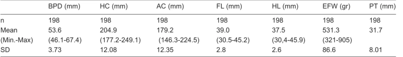
The mean values of fetal biometric measurements and PT are shown in table I.
A weak positive correlation was found between the PT and EFW (r=0.151; p=0.036). The placenta was located in the an-terior uterine wall in 92 patients (46.23%), in the posan-terior uterine wall in 76 patients (38.2%), in the right uterine wall in 13 patients (6.53%), and in the left uterine wall in 18 patients (9%). The PT did not change significantly according to the lo-cation.
There were no statistically significant relationships be-tween the PT and umbilical artery PI, RI, and S/D (p=0.194,
Table I: Evaluation of fetal biometric measurements and placental thickness
BPD (mm) HC (mm) AC (mm) FL (mm) HL (mm) EFW (gr) PT (mm)
n 198 198 198 198 198 198 198
Mean 53.6 204.9 179.2 39.0 37.5 531.3 31.7
(Min.-Max) (46.1-67.4) (177.2-249.1) (146.3-224.5) (30.5-45.2) (30,4-45.9) (321-905)
19 Aydin E. and Bulut AN.
p=0.075, p=0.15, respectively). There were weak positive
cor-relations between the PT and uterine artery PI (r=0.253;
p=0.002), RI (r=0.236; p=0.004), and S/D (r=0.231; p=0.005); however, these associations were not statistically
significant.
Discussion
The placenta is an indispensable component for the con-tinuation of pregnancy and fetal health from the very first day of pregnancy. To date, many studies have been conducted ex-amining both the structure of the placenta and placental hor-mones. Generally, the aim of the placental evaluation is to de-termine structural anomalies; such as the presence of acces-sory lobes, placental localization anomalies, determination of placental insertion localization of umbilical cord or placental bleeding area or vascular pooling. These evaluations were made by various ultrasonographic methods, and some of them also included Doppler evaluations (13,14).
In a previous study, the placental length and thickness were measured by three different methods (linear, curvilinear, and panoramic), and the most reproducible approach in the second trimester for the measurement of placental length was evaluated (13). Some previous studies also aimed to determine the nomogram for the PT by determining the mean PT during the first and second trimesters (2,6). One of these studies re-ported that the PT was directly related to the gestational age and that the optimum PT can be determined with the follow-ing formula, usfollow-ing a linear regression analysis model: gesta-tional age (in weeks)×1.4-5.6 (r=0.82) (2). In another study, the mean PT was reported to be 23.2 (2.8) mm in the second trimester and 36.1 (3.6) mm in the third trimester (6).
In the current study, the same methodology was used for determining PT. We found that the mean PT was 31.72 mm (SD±8.01 mm). We suggest that the positive relationship be-tween the EFW and PT, noted in our study, was due to the pos-itive relationship between the PT and gestational age, which was reported previously (6). In another study, it was hypothe-sized that placentas present at the anterior uterine wall may be thinner than the placentas present at the posterior uterine wall; the study also reported that the mean PT was 24.6 mm (SD±7.29) (5). This PT thickness was consistent with our PT thickness; however, we did not observe thickness differences according to the placental location. In this study, the investiga-tors claimed that anterior placentas of thickness >33 mm and posterior placentas of thickness >40 mm should be considered abnormally thick (5). With respect to these findings, placentas found to be thick on sonography have been reported to be as-sociated with adverse pregnancy outcomes, such as mortality related to fetal anomalies and higher proportions of infants who are either small for gestational age or large for gestational age at term (14,15). In our study, we did not observe abnor-mally thick placentas, according to these upper limits.
Previous work analyzing the correlation between the PT in the second and third trimester and fetal weight claimed that birth weight had a positive relationship with both second and third trimester PT (16). However, the study also suggested that the PT change could not predict low birth weight. In that study, the subjects were examined two times, at 15-20 and 30-34 weeks of gestation. Thus, placental and fetal growth were eval-uated in the same patients and growth curves were obtained (16). In our study, only one evaluation of each subject per-formed during the second trimester was included. This was conducted with the goal of providing a practical prediction model, with only one evaluation rather than repetitive imaging. Additional data recorded during our evaluations included the umbilical and uterine artery Doppler findings. A previous study examined the morphology of the placentas of the fetuses diagnosed with intrauterine growth retardation (17,18). In that study, it was stated that placentas from fetuses with an in-creased S/D ratio (>+2SD) were large, thin, and had a high maximum diameter/maximum thickness ratio. In our study, we did not find any relationship between the umbilical artery Doppler parameters and the PT in the second trimester.
However, in our study, we found a weak positive correla-tion between the uterine artery S/D ratio and the PT; however, this result was not statistically significant. The uterine artery RI and PI were also positively correlated, but these correla-tions were also not statistically significant. We thought that our limitations, which were effective in that situation, were the small number of subjects, a single evaluation, and the absence of the evaluation of the postpartum outcomes.
During pregnancy follow-ups, placental evaluation is con-sidered secondary to the fetus and amniotic fluid evaluation. However, as can be seen from this study and other studies in the literature, the placenta is a structure that can give us clues about the infant and the course of pregnancy and should not be ignored.
Acknowledgments: None. Conflict of interest: None.
Author contributions: EA: Protocol/project development, data collection, management, manuscript writing/editing, ANB: Manuscript writing/editing
References
1. Thame M, Osmond C, Wilks R, Bennett FI, Forrester TE. Second-trimester placental volume and infant size at birth. Obstet Gynecol. 2001;98(2):279-83.
2. Tongsong T, Boonyanurak P. Placental thickness in the first half of pregnancy. J Clin Ultrasound. 2004;32(5):231-4. 3. Nordenvall M, Ullberg U, Laurin J, Lingman G, Sandstedt
B, Ulmsten U. Placental morphology in relation to umbil-ical artery blood velocity waveforms. Eur J Obstet Gynecol Reprod Biol. 1991;40(3):179-90.
Gynecology Obstetrics & Reproductive Medicine 2020;29(1):17-20 20
4. Gilio DB, Miranda Correa RR, De Oliveira Guimarães CS, Peres LC, Marques Salge AK, Cavellani CL, et al. Analysis of placenta vascularization in patients with uter-ine altered artery Doppler flow velocity exams. J Obstet Gynaecol Res. 2009;35(4):648-53.
5. Lee AJ, Bethune M, Hıscock RJ. Placental thickness in the second trimester: a pilot study to determine the normal range. J Ultrasound Med. 2012;31(2):213-8.
6. Agwuna KK, Eze CU, Ukoha PO, Umeh UA. Relationship between sonographic placental thickness and gestational age in normal singleton fetuses in Enugu, Southeast Nigeria. Ann Med Health Sci Res. 2016;6 (6):335-40.
7. Mital P, Hooja N, Mehndiratta K. Placental thickness. A sonographic parameter for estimating gestational age of the fetus. Indian J Radiol Imaging. 2002;12(4):553-4.
8. Hanretty KP. Obstetrics Illustrated. 6th ed. Edinburgh:
Churchill Livingstone; 2003. p. 9-12.
9. Hadlock FP, Deter RL, Harrist RB, Park SK. Fetal bipari-etal diameter: Rational choice of plane of section for sono-graphic measurement. AJR Am J Roentgenol. 1982; 138(5):871-4.
10. Chitty LS, Altman DG, Henderson A, Campbell S. Charts of fetal size: 2. Head measurements. Br J Obstet Gynaecol. 1994;101(1):35-43.
11. Campbell S, Wilkin D. Ultrasonic measurement of fetal abdomen circumference in the estimation of fetal weight. Br J Obstet Gynaecol. 1975;82(9):689-97.
12. Chitty LS, Altman DG, Henderson A, Campbell S. Charts of fetal size: 4. Femur length. Br J Obstet Gynaecol. 1994;101(2):132-5.
13. Bhide A, Acharya G, Bilardo CM, Brezinka C, Cafici D, Hernandez-Andrade E, et al. ISUOG practice guidelines: use of Doppler ultrasonography in obstetrics. Ultrasound Obstet Gynecol.. 2013;41(2):233-9.
14. Milligan N, Rowden M, Wright E, Melamed N, Lee YM, Windrim RC, et al. Two-dimensional sonographic assess-ment of maximum placental length and thickness in the second trimester: a reproducibility study. J Matern Fetal Neonatal Med. 2015;28(14):1653-9.
15. Elchalal U, Ezra Y, Levi Y, Bar-Oz B, Yanai N, Intrator O, et al. Sonographically thick placenta: a marker for in-creased perinatal risk-a prospective cross-sectional study. Placenta. 2000;21(2-3):268-72.
16. Dombrowski MP, Wolfe HM, Saleh A, Evans MI, O'Brien J. The sonographically thick placenta: a predictor of in-creased perinatal morbidity and mortality. Ultrasound Obstet Gynecol. 1992;2(4):252-5.
17. Afrakhteh M, Moeini A, Taheri MS, Haghighatkhah HR. Correlation between placental thickness in the second and third trimester and fetal weight. Rev Bras Ginecol Obstet. 2013;35(7):317-22.
18. Nardozza LMM, Araujo Júnior E, Barbosa MM, Caetano AC, Lee DJ, Moron AF. Fetal growth restriction: current knowledge to the general Obs/Gyn. Arch Gynecol Obstet. 2012;286(1):1-13.