DOI: 10.5455/jtomc.2018.02.036 2018;25(2):252-7
Evaluation of neutrophil-to-lymphocyte ratio as a marker
of inflammatory response in spondylodiscitis
Numan Karaarslan1, Ibrahim Yilmaz2, Feride Sinem Akgun3, Tezcan Caliskan1, Mustafa Dogan4, Bulent Bilir5, Ozkan Ates6
1Namik Kemal University Faculty of Medicine, Department of Neurosurgery, Tekirdag, Turkey
2Medical Pharmacologist, pharmacist, Istanbul Medipol University Faculty of Medicine, Department of Pharmacology, Istanbul, Turkey 3Istanbul Maltepe University Faculty of Medicine, Department of Emergency, Istanbul, Turkey
4Namik Kemal University Faculty of Medicine, Department of Infectious Diseases, Tekirdag, Turkey 5Namik Kemal University Faculty of Medicine, Department of Internal, Tekirdag, Turkey
6Istanbul Esenyurt University, Esencan Hospital, Department of Neurosurgery, Istanbul, Turkey Copyright © 2018 by authors and Annals of Medical Resarch Publishing Inc.
Abstract
Aim: Spondylodiscitis, if not diagnosed on time, can cause morbidity or mortality at high rates. This study was carried out with the aim of testing the hypothesis that “neutrophil-to-lymphocyte ratio can be used” especially in cases where it is difficult to diagnose spondylodiscitis.
Material and Methods: This study involved 24 patients admitted to the State Hospital of Ministry of Health and Namik Kemal University for spondylodiscitis between January2014 and June2017. After excluding the cases that did not meet the inclusion criteria (n=6), the remaining cases (n=24) were referred to as the study group. A control group was created from healthy volunteers (n=24) who applied for routine physical checkups at the clinic between the same dates and who were similar in terms of age, sex, and body mass index to the study group. Hemogram parameters of the cases in both groups; white blood cell, C-reactive protein, erythrocyte sedimentation rate, and neutrophil-to-lymphocyte ratio were statistically compared.
Results: Patients in the spondylodiscitis group, compared to healthy volunteers had statistically significant neutrophil-to-lymphocyte ratio value.
Conclusion: Especially in cases where the diagnosis of spondylodiscitis is not assured, the neutrophil-to-lymphocyte ratio parameter, which is less costly than other diagnostic methods and the analysis results of which can be obtained in a shorter time, may be used to support clinical diagnosis.
Keywords: Low Cost Diagnosis Method; Neutrophil-To-Lymphocyte Ratio; Spondylodiscitis.
Received: 25.02.2018 Accepted: 03.04.2018 Available online: 06.04.2018
Corresponding Author: Numan Karaarslan, Namik Kemal University Faculty of Medicine, Department of Neurosurgery, Tekirdag,
Turkey, E-mail: [email protected]
INTRODUCTION
Spondylodiscitis is a serious medical condition that is known to cause vertebral infection with intervertebral disc infection and osteomyelitis in the spinal column (1). The inflammation is observed in the lower and upper vertebral plates and the adjacent intervertebral disc. This is often accompanied by spondylitis (2). The epidural space, muscle fascia and adjacent soft tissues are damaged due to the spread of the infection (3). Nowadays, many diagnostic classification systems, such as histological response, location, route of disease transmission or the causative agents, have been used to be able to diagnose spondylodiscitis (4,5).
The incidence of Spondylodiscitis is 2.4 per 100,000 people (6). Diagnosis is made with one or more imaging and laboratory
examinations in cases with spondylitis-compatible clinical symptoms, such as spinal pain, tenderness and fever. The narrowing of the intervertebral disc space and osteolysis of the end plates or adjacent vertebral corpus are detected in direct radiography.
The presence of the same findings and/or soft tissue mass may be detected in computed tomography (CT). Bone scintigraphy reveals a signal increase in the vertebral bodies. Magnetic resonance imaging (MRI) may show decreased signal intensity in the vertebra and intervertebral disc space on T1 weighted sequences, and increased signal intensity in the vertebra and disc on T2 weighted sequences (7,8,9). In addition, to make a definite etiologic diagnosis; CT-guided percutaneous needle biopsy, open biopsy may be carried
out, and in the cases, where the fistula from the epidural area to the skin tissue, associated with abscess formation is also observed, a definitive diagnosis may be made by identifying the causative microorganisms in culture antibiograms made from wound site swabs (10,11). However, adverse outcomes such as high rate of the Gram staining and post-culture failure to isolate the agent, or high false-negative rate in bacteriological and histological tests may be encountered (11,12). The systemic symptoms, such as fever, fatigue are rare, but the pain is often observed in this disease, and it has also sneaky and silent clinical course. Therefore, it is reported that the early diagnosis is difficult and that the average delay time of diagnosis is four months (7,13).
Recently neutrophil-to-lymphocyte ratio (NLR) -the level of neutrophil reflecting the severity of inflammation and lymphocyte occurring after physiological stress has been gaining popularity, which was, along with other inflammatory markers, commonly accepted as an accurate marker of the inflammatory status (14).
In the field of neurosurgery, there are some studies in which NLR has been reported to be a new marker for the detection of severe traumatic brain injury prognosis (sensitivity and specificity, statistically significant) (15). In addition, in a study with high evidence, NLR is reported to be prognostic value in glioblastomas (16) and aneurysmal subarachnoid hemorrhage (17).
NLR, which is not routinely, utilized like other infection markers, such as leukocyte count, sedimentation, and C-reactive protein (CRP), has not been investigated spondylodiscitis cases in the literature.
This study has been conducted to evaluate the usefulness of the NLR parameter, which is inexpensive and results in a short period of time, as support for clinical diagnosis in spondylodiscitis-suspicious cases and spondylodiscitis cases that can be encountered in the practice of branches related to spinal diseases.
MATERIAL and METHODS
Ethical approval was obtained from the hospital administration to use the patients’ data.
This retrospective, controlled and multicentered study involved 24 patients admitted to the State Hospital of Ministry of Health and Namik Kemal University for spondylodiscitis between January2014 and June2017. The patients’ demographic and clinical data were retrieved from the hospitals’ electronic database.
Study Design Eligibility criteria
The data obtained from a total of four cases with malignancy, rheumatic disease (n = 1), any other infectious disease such as tooth and mouth health (n = 2) were not included in the study. Then, patients who received treatment (n = 2) that could affect laboratory tests were excluded from the study.
Physical and neurological examinations of cases with severe back pain, fever and / or neurological deficit were carried out. Laboratory and imaging methods were used to make pre-diagnosis of spondylodiscitis. Then in the archive scan; the cases with spondylodiscitis, diagnosed after microbiological evaluation of the specimens obtained by performing subcutaneous or open biopsy, were included in the study (18).
Accumulation of data
Demographic and clinical features of the patients from all centers were incorporated into the analyses. Patients with any other condition that may potentially contaminate erythrocyte sedimentation rate (ESR), CRP or white blood cell (WBC) data and those with incomplete lab results were excluded (Figure 1).
Control group included patients admitted to either of the hospitals for a routine physical checkup, who did not have any serious disease or malignancy and no history of glucocorticoid using. The control group was compatible with the spondylodiscitis groups and gender distribution (n= 24).
In the cases involved in the study group, it was determined that the samples to be used for microbiological examination were obtained by percutaneous needle biopsy, open biopsy and wound swap. Also in these cases, preoperative complete blood cell count, ESR and NLR levels were registered.
To perform percutaneous needle biopsy under imaging guidance; the cases were placed under local anesthesia in a prone position, then the samples were taken by entering the infected intervertebral disc space, using the paramedian approach, with a biopsy needle under C-arm scopy. To perform an open biopsy; again, in the same position, but under general anesthesia, the samples were taken by penetrating the infected intervertebral disc space using the median approach.
Statistical Analyses
The data were analyzed using SPSS version 20.0 descriptive statistics were presented as mean ± standard deviation, median±min-max or frequency (%). Paired Sample t-Test was used to compare the averages of the observed values of a variable in two different cases. The alpha significance value was accepted as 0.05. Correlation and simple linear regression analysis were used to test whether the collected data differed between the groups, whether the observed differences were random in terms of probability calculations, and whether the spondylodiscitis group differed from the control group. The strength of the relationship between spondylodiscitis and NLR was shown by the correlation coefficient (r). It was evaluated that the closer it was to 0, the weaker the relationship was, and that the closer it was to 1, the stronger the relationship was. Since our data were not fit to parametric test assumptions, the (non-parametric) Spearman Rank Correlation Coefficient (rs) was used instead of Pearson Correlation analysis. The alpha significance value, used here, was accepted as 0.01.
RESULTS
The number of male volunteers in the control group (n = 24) was 14 (58.33%) and the number of male cases in the study group (n = 24) was 12 (50.00%). The average age of the male and female volunteers in the control group were; 42.36 ± 10.94 and 44.96 ± 14.36 years, respectively, the average age of male and female cases in the study group were; 50.5 ± 5.09 and 47.17 ± 11.41 years (Table 1).
Table 1. Comparison of NLR and age between groups
CONTROL GROUP (N=24) (HEALTHY INDIVIDUALS) MEAN ± SD STUDY GROUP (N=24) (SPONDYLODISCITIS) MEAN ± SD 49.43 ± 7.22 4.92 ± 2.62 AGE (YEAR) 46.38 ± 12.76 NLR 1.99 ± 0.84
Laboratory results of the patient group and the control group were compared (Figure 1, Table 2.).
Figure 1. Graphical comparison of vein parameters.
Table 2. Comparison of laboratory parameters between healthy volunteers and the cases with spondylodiscitis
Pair
Mean ± S.D. SEM 95% CI of the difference t df tailed)Sig. 2-*
Lower Upper ESR -58.38±39.87 8.14 -75.21 -41.54 -7.17 24 0.000 WBC -2.33±3.64 0.74 -3.86 -0.79 -3.13 24 0.005 CRP -104.03±75.61 15.43 -135.95 -72.10 -6.74 24 0.000 Neutrophil -2.94±3.31 0.68 -4.33 -1.55 -4.35 24 0.000 Lymphocyte 0.71±0.92 0.19 0.32 1.09 3.77 24 0.001
*Paired Samples t-Test. SD: Standard deviation; ESR: Erythrocyte sedimentation
rate, WBC: white blood cell, and CRP: C-reactive protein symbolized
Twelve of the cases were opened and eight of them were percutaneously biopsied. Three cases, where it was obtained via wound swab culture specimen, were also reported (Table 3).
Infection was most frequently observed at L4-L5 level. In males, spondylodiscitis was seen at L4-L5 and L3-L4 levels, and in women, it was seen at L3-L4 level (Table-4). It was calculated that the length of hospitalization was at least seven days and a maximum of 92 days, and the
average of this period was 37.35 ± 20.42 days. It was observed the reproduction of the extended-spectrum-β-lactamase and Escherichia coli pathogens in the culture result of the case that had the longest length of hospitalization.
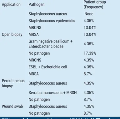
Table 3. Culture samples and Gram staining results of the study group
Application Pathogen Patient group (Frequency) Staphylococcus aureus None Staphylococcus epidermidis 4.35%
MRCNS 13.04%
Open biopsy MRSA 13.04%
Gram negative basilicum +
Enterobacter cloacae 4.35%
No pathogen 17.39%
MRCNS 4.35%
ESBL + Escherichia coli 4.35%
MRSA 8.7%
Percutaneous
biopsy Staphylococcus aureus 4.35% Serratia marcescens + MRSH 4.35%
No pathogen 8.7%
Wound swab Staphylococcus aureus 4.35%
No pathogen 8.7%
ESBL: extended-spectrum-β-lactamase; MRCNS: Methicillin-resistant coagulase-negative staphylococci; MRSA: Methicillin-resistant Staphylococcus aureus; MRSH: methicillin resistant Staphylococcus haemolyticus
Table 4. Spinal levels where it is observed infection involvement
Localization of Infection Male Female Only L2-L3 Spondylodiscitis None None
L2-L3 + L3-L4 Spondylodiscitis 1 1
Only L3-L4 Spondylodiscitis 1 2
L3-L4 Spondylodiscitis + Psoas abscess 1 None
Only L4 Spondylodiscitis 1 None
L3-L4 + L4-L5 Spondylodiscitis 2 1
Only L4-L5 Spondylodiscitis 4 4
L4-L5 + L5-S1 Spondylodiscitis None 1
L5-S1 Spondylodiscitis 1 2
C3-C6 Spondylodiscitis 1 None
L: Lumber, C: cervical, S: sacral vertebrate symbolized
However, it was not observed any pathogen in the culture results of the case that had the shortest length of hospitalization. It was noted that ampicillin sulbactam was the most preferred antibiotic used in the treatment and this was followed by cefazolin sodium active drug. The average duration of antibiotherapy was recorded as 38.13 ± 22.11 days. The longest period of antibiotherapy was found to be imipenem active drug, which was administered for 92 days (Figure 2).
Figure 2. Comparison of the periods of antibiotic use.
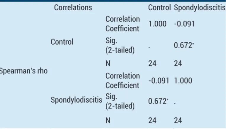
The shortest period of antibiotherapy was found to be cefazolin or ciprofloxacin or clindamycin, which were applied for 21 days. In addition, it was reported that NLR showed a significant positive correlation in the spondylodiscitis group (Table 5).
Table 5. Statistical evaluation of NLR and Spearman’s rho in cases with spondylodiscitis
Correlations Control Spondylodiscitis
Spearman’s rho Control Correlation Coefficient 1.000 -0.091 Sig. (2-tailed) . 0.672* N 24 24 Spondylodiscitis Correlation Coefficient -0.091 1.000 Sig. (2-tailed) 0.672* . N 24 24
*Correlation is significant at the 0.01level (2-tailed), rs=0.672
(p=0.000).
DISCUSSION
The diagnosis of spondylodiscitis in patients admitted to the clinic requires some tests that can be performed at excessive cost and the results of which can be obtained in a long time. In addition to these tests, the laboratory findings showing increased levels of WBC, ESR and CRP in peripheral blood are used.
However, in the diagnosis of spondylodiscitis, especially in the case of suspicious spondylodiscitis, increase in ESR is generally regarded as clinically meaningless, since it is affected by many clinical situations (14).
In this study, it was aimed to investigate whether NLR was the clinical significance to be able to start treatment by making early diagnosis, especially in cases where the diagnosis of spondylodiscitis was made with difficulty. In this study, it was reported that more than 90% of the cases described in the literature were successful spinal fusion of the affected vertebral segments, including infection treatment. It was also reported that Staphylococcus pathogen was the causative agent in 34% of the cases (19).
Fransen et al. (20); in a six-year, single-center, retrospective cohort study of 49 cases over the age of 18 who received treatment for spondylodiscitis, reported that the average age of the cases was 69 (40-89) years. They performed MRI scanning to confirm diagnosis in many cases. They also emphasized that the most frequent pathogens were Staphylococcus aureus (n = 14), Streptococcus spp. and related bacteria (n = 11), and Gram-negative bacteria (n = 11) in culture samples after laboratory test evaluations. It was reported that the antibiotherapy was applied to the all cases and the antibiotics were used for at least six weeks. 17 of the cases were indicated to be hospitalized for 90 days or longer. While considering spondylodiscitis as a possible diagnosis, all patients should undergo physical examination and neurological screening and blood tests should be carried out for infection and blood cultures. If the results are not definitive, an MRI scan should be performed and then a positron emission tomography-CT scan should be done. Ideally, broad spectrum antibiotic treatment should be applied to all patients until the culture results are obtained after a CT scan guided biopsy has been performed before starting treatment. Targeting only Gram-positive microorganisms in empirical therapy would lead to delay adequate treatment in an important group of patients. In this cohort study where the multidisciplinary approach in the treatment of spondylodiscitis was emphasized; they also underlined the diagnosis of spondylodiscitis, a destructive disease with high morbidity and mortality, which was also called vertebral osteomyelitis, was often delayed since the rare and early symptoms of the disease were mostly non-specific (20).
Postoperative spondylodiscitis is relatively rare. This complication is associated with increased cost and long-term work loss and even morbidity. Although most cases of postoperative spondylodiscitis are well-managed with conservative treatment, it may not respond to conservative treatment and may present difficulties for the surgeon in the cases where it was performed instrumentation (21). Therefore, it is important to make an effective and quick diagnosis in such cases.
When the literature is examined; there are studies reporting that the incidence of spondylodiscitis has increased and accordingly the intravenous antibiotics have been used (22). However, the rational use of drugs is important, and it is also important to apply the right drug at the right time. This can only be achieved by making an effective and right diagnosis in a short time. It is important that not only antibiotics, but also other drugs be used rationally.
In a double blind, randomized, prospective study; the affected intervertebral disc in many cases with spondylodiscitis has been reported to be detectable by radiological examination, but it has also reported that clinical significance of these findings is not clear (23). In a multi-center, case-control study in which pyogenic vertebral osteomyelitis was examined; antibiotic usage durations were evaluated in 97 cases at the end of 3 months follow-up period. It was reported that the
prognosis of cases with cervical and / or thoracic involvement was worse and the most frequently detected pathogen was Staphylococcus aureus. In this study, where the average level of CRP was reported to be 150 mg / L, it was indicated that the duration of antibiotic treatment was not associated with functional improvement. It was also pointed out that the exact effect of optimal surgery and / or corticosteroid therapy should be demonstrated in further studies (24).
In a similar study, it was reported that pyogenic spondylodiscitis was most commonly seen in the lumbar region, and this was followed by thoracic and cervical spinal regions (25).
Research on inflammatory biomarkers such as NLR has gained popularity in infectious diseases such as brucellosis which is also present in the etiology of spondylodiscitis. In their study (26), Aktar et al. identified the mean platelet volume (MPV), platelet distribution width (PDW), red cell distribution width (RDW), neutrophil to lymphocyteratio (NLR) and platelet to lymphocyte ratio (PLR) as markers of inflammation in brusella arthritis. The study comprised 64 children with Brusella arthritis and 66 healthy control subjects. They reported that the most commonly affected joint was ankle and that synovial fluid puncture-brucella agglutination test was positive in 34.3% patients. They indicated that the patients were treated with a combination of rifampicin plus sulfamethoxazole/trimethoprim and gentamicin. They emphasized that PDW, RDW, MPV, NLR and PLR values were statistically significant in children with brusella arthritis. In addition, they noted that there was a positive relationship between MPV and NLR values (26).
The mean age was 49.43 ± 7.22 years in the cases we examined. After performing biopsy or obtaining samples from wound swab culture, it was observed that methicillin-resistant coagulase-negative Staphylococci was the most common pathogen in 13.4% of the cases. This was followed by Methicillin-resistant Staphylococcus aureus with 8.7% and Staphylococcus aureus with 4.35%. The most frequent infection involvement was observed at L4-L5 level. The average hospitalization period was estimated to be 37.35 ± 20.42 days. It was observed the reproduction of the extended-spectrum-β-lactamase and Escherichia coli pathogens in the culture result of the case that had the longest length of hospitalization. However, it was not observed any pathogen in the culture results of the case that had the shortest length of hospitalization. The average duration of antibiotherapy was recorded as 38.13 ± 22.11 days. It was noted that ampicillin sulbactam was the most preferred antibiotic used in the treatment and this was followed by cefazolin sodium active drug. It was reported that NLR showed a significant positive correlation in the spondylodiscitis group [(p=0.000; Spearman’s rho (rs ) =0.672].
Although the small number of cases (n=24) in this study
may seem to be a limitation of the research, this is not the real limitation, since the incidence of spondylodiscitis is 2.4 per 100,000 people (6). The real limitation of this research is that it has a retrospective design.
CONCLUSION
here the diagnosis of spondylodiscitis is not assured, in order to start treatment immediately, the NLR parameter, which is less costly than other diagnostic methods and the analysis results of which can be obtained in a shorter time, may be used to support clinical diagnosis. To enter NLR into routine clinical practice, it is necessary to perform prospective studies comparing the control group with other infectious events that are related to joint in the first stage and those with distant infections in the next stage.
Competing interests: The authors declare that they have no competing interest.
Funding: No financial disclosure was declared by the authors. Ethical approval: Ethical approval was obtained from the hospital administration to use the patients’ data.
REFERENCES
1. Fantoni M, Trecarichi EM, Rossi B, Mazzotta V, Di Giacomo G, Nasto LA, et al. Epidemiological and clinical features of pyogenic spondylodiscitis. Eur Rev Med Pharmacol Sci 2012;16 Suppl 2:2-7.
2. Tsujimoto T, Takahata M, Kokabu T, Matsuhashi M, Iwasaki N. Pyogenic spondylodiscitis following anti-interleukin-6 therapy in a patient with rheumatoid arthritis (implication of hematogenous infection risk in degenerative intervertebral discs): a case report and review of the literature. J Orthop Sci 2016;21(5):694-7.
3. Quesnele J, Dufton J, Stern P. Spinal infection: a case report. J Can Chiropr Assoc 2012;56(3):209-15.
4. Beronius M, Bergman B, Andersson R. Vertebral osteomyelitis in Göteborg, Sweden: a retrospective study of patients during 1990-95. Scand J Infect Dis 2001;33(7):527-32. 5. Homagk L, Homagk N, Klauss JR, Roehl K, Hofmann GO,
Marmelstein D. Spondylodiscitis severity code: scoring system for the classification and treatment of non-specific spondylodiscitis. Eur Spine J 2016;25(4):1012-20.
6. Zimmerly W. Clinical practice. vertebral osteomyelitis. N Eng J Med 2010;362(11):1022-9.
7. Colmenero JD, Jiménez-Mejías ME, Sánchez-Lora FJ, Reguera JM, Palomino-Nicás J, Martos F, et al. Pyogenic, tuberculous, and brucellar vertebral osteomyelitis: a descriptive and comparative study of 219 cases. Ann Rheum Dis 1997;56(12):709-15.
8. Frel M, Białecki J, Wieczorek J, Paluch Ł, Dąbrowska-Thing A, Walecki J. Magnetic resonance imaging in differentatial diagnosis of pyogenic spondylodiscitis and tuberculous spondylodiscitis. Pol J Radiol 2017;82:71-87
9. Rehm J, Veith S, Akbar M, Kauczor HU, Weber MA. CT-guided percutaneous spine biopsy in suspected infection or malignancy: a study of 214 patients. Rofo. 2016;188(12):1156-62.
10. McNamara AL, Dickerson EC, Gomez-Hassan DM, Cinti SK, Srinivasan A. Yield of image-guided needle biopsy for infectious discitis: a systematic review and meta-analysis. AJNR Am J Neuroradiol 2017;38(10): 2021-7.
11. Gupta P, Ehlert M, Bartley J, Gilleran J, Killinger KA, Boura JA, et al. Perioperative outcomes, complications, and efficacy of robotic-assisted prolapse repair: a single institution study of 196 patients. Female Pelvic Med Reconstr Surg 2017;15.
12. Rankine JJ, Barron DA, Robinson P, Millner PA, Dickson RA. Therapeutic impact of percutaneous spinal biopsy in spinal infection. Postgrad Med J 2004;80(948):607-9.
13. Akcam FZ, Kaya O, Ceylan T. Comment on: Spondylodiscitis: update on diagnosis and management. J Antimicrob Chemother 2011;66(5):1199-200.
14. Bilir B, Isyar M, Yilmaz I, Saracoglu GV, Cakmak S, Dogan M, et al. Evaluation of neutrophil-to-lymphocyte ratio as a marker of inflammatory response in septic arthritis. European Journal of Inflammation 2015;13(3):196-203. 15. Chen W, Yang J, Li B, Peng G, Li T, Li L, et al. Neutrophil to
lymphocyte ratio as a novel predictor of outcome in patients with severe traumatic brain injury. J Head Trauma Rehabil 2018;33(1):53-9.
16. Wang PF, Song HW, Cai HQ, Kong LW, Yao K, Jiang T, et al. Preoperative inflammation markers and IDH mutation status predict glioblastoma patient survival. Oncotarget 2017;8(30):50117-23.
17. Tao C, Wang J, Hu X, Ma J, Li H, You C. Clinical value of neutrophil to lymphocyte and platelet to lymphocyte ratio after aneurysmal subarachnoid hemorrhage. Neurocrit Care 2017;26(3):393-401.
18. Pola E, Autore G, Formica VM, Pambianco V, Colangelo D, Cauda R, et al. New classification for the treatment of pyogenic spondylodiscitis: validation study on a population of 250 patients with a follow-up of 2 years. Eur Spine J 2017;26(9):2476-77.
19. Madert J, Liem M, Frosch KH, Niemeyer T. Dorsolateral
access and interbody spinal fusion in spondylodiscitis of the thoracolumbar spine (TLIF technique). Oper Orthop Traumatol 2013;25(3):262-72.
20. Fransen BL, de Visser E, Lenting A, Rodenburg G, van Zwet AA, Gisolf EH. Recommendations for diagnosis and treatment of spondylodiscitis. Neth J Med 2014;72(3):135-8.
21. Wang X, Tao H, Zhu Y, Lu X, Hu X. Management of postoperative spondylodiscitis with and without internal fixation. Turk Neurosurg 2015;25(4):513-8.
22. Sur A, Tsang K, Brown M, Tzerakis N. Management of adult spontaneous spondylodiscitis and its rising incidence. Ann R Coll Surg Engl 2015;97(6):451-5.
23. Niemier K, Schindler M, Volk T, Baum K, Wolf B, Eberitsch J, et al. Efficacy of epidural steroid injections for chronic lumbar pain syndromes without neurological deficits. A randomized, double blind study as part of a multimodal treatment concept. Schmerz 2015;29(3):300-7.
24. Lemaignen A, Ghout I, Dinh A, Gras G, Fantin B, Zarrouk V, et al. Characteristics of and risk factors for severe neurological deficit in patients with pyogenic vertebral osteomyelitis: a case-control study. Medicine (Baltimore) 2017;96(21):e6387.
25. Mylona E, Samarkos M, Kakalou E, Fanourgiakis P, Skoutelis A. Pyogenic vertebral osteomyelitis: a systematic review of clinical characteristics. Semin Arthritis Rheum 2009;39(1):10-7.
26. Aktar F, Tekin R, Bektaş MS, Güneş A, Köşker M, Ertuğrul S, et al. Diagnostic role of inflammatory markers in pediatric