HEPATOCELLULAR CARCINOMA
VIRAL ETIOLOGY AND CELLULAR MECHANISMS
A THESIS SUBMITTED TO
THE DEPARTMENT OF MOLECULAR BIOLOGY AND GENETICS AND THE INSTITUTE OF ENGINEERING AND SCIENCE OF
BİLKENT UNIVERSITY
IN PARTIAL FULFILLMENT OF THE REQUIREMENTS FOR THE DEGREE OF DOCTOR OF PHILOSOPHY
BY ESRA YILDIZ
TO MY SON AND MOM
MY FUTURE AND PAST,I certify that I have read this thesis and that in my opinion it is fully adequate, in scope and in quality, as a thesis for the degree of Doctor of Philosophy.
________________________________ Prof. Dr. Mehmet Öztürk
I certify that I have read this thesis and that in my opinion it is fully adequate, in scope and in quality, as a thesis for the degree of Doctor of Philosophy.
________________________________ Prof. Dr. Meral Özgüç
I certify that I have read this thesis and that in my opinion it is fully adequate, in scope and in quality, as a thesis for the degree of Doctor of Philosophy.
________________________________ Prof. Dr. Ahmet Koman
I certify that I have read this thesis and that in my opinion it is fully adequate, in scope and in quality, as a thesis for the degree of Doctor of Philosophy.
________________________________ Assist. Prof. Rengül Çetin-Atalay
I certify that I have read this thesis and that in my opinion it is fully adequate, in scope and in quality, as a thesis for the degree of Doctor of Philosophy.
________________________________ Assist. Prof. Can Akçalı
Approved for the Institute of Engineering and Science
_______________________________ Prof. Dr. Mehmet Baray
ABSTRACT
Hepatocellular Carcinoma
Viral Etiology and Cellular Mechanisms Esra Erdal-Yıldız
Ph.D. in Molecular Biology and Genetics Supervisor: Prof. Dr. Mehmet Öztürk
2002, 95 pages
Hepatocellular carcinoma (HCC) is one of the most frequent carcinomas throughout the world, being responsible for more than 1 million deaths annually and has a strong association with several etiological factors including aflatoxinB1, alcohol and Hepatitis virus B and C. Several studies suggested that HCV subtype 1b causes more severe liver diseases including HCC in a high manner and resistance to antiviral therapy. So, it is important to know genotype and some characteristics of HCV which are unique for the countries to develop better strategies regarding public health. By using direct sequencing information from 5`UTR and NS5B regions we identified subtype 1b as a predominant hepatitis C virus genome in Turkey. Next, the full genome sequence of a Turkish 1b isolate (HCV-TR1) was obtained by cloning of polypeptide-encoding region into 7 overlapping fragments. Although major structural and functional motifs of HCV proteins were maintained in HCV-TR1, it displayed amino acid substitutions in 6 out of 9 major cytotoxic T-cell epitopes. Several HCV proteins have been reported to contribute hepatocellular malignancy by interaction with critical cellular proteins involved in hepatocyte proliferation and survavil. Such studies often use HCC-derived cell lines as experimental models. As a prerequisite to future studies about the Turkish HCV 1b isolate in term of its contribution to HCC developments we investigated on phenotypic characterization of HCC cell lines. We provide experimental evidence that α-fetoprotein-producing HCC lines display in
vitro liver stem cell-like properties with self-renewing capability and multi-lineage
differentiation potential, even after single-cell cloning. However, their ability to generate fully differentiated normal progeny was disrupted, even if they modulate their differentiation program in response to external factors. These features qualify AFP-producing HCCs as “mis-specified” liver stem cell cancers whose cellular programs are deviated from repopulating liver to forming malignant tumors. Interestingly, stem-like cells described here have been used extensively to study the role HCV proteins. Our observations offer new opportunities for addressing the potential role of HCV in the misspecification of liver stem cells in relation with viral hepatocellular carcinogenesis.
ÖZET
Karaciğer kanseri
Viral Etiyoloji ve Hücresel Mekanizmalar Esra Erdal-Yıldız
Doktora Tezi, Moleküler Biyoloji ve Genetik Bölümü Tez yöneticisi: Prof. Dr. Mehmet Öztürk
2002, 95 sayfa
Hepatosellular karsinoma (HCC), yılda 500.000’den fazla kişinin ölümünden sorumlu olan ve aflatoksin B1, alkol, Hepatit C ve B virusları gibi birçok etiyolojik faktörle ilişkili, tüm dünyada en sık görülen karsinomalardan biridir. HCC patogenezinde HCV’nin rolü tüm olarak aydınlatılamamışsa da, birçok çalışma HCV altgrup 1b’nin büyük oranda HCC olmak üzere, ağır karaciğer hastalıklarına ve antiviral terapiye dirence sebep olduğunu öngörmektedir. Bundan dolayı, HCV’nın ülkelere özgün bazı yapılarının ve genotipinin bilinmesi toplum sağlığı ile ilgili stratejilerin daha iyi geliştirilmesi için önemlidir. 5’UTR ve NS5B bölgelerinden direk sekanslama bilgisi kullanılarak Türkiye’de genotip 1b’nin baskın Hepatit C virus genomu olduğunu tespit ettik. Daha sonra proteın 7 birbirini takip eden parçalar şeklinde klonlanması ile Türk 1b isolatının (HCV-TR1) tüm genom sekansı elde edildi. HCV-TR1’de HCV proteinlerinin temel yapısal ve işlevsel motifleri bulunsa da 9 temel sitotoksik T-hücre epitoplarından 6’sında amino asit değişiklikleri görülmüştür. Birçok HCV proteininin hepatosit çoğalması ve yaşamasında rol alan önemli hücresel proteinlerle ilişkiye girerek hepatosellular malignansı oluşturduğu rapor edilmiştir. Bu çalışmalarda sıklıkla deneysel model olarak HCC kaynaklı hücre hatları kullanılmıştır. HCC gelişimindeki rolü ile ilgili Türk HCV izolatının gelecekteki çalışmalarına ön hazırlık olarak, HCC hücre hatlarının fenotipik karakterizasyonu incelendi. Alfafeto protein üreten HCC hatlarının in vitro koşullarda, tek hücre
klonlamasından sonra bile, farklı hücre tipleri verebilme ve kendini yenileyebilme gibi karaciğer kök hücre benzeri özellikler gösterdiği deneysel olarak kanıtlandı. Fakat, dış faktörlerle module edilebilmesine rağmen, tamamen farklılaşmış progeni oluşturma özelliği kaybolmuştur. Bu özellikler AFP üreten HCC hücre hatlarını, hücresel programları habis tümor oluşturmak üzere tekrar popule olabilen “yanlış özelleştirilmiş (mis-specified)” karaciğer kök hücre kanserleri olarak belirlemiştir. İlginç olanı, burada tanımlanan kök hücre benzeri hücreler çoğunlukla HCV proteinlerinin rolünü çalışmak için kullanılmıştır. Bizim gözlemlerimiz, viral hepatosellular karsinogenez ile ilgili karaciğer kök hücrelerinin, yanlış özelleşmesinde HCV’nin muhtemel rolünü göstermek için yeni ufuklar açmıştır.
ACKNOWLEDGEMENT
İlk olarak, öğrencisi olmaktan hep gurur duyacağım ve örnek alacağım, Prof.Dr. Mehmet Öztürk’e bana, daima yanımda olduğunu hissettirdiği için teşekkür etmek istiyorum. O çok öğretici ve yönlendirici bir muallim. Onu tanımayan arkadaşlarıma da söylediğim gibi “Mehmet Öztürk benim başıma gelen çok az sayıdaki en güzellerden biri”.
Anne ve babam, ben lisedeyken ikiside üniversite öğretim üyesi idiler ve ödev hazırlarken bana önce kütüphaneye sonra onlara gelmemi söylerlerdi. Araştırmacı olma yolundaki bu ilkleri yaşattıkları, her zaman arkamda oldukları için, ve sevgili eşim Ahmet Yıldız’a bana güçlü olmayı öğrettiğin için teşekkür ederim.
En hızlı ve doğru bilgiye ulaşmanın yolu Dr. Rengül Çetin Atalay’a, kritikleri ve yardımları için,
Birlikte çalıştığımız , kalbimde çok özel yerleri olan Aslı, Funda ve Çağla’ya,
Her başım sıkıştığımda yanımda bulduğum Tolga, Hani ve Nuri’ye,
Dr. Uğur Yavuzer, Sevim Hanım, Kezi, Emre, Berna, Tuba, Arzu, Cemo, ... tüm MBG ailesine,
Dr. Ergün Pınarbaşı’na,
Ve son olarak cancer araştırmaları yapabilmek için bu labaratuvarları kuran Prof. Dr. İhsan Doğramacı’ya
TABLE OF CONTENTS
PAGE
ABSTRACT... ii
ÖZET... iv
ACKNOWLEDGEMENT……… vi
TABLE OF CONTENTS... vii
LIST OF TABLES... x
LIST OF FIGURES... xi
ABBREVIATIONS... xii
CHAPTER 1. INTRODUCTION 1
1-1 HEPATOCELLULAR CARCINOMA 1
1-1.1 Genetic mechanisms of hepatocarcinogenesis 2
1-1.2 Significance of Hepatitis B and C viruses in HCC 7 1-2 HEPATITIS C VIRUS 9
1-2.1 Epidemiology 10
1-2.2 Genomic organization of HCV 10
1-2.3 Genetic heterogeneity and classification systems 12 1-2.4 Importance of HCV genotypes on disease progression and treatment 13 1-2.5 Geographic distribution of HCV genotypes 15 1-2.6 Methods for HCV genotyping 16
1-2.7 Role of genomic heterogeneity in HCV persistence and vaccine development 18 1-2.8 Molecular mechanisms of HCV related hepatocarcinogenesis 19
1-3 LIVER CANCER STEM CELLS 21
1-3.1 Stem cell, cancer stem cells 21
1-3.2 Origin of Hepatocellular carcinoma 22 1-3.3 Stem Cells during liver embryonic development 24 1-3.4 Identification of stem-like cells in animal and
CHAPTER 2. AIM AND STRATEGY OF THE STUDY 29
CHAPTER 3. MATERIALS AND METHODS 31
3-1 Genotyping of Hepatitis C Virus and Analysis of Full Genome Turkish HCV 1b Isolate 31
3-1.1 Patients 31
3-1.2 Viral RNA Extraction from serum and cDNA
synthesis 31 3-1.3 PCR amplification of 5`UTR and NS5B regions of HCV for genotyping studies 32
3-1.4 Agarose Gel Electrophoresis 33
3-1.5 HCV genotype identification and phylogenetic
sequence analysis 33 3-1.6 Molecular cloning and characterization of a Turkish HCV 1b isolate (HCV-TR1) 34
3-2 Identification of Stem-like Cells in HCC Lines 36 3-2.1 Maintenance and Subculturing of Cells 36
3-2.2 In vitro Colony assays 38
3-2.3 Immunofluorescence staining 38
3-2.4 Preparation of Huh7-derived stable clones 39 3-2.5 External modulation of cell differentiation fate 39
3-2.6 Immunoblotting 40
3-2.7 Expression analysis of a gene by
semi-quantitative PCR 47 3-2.7.1 Extraction of total RNA from tissue
culture cells 47 3-2.7.2 Formaldehyde Containing RNA Gel and
RNA Electrophoresis 47
3-2.7.3 First strand cDNA sythesis 48
` 3-2.7.4 Primer design for expression analysis by semi-quantitative PCR 48
3-2.7.5 Fidelity and DNA Contamination control in first strand cDNA 49 3-2.7.6 PCR Amplification using cDNA 49
3-2.7.7 Determination of optimal cycle for semi- quantitative PCR of a gene 50
3-2.7.8 GAPDH normalization 50
3-2.7.9 PCR amplification of selected transcripts using the optimized cDNA 50
CHAPTER 4. RESULTS AND DISCUSSION 52
4-1 Molecular characterization of a full genome Turkish Hepatitis C virus 1b isolate (HCV-TR1): a predominant
viral form in Turkey 52
4-1.1 Hepatitis C virus genotyping in Turkish patients 52 4-1.2 Molecular characteristics of Turkish isolate of
HCV 1b (HCV-TR1) 55 4-1.3 Main features of HCV-TR1 polyprotein primary
structure
4-1.4 Discussion on Turkish HCV 1b viral genome 63 4-2 α-Fetoprotein-producing Hepatocellular carcinomas as
liver stem cell cancers 67 4-2.1 AFP-producing HCC cell lines express early and
late hepatic lineage markers 67 4-2.2 Self-renewal and multi-lineage differentiation
potential of AFP-producing HCC cell lines 71 4-2.3 In vitro modulation of differentiation program of
AFP-producing Huh7 cell line 74 4-2.4 Discussion about AFP-producing HCC cells 76
CHAPTER 5. CONCLUSION AND FUTURE PERSPECTIVES 79
CHAPTER 6. REFERENCES 80
LIST OF TABLES
NUMBER/NAME PAGE
Table 1.1: Terminology commonly used in studies related to HCV
genomic heterogeneity 13
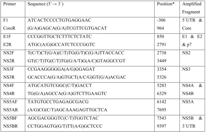
Table 3.1: Sequences of primers used for PCR amplification of
overlapping cDNA regions of the genome of HCV isolate
HCV-TR1 35
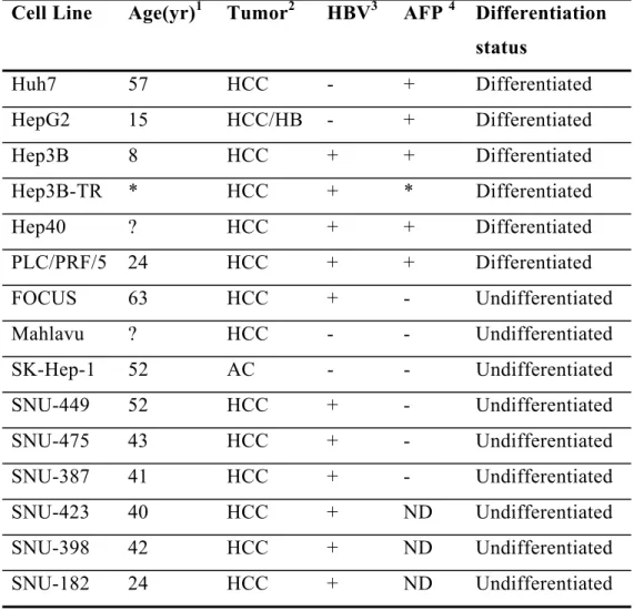
Table 3.2: The cell lines used in this thesis 37
Table 3.3: Effective range of separation of SDS-PAGE gels 42
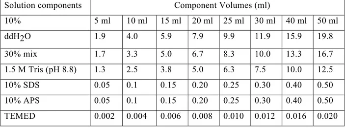
Table 3.4: Solution of preparing 10% resolving gel for Tris-glycine
SDS-PAGE 44
Table 3.5: Solution of preparing 5% stacking gels for Tris-glycine
SDS-PAGE 44
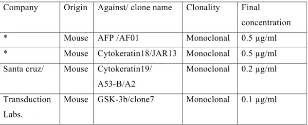
Table 3.6: The primary antibodies used in this thesis for the western blotting 46 Table 3.7: Primers Used In the semi-quantitative RT-PCR analysis of
hepatocyte nuclear factors 51
Table 4.1: Comparison of inferred amino acids at proteolytic cleavage
sites between HCV-TR1, HCV-J and HCV-BK 60
Table 4.2: Summary of amino acid differences between the HCV-TR1
Turkish isolate and other characterized HCV-1b genomes 61
Table 4.3: Comparisons of immunodominant Cytotoxic T cell epitopes
of HCV with corresponding amino acid residues in Turkish
isolate HCV-TR1 63
Table 4.4: Multilineage colony formation from AFP-producing human HCC
LIST OF FIGURES
NUMBER/NAME PAGE
Figure 1.1: Multistage process of carcinogenesis. 2
Figure 1.2: Schematic representation of the cell cycle and G1/S
controlling elements. 4
Figure 1.3: Genomic map of the Hepatitis C virus. 11
Figure 1.4: Schematic diagram of fetal liver development in the mouse. 25
Figure 1.5: Key stages in liver development. 26
Figure 3.1: Amplification strategy of HCV genome. 35
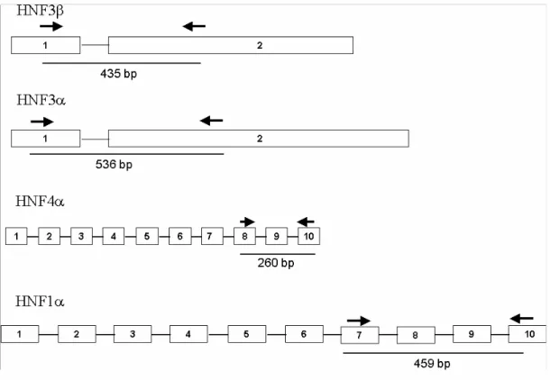
Figure 3.2: Schematic representation of the primers position on the
exonic structure of hepatocyte nuclear factors 3β, 3α, 1α
and 4α, respectively. 51
Figure 4.1: Pylogenetic tree of the 5`UTR sequences from 70 isolates
of HCV from Turkey. 54
Figure 4.2: Complete sequence of HCV-TR1. 60
Figure 4.3: Hepatic transcription factor and lineage marker expression
in liver cancer cell lines. 68
Figure 4.4: Heterogeneously staining cell types in AFP-producing human
HCC cell lines. 69
Figure 4.5: Multilineage colony formation from AFP-producing human
HCC cell lines. 72
Figure 4.6: Clonal expansion and self-renewal capability of Huh7 cells. 74
ABBREVIATIONS
ARF Alternative Reading Frame
BSA Bovine Serum Albumin
C Capsid
CDK Cyclin Dependent Kinase
C-terminus Carboxy terminus
DNA Deoxyribonucleic acid
E Envelope
ED Embriyonal day
HRP Horse Reddish Peroxidase
MDM2 Mouse Double Minute 2
MMLV Murine Maloney Leukemia Virus
NS Nonstructural
N-terminus Amino terminus
O/N Over Night
OD Optical Density
PAGE Polyacrylamide gel electrophoresis
PBS Phosphate Buffered Saline
PBS-T Phosphate Buffered Saline with Tween-20
PCR Polymearase chain reaction
pRb Retinoblastoma protein
RNA Ribonucleic acid
S/N Supernatant
SDS Sodium Dodecyl Sulfate
SDS-PAGE SDS- Polyacrylamide Gel Electrophoresis
TBS Tris Buffered Saline
TBS-T Tris Buffered Saline with Tween-20
TEMED N,N,N,N-tetramethyl-1,2 diaminoethane
Tris Tris (hydroxymethyl)-methylamine
CHAPTER 1. INTRODUCTION
1-1 HEPATOCELLULAR CARCINOMA
Hepatocellular carcinoma (HCC) accounts for 80-90% of liver cancers and is one of the most frequent carcinomas worldwide, with an estimated 564,000 new cases per year and almost as many deaths in 2000 (Parkin, 2001). In developing countries the incidence rates are two to threefold higher than in developed countries. The disease is more prevalent in parts of Africa and Asia than in continental America and Europe with a strong etiological association with viral hepatitis, hemochromatosis, known liver (hepatic) carcinogens, and toxins (aflatoxin).
Hepatocarcinogenesis is a slow process during which genomic changes progressively alter the hepatocellular phenotype to produce cellular intermediates that evolve into hepatocellular carcinoma. During the long preneoplastic stage, in which the liver is often the site of chronic hepatitis, cirrhosis, or both, hepatocyte cycling is accelerated by upregulation of mitogenic pathways. It is believed that this chronic regeneration process leads to the production of aberrant and dysplastic hepatocytes that have telomere erosion and telomerase re-expression, sometimes aberrant methylation or occasionally structural changes in genes and chromosomes. Development of dysplastic nodules and hepatocellular carcinoma are associated with the accumulation of irreversible structural alterations in genes and chromosomes, but the genomic basis of the malignant phenotype is heterogeneous. The malignant hepatocyte phenotype may be produced by the disruption of a number of genes that function in different regulatory pathways, producing several molecular variants of hepatocellular carcinoma (Thorgeirsson and Grisham, 2002).
1-1.1 Genetic Mechanisms of Hepatocarcinogenesis
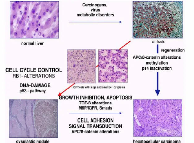
As with other kinds of cancer, the etiology and carcinogenesis of HCC are multifactorial and multistage. The multistep process of HCC may be divided into chronic liver injury that produces inflammation, cell death, cirrhosis and regeneration, dysplasia, and finally HCC (Figure 1.1).
Figure 1.1. Multistage process of carcinogenesis (Tannapfel and Wittekind, 2002)
It has been proposed that six essential alterations in normal cell physiology state progression of liver malignancy, including independence towards growth, anti growth and apoptotic signals, unlimited division, and angiogenetic and metastatic capacities (Ozturk and Cetin-Atalay, in press ). Mutations in critical genes may range from subtle sequence changes at a few nucleotides to gross chromosomal abnormalities including deletions, amplifications, and translocations of large DNA fragments.
Allelic imbalance and microsatellite instability
Most of the genes mutated in HCC are tumor suppressor genes, and frequent allelic loses (loss of heterozygosity, LOH) have been described. By comparative genomic hybridization, chromosomes 1q, 8q, and 17q show gene dose increase while chromosomes 1p, 4q, 8p, 9p, 13q, 16p, 16q, and 17p show gene dose loss. Frequent LOH, or more comprehensive, allelic imbalance (AI), is consistently observed on chromosomes 1p, 4q, 6p, 8p, 13q, 16q, and 17p by whole-genome allelotyping (Tannapfel and Wittekind, 2002). The chromosome regions with gene dose increase may contain critical oncogenes while those with gene dose loss may contain tumor-suppressor genes. For chromosomes 17p, 13q, 9p, 6q, and 16p, LOH could be related to p53, Retinoblastoma 1(RB1), p16, Insulin-like growth factor-2 receptor (IGF2R) and E-cadherin inactivation (Feitelson et al., 2002). In dysplastic nodules LOH has been observed with a prevalence of 50-80%. (Thorgeirsson and Grisham, 2002).
In HCC, chromosome 2 and 3 on which DNA mismatch repair genes are located are not frequently affected by allelic loses. But mutations in a mismatch repair gene known as Human Mut S homolog-2 (hMSH2) have been reported at about 30% of HCCs examined(Yano et al., 1999).
Cell cycle regulation
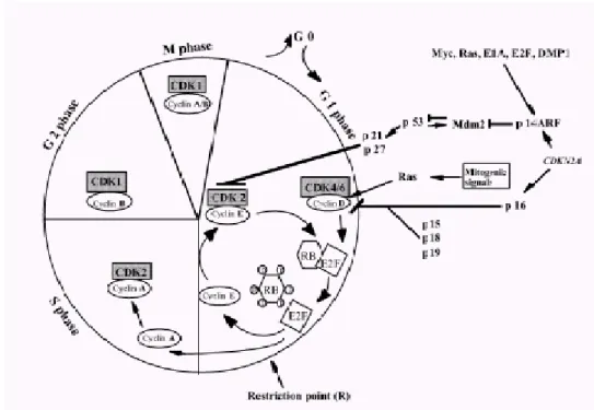
Cells respond to proliferative or antiproliferative signals through the cyclin D1-RB-CDK4/6 and the p14ARF/mdm2/p53 pathways. When quiescent cells in G0 are stimulated to enter cell cycle, genes encoding cyclin-D type cyclins are induced in response to mitogenic signals (Figure 1.2). These cyclins associate with either CDK4 or CDK6 subunits and the complex becomes activated by phosphorylation. Active cyclin/CDK complexes drive the cell cycle forward via phosphorylation of substrates such as Rb in early G1 phase (Weinberg, 1995). Rb is thereby inactivated, and its growth repressive functions abolished, resulting in release of a class of associated transcription factors known as E2Fs. Then “free” E2F transactivate cyclin E gene and promote DNA synthesis necessary for cell cycle progression. According to this, loss of Rb or its aberrant phosphorylation leads to a loss of growth control at the G1 phase. To maintain Rb protein in its active, anti-proliferative state, p16
(INK4a) inhibits the activity of CDK4 by specific binding thus preventing its association with cyclin D and/or blocking the catalytic activity of the kinase (Hirai et al., 1995). The p14ARF tumor suppressor, encoded by an alternative reading frame of the INK4a-ARF locus (9q21), senses "mitogenic current" flowing through the Rb pathway and is induced by abnormal growth promoting signals. By antagonizing Mdm2, a negative regulator of the p53 tumor suppressor, ARF triggers a p53-dependent transcriptional response that diverts incipient cancer cells to undergo growth arrest or apoptosis. Although ARF is not directly activated by signals that damage DNA, its loss not only dampens the p53 response to abnormal mitogenic signals but also renders tumor cells resistant to treatment by cytotoxic drugs and irradiation.
Figure 1.2 Schematic representation of the cell cycle and G1/S controlling elements
(Hashemi, 2002)
→ activation I inhibition
Taken together, disturbances in the p16-cyclin D-CDK4-Rb and p14-Mdm2-p53 pathways could be a main axis of genetic events in HCC because all players in these pathways seem to be altered in HCC. The Rb gene, one of the main player, is localized to chromosome 13q, which is a common deletion site for HCC and Rb
mutations are also observed in 15% of HCCs (Ozturk M., 1999). Moreover, Rb protein is a target for ubiquitin-dependent degradation and this degradation mechanism was shown to be dysregulated in HCCs by overexpression of a pRb specific ubiquitin ligase, gankyrin (Higashitsuji et al., 2000). Also, overexpression of cyclin D1 has been observed in about 10-13% of HCC cases (Ozturk, 1999). It has recently been shown in a transgenic mouse model that overexpression of cyclin D1 is sufficient to initiate hepatocellular carcinogenesis (Deane et al., 2001). The transduction of antisense cyclin D1 inhibits tumor growth in a xenograft hepatoma model. Correcting alterations that have occurred in the G1 phase regulatory machinery may therefore provide a novel weapon to treat and prevent HCC (Deane et al., 2001). Also, it was reported that about 50% of HCC displays de novo methylation of INK4a-ARF locus that encodes p16INK4 and p14ARF and LOH at the same locus was 20 % (Ozturk, 1999; Liew, 1999)
p53 and homologues
The protein product of p53 gene is activated by different stimuli such as oncogenic activation, DNA damage, decrease in nucleotide pools and oxidative stress and induces cell cycle arrest or apoptosis, depending on the cell context (Blagosklonny, 2002). p53 mutations are found in about 30% of HCC cases worldwide. Until now all reported mutations (mostly missense, leading to stabilization of protein) have been somatic, indicating that germline p53 mutations do not appear to be predisposition for HCC. Tumor-specific p53 mutations have been identified in several studies, linking the mutation pattern to suspected etiological factors. A selective guanine-to-thymine transversion mutation in codon 249 AGG to AGT (transversion italicized) leading to an arginine-to-serine substitution of the p53 gene has been identified as a "hotspot" mutation for HCC. Epidemiological and experimental evidence suggests that in HCC this mutation is strongly associated with exposure to aflatoxin B1 in combination with a high level of chronic hepatitis B virus infection in the population (Bressac et al., 1991; Hsu et al., 1991). Moreover, there is a strong correlation between p53 mutations, large tumor size, and poor differentiation state.
No specific mutations or interactions have yet been described for p73 and p63 which are homologs of p53. However, overexpression of p73 (wild type) has been described in a subset a HCC, indicating a poor prognosis in these patients (Tannapfel et al., 1999). More recently Sayan et al., identified that it is the transcriptionally active (TA) form of p73 which is upregulated in HCC, probably because of Rb pathway dysregulation (Sayan et al., 2001).
Wnt pathway: APC, β-catenin, axin 1, and E-cadherin
Somatic mutations of β-catenin have been observed in 19-26% of HCC cases, mostly missense mutations and interstitial deletions of exon 3 (Tannapfel and Wittekind, 2002). These mutations cause nuclear accumulation of aberrant β-catenin proteins that stimulates the activity of LEF-TCF family of transcription factors which in turn transactivate a series of cell cycle progression genes such as cyclin D and myc as shown in colorectal cancers (Calvisi et al., 2001). Axin, an important regulator of β-catenin, is mutated in about 10% of HCC cases, leading to an activation of the Wnt pathway. Axin1 and β-catenin mutations are mutually exclusive in HCC, suggesting that they affect the same (presumably Wnt) pathway (Morin et al., 1997; Satoh et al., 2000). It has recently been shown that ectopic expression of the wild-type axin gene (AXIN1) induces apoptosis in HCC cells, indicating that axin 1 may be an effective growth suppressor of hepatocytes (Satoh et al., 2000)
Somatic APC mutations are rare events in HCC, but it was recently reported that biallelic inactivation of the APC gene contributed to the development of HCC in a patient with familial adenomatous polyposis and a known germline mutation of the APC gene at codon 208 (Su et al., 2001). E-cadherin, a receptor in adherence junctions, which is essential both for maintenance of tissue structure and regulation of free cytoplasmic β-catenin level, is rarely mutated in HCC. However, loss of function due to LOH or de novo methylation occurs in about 30% of HCC cases (Tannapfel and Wittekind, 2002).
Alterations of the TGF-β /IGF-axis
Transforming growth factor (TGF) β initiates signaling through heteromeric complexes of transmembrane type I and type II serine/threonine kinase receptors. Activated TGF-β receptors phosphorylate receptor-regulated Smads which induces both inhibition and apoptosis in hepatocytes. Genetic alterations of the TGF-β pathway are mediated by mutations of the Smad2 and Smad4 gene, which occur in about 10% of HCC cases (Yakicier et al., 1999). Mutations of the TGF-β receptor (TGF-β1RII) gene itself are detected in patients with HCC and may also abrogate TGF-β signaling (Enomoto et al., 2001).
A potent activator of TGF-β is the mannose-6-phosphate/insulin-like growth factor 2 receptor (M6P/IGF2R) which suppresses cell growth through binding to the insulin-like growth factor (IGF) 2 and latent complex of TGF-β. The deregulation of the IGF axis, including the autocrine production of IGFs, IGF binding proteins (IGFBPs), IGFBP proteases, and the expression of the IGF receptors, has also been identified in the development of HCC. Also, both LOH and mutations of the M6P/IGF2R have been reported in about 30% HCC patients (Oka et al., 2002; De Souza et al., 1995; Piao et al., 1997).
PTEN/MMAC1/TEP1 (PTEN) tumor suppressor gene has recently been shown to block growth-stimulatory and survival signals mediated by PI-3 kinase and to converge the activation of protein kinase B/Akt. Alterations, mainly mutations but also LOH, of PTEN have been reported in about 27% of HCC cases (Kawamura et al., 1999). Recently it was demonstrated that PTEN significantly lowers IGF secretion and also expression of secretory and cellular vascular endothelial growth factor proteins in HCC cell lines and could therefore inhibit tumorigenicity (Kawamura et al., 1999).
1-1.2 Significance of Hepatitis B and C viruses in HCC
From the information released by the World Health Organization, it is estimated that the numbers infected with HBV and HCV worldwide are 180 and 300
millions, respectively. Together, the two viruses contribute to the etiology of about 80% of global HCC (World Health Organization., 2000). They contribute to hepatocarcinogenesis indirectly, by causing chronic necroinflammatory hepatic disease. They may also display direct hepatocarcinogenic activity.
Hepatitis B Virus (HBV) is a partially double-stranded DNA virus belonging to the Hepadnaviridae and approximately 25% of chronic carriers of the virus develop the tumor (Beasley and Hwang, 1984). Oncogenic mechanism of HBV infection may be simply defined as releasing the growth control of hepatocytes by coding for a factor that activates otherwise dormant genes or activates proto-oncogenes or silences anti-proto-oncogenes; by inserting its DNA sequences that can activate and influence the transcription of cellular genes; by causing chronic inflammation with cell death and hepatocyte regeneration with fibrosis; as well as by activation of the immune system liberating cytokines at the wrong time in the wrong place. Transcriptional activation of a wide range of viral, as well as cellular genes such as c-fos, c-myc, IGF2, insulin-like growth factor I receptor (IGFR1) and β-interferon, by HBV encoded X antigen (HBxAg) was shown in many studies (Caselmann, 1996; Colgrove et al., 1989; D'Arville et al., 1991; Kim et al., 1996; Twu and Schloemer, 1987). In chronic HBV infection, it has been shown that HBxAg binds and functionally inactivates the tumor suppressor p53 (Huo et al., 2001; Ueda et al., 1995) and the negative growth regulator p55sen (Feitelson, 1999; Ueda et al., 1995), both of which are involved in senescence-related pathways. The activation of the Rb tumor suppressor by hyperphosphorylation resulting in the activation of E2F1 has been reported in HBxAg positive HCC cells (Sirma et al., 1999). It has also been shown that HBxAg can down regulate the expression of translational factor, sui1, and cyclin dependent kinase inhibitor, p21WAF1/CIP1/SDI1(Feitelson et al., 1999; Sirma et al., 1999). As with HBxAg, carboxyterminal truncated middle hepatitis B surface protein (MHBSt) can activate various viral and cellular gene promoters (Caselmann et al., 1990; Kekule et al., 1990). Recent data suggests that HBxAg contributes to HCC development also by mechanisms other than transactivation. HBxAg binds to the X-associated protein 1 and possibly disturbs its function in nucleotide excision repair mechanism (Becker et al., 1998) and also it has been show that HBxAg stimulated cell growth is associated with constitutive activation of the ras/raf/MAPK and NKκ-B signal transduction
pathways (Benn and Schneider, 1994; Lucito and Schneider, 1992; Shirota et al., 2001).
Hepatitis C Virus is a more important causal association of the tumor than is HBV, and in Japan, Italy, and Spain the virus accounts for as much as 80% of HCC (Kew, 1998). It has been postulated that HCC largely develops indirectly as a result of the inflammatory responses that lead to hepatocyte destruction, regeneration and fibrosis. Since there is no evidence that HCV RNA is integrated into host genome as stated in HBV, the virus may play a more direct role in neoplastic transformation of hepatocytes. HCV proteins are shown to interact with various cellular proteins: 14-3-3 protein, apolipoprotein AII, Tumor necrosis factor (TNF) receptor, lymphotoxin-ß receptor, DEAD domain of RNA helicase, nuclear ribonucleoprotein K for core protein, double stranded RNA protein kinase (PKR) for E2 and NS5A, p53 for NS3 and possibly core, and SNARE-like protein for NS5A. (Ghosh et al., 1999; Ray and Ray, 2001; Shimotohno, 2000). Interactions with these cellular proteins which seem to be important for the function of HCV proteins in the regulation of cell proliferation will be summarized in 1.2.3.
Recently developed transgenic mice model for the viral hepatocarcinogenesis showed that HBx protein and HCV core protein may be capable of inducing HCC in the absence of a complete set of genetic aberrations which are necessary for a multistage development of all cancer types as proposed by Vogelstein. (Koike et al., 2002;Moriya et al., 1998)
1-2 HEPATITIS C VIRUS
Hepatitis C was first recognized as a separate disease entity in 1975 when the majority of cases of transfusion-associated hepatitis were found not to be caused by the only two hepatitis viruses recognized at the time, hepatitis A virus and hepatitis B virus. The disease was called "non-A non-B hepatitis," and it was demonstrated to be
sequencing of the viral genome of the non-A non-B hepatitis virus was first reported and the virus was renamed "hepatitis C virus" (Choo et al.,1991).
Hepatitis C virus is a positive strand RNA virus of Filaviviridae, genus hepacivirus, approximately 9.6 kb in length, and is most closely related to the pestiviruses.
1-2.1 Epidemiology
Hepatitis C is a major cause of acute hepatitis and chronic liver disease, including cirrhosis and liver cancer. An estimated 170 millions of persons are chronically infected with HCV worldwide and 3-4 million persons are newly infected each year (World Health Organization, 2000).
HCV infection appears to be endemic in most parts of the world, with an estimated overall prevalence of 3%. However, there is considerable geographic and temporal variation in the incidence and prevalence of HCV infection. In United States, 1.8% of population is infected with HCV (Alter, 1997) while the HCV prevalence rate is in between 0.2-3% in Europe (Memon and Memon, 2002). In Turkey the prevalence of HCV is % 0.3-1.8(Sharara et al., 1996).
There are a few countries with very high HCV prevalence rate, such as Egypt, where 10-30% of population is infected with HCV (Arthur et al., 1997; el-Sayed et al., 1996). The nationwide campaing to treat schistosomiasis infections by inoculation of needles in 1970s is hypothesized to be responsible for this high prevalence, as lower HCV prevalence among individuals born after the end of campaign was observed as a preliminary evidence (el-Zayadi et al., 1997).
1-2.2 Genomic Organization of HCV
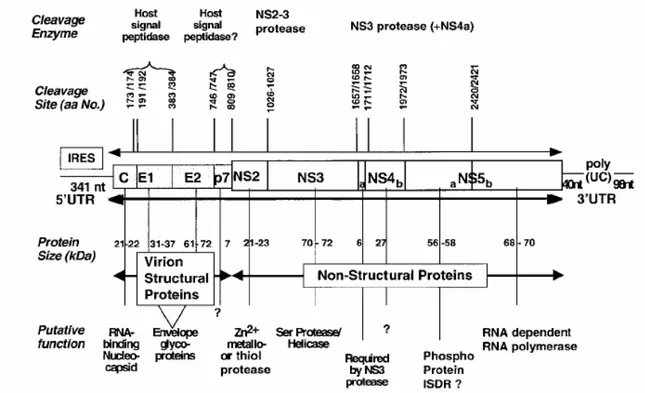
The genome of HCV is a single-strand linear RNA of positive sense. The functional and structural units of the HCV genome are schematically depicted in
Figure1.3. A 5' untranslated (UTR) region consists of approximately 340 nucleotides, which has stem-loop structure and contains an apparent internal ribosomal entry site (IRES). Immediately downstream is a single large open reading frame (ORF) of approximately 9,600 nucleotides, encoding a large polyprotein precursor of approximately 3,200 amino acids that is cotranslationally or posttranslationally cleaved into separate proteins by a combination of host and viral proteases. The genomic order of HCV has been shown to be C-E1-E2-p7-NS2-NS3-NS4A-NS4B-NS5A-NS5B. Capsid protein (C), two envelope proteins (E1 and E2) are the virion structural proteins. The function of p7 is currently unknown. These proteins have been shown to arise from the viral polyprotein via proteolytic processing by the host signal peptidases.
Figure 1.3 Genomic map of the Hepatitis C virus (Rosenberg, 2001).
Generation of the mature nonstructural protein, NS2 to NS5B, relies on the activity of viral proteinases. Cleavage at the NS2/NS3 junction is accomplished by metal-dependent autocatalytic proteinases encoded within NS2 and the N-terminus of NS3. The remaining cleavages downstream from this site are affected by a serine proteinase also contained within the N-terminal region of NS3. NS3 also contains an
RNA helicase domain at its C-terminus. NS3 forms a heterodimeric complex with NS4A. The latter is a membrane protein that has been shown to act as a cofactor of the proteinase. While no function has yet been attributed to NS4B, it has been suggested that NS5A is involved in mediating the resistance of the HCV to interferon. And the NS5B protein has been shown to be the viral RNA-dependent RNA polymerase. Finally, there is a 3' UTR region that consists of approximately 40 nucleotides, a polypyrimidine track and a highly conserved terminal sequence of approximately 90 nucleotides.
1-2.3 Genetic Heterogeneity and Classification systems
After the complete HCV genome was determined by Choo et al. (1991), several HCV isolates from different parts of the world were obtained and sequenced. Comparison of the published sequences of HCV has led to the identification of several distinct types that may differ from each other by as much as 33% over the whole viral genome (Okamoto et al., 1992). Sequence variability is distributed equally throughout the viral genome, apart from the highly conserved 5` UTR and core regions and the hypervariable envelope (E) region that is the most heterogeneous portion of the genome.
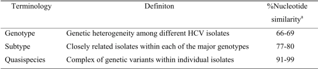
As different investigators developed and used their own classification for HCV strains, a confusing literature developed by Okamoto, Simmonds, Enomato, Choo. However, at the 2nd International Conference of HCV and Related Viruses, a consensus nomenclature system was proposed to be used in future studies of HCV genotypes and subtypes (Simmonds et al., 1994). According to this system, HCV is classified on the basis of the similarity of nucleotide sequence into major genetic groups designated genotypes. HCV genotypes are numbered (arabic numericals) in the order of their discovery. The more closely related HCV strains within types are designated subtypes, which are assigned lowercase letters (in alphabetic order) in the order of their discovery. The complex of genetic variants found within an individual isolate is termed the quasispecies (Table 1.1).
The heterogeneity of HCV within an individual can be attributed to error prone RNA-dependent RNA polymerase (RdRp) of HCV, NS5B. The absence of proof reading activity of NS5B creates de novo mutations. Recently, HCV RNA turnover rate is calculated in humans as 4-7 hours and this high turnover rate leads to production of 300 billion HCV RNA molecules per day (Neumann et al., 1998), which contributes significantly to the heterogeneity of HCV. It was hypothesized that the existence of HCV as quasispecies may be the basis of the mechanism for viral persistence. With high numbers of RNA produced per day and NS5B without proofreading activity would lead to production of escape mutants, therefore, HCV establishes chronic infection.
According to this classification, there are 6 main genotype and approximately 70 subtypes and some of the recently identified isolates from Vietnam, Thailand and Indonesia were identified as subtypes of HCV genotype 6.
TABLE 1.1. Terminology commonly used in studies related to HCV genomic heterogeneity
Terminology Definiton %Nucleotide similaritya
Genotype Genetic heterogeneity among different HCV isolates 66-69 Subtype Closely related isolates within each of the major genotypes 77-80 Quasispecies Complex of genetic variants within individual isolates 91-99
a % Nucleotide similarity refers to the nucleotide sequence identities of the full-length sequence of the
HCV genome
1-2.4 Importance of HCV genotypes on disease progression and treatment
The role of HCV genotypes in the progression of liver disease is one of the most controversial areas of HCV research. Several studies showed that subtype 1b infections proceed much faster to severe forms of chronic hepatitis, cirrhosis (Watson et al., 1996) and hepatocellular carcinoma (Silini et al., 1996; Zein et al., 1996). However, there are also reports that failed to show the association between subtype 1b infections and faster progression into cirrhosis (Mita et al., 1994) or HCC
(Lee et al., 1996; Yotsuyanagi et al., 1995). Moreover, in the patients with chronic HCV, infection with genotype 1b is reportedly associated with a more severe liver disease and a more aggressive course than is infection with other HCV genotypes (Nousbaum et al., 1995; Pozzato et al., 1995). Similar to these studies (Silini et al., 1995; Zein et al., 1995), HCV genotype 1b was significantly more prevalent among patients with liver cirrhosis and those with decompensated liver disease requiring liver transplantation than among those with chronic active hepatitis C (Zein et al., 1995; Belli et al., 1996) and was associated with earlier recurrence and more severe hepatitis than other genotypes in liver transplant recipients (Pageaux et al., 1997). Although these are indirect evidence, they suggest an association between HCV genotype 1b and the development of these complications. Furthermore, HCV genotype 1b was shown to be present in most of the patients in approximately 60 to 70 % with HCV associated hepatocellular carcinoma (Zein et al., 1996; Reid, 1994). However, some reports refute the associations related with HCV subtype 1b, which was mentioned above (Benvegnu et al., 1997;Brechot, 1997; Naoumov et al., 1997; Yamada et al., 1994). Since the patients infected HCV 1b generally were older than those infected with other genotypes and genotype 1b may have been present before the other genotypes, whether HCV genotype 1b is a marker for severe HCV-associated liver disease remains unclear, because it may have been a reflection of a longer time of infection rather than a more aggressive form of hepatitis C.
Enomoto et al by comparing full length genome sequences of HCV isolates obtained from different Japanese patients, identified a 39 amino acid long region (2209-2248) in carboxyl terminus of N55A, which is associated with sensitivity to interferon and referred as interferon sensitivity determining region (ISDR) (Enomoto et al., 1996). Other groups working with Japanese patients infected with genotype 1b, 2a, or 2b HCV strains confirmed that the IFN-α resistant strains had same sequences with HCV- 1b prototype strains in the ISDR region (Chayama et al., 1997; Kurosaki et al., 1997; Halfon et al., 2000). However, the correlation is substantially weaker or lacking in patients infected with genotype 1a HCV strains or European patients infected with HCV strains of 1b, 2b, or 3a (Casato et al., 1997; Pawlotsky et al., 1998). The reason for these discrepancies are not clear, but differences in the interferon doses of these patients and the lower mutation rate of the ISDR in European HCV-1b patients relative to their Japanese counterparts are likely to be
contributing factors. In summary, ISDR sequences may affect the response of the individual HCV isolates to interferon treatment.
1-2.5 Geographic Distribution of HCV Genotypes
At least six major genotypes of HCV, each comprising multiple subtypes, have been identified worldwide. The geographical distribution of different genotypes and subtypes differs greatly from one region to the other. The reasons of this differential distribution are ill known, but the profile of geographical distribution could reflect the different modes of viral transmission as well as the host immune response variations. For example, HCV 1a subtype, which is seen frequently in North America, could have been transmitted to other regions of the world, especially to Europe, by contaminated blood-derived products (Brechot et al., 1998). In contrast, subtype 1b appears to be dominant in Japan and Southern Europe. In Europe, HCV 1 is the major genotype and there is a south-north gradient for 1a and 1b subtypes, the prevalence of subtype 1b being increasingly higher in southern Europe. The data on HCV subtypes in the Middle Eastern countries is limited. In Egypt, Hepatitis C is an endemic disease that is associated with genotype 4, almost exclusively (Ray et al., 2000). Similarly, genotype 4 is also predominant in the Gaza region, but not in Israel where subtype 1b is predominant (Shemer-Avni et al., 1998). Moreover, genotypes 5 and 6 seem to be confined to South Africa and Hong Kong, respectively. HCV genotype 7, 8, and 9 have been identified only in the Vietnamese patients, and genotypes 10 and 11 were identified in patients from Indonesia.
The HCV genotype distribution of patients living in Turkey is not well known. To our knowledge, there are only two published reports concerning Turkish patients, which indicated a high frequency (75-87 %) of subtype 1b (Abacioglu et al., 1995; Simsek et al., 1996). Regarding the distribution of HCV genotypes in neighbors of Turkey, subtype 4a and 1b are predominant in Syria and there is no data for Iraq and Iran (Abdulkarim et al., 1998). On the other hand, in countries located on the northern frontiers of Turkey as well as in Greece, subtype 1b appears to be the dominant form (Viazov et al., 1997; Andonov et al., 1996).
The geographical distribution and diversity of HCV genotypes may provide clues about the historical origin of HCV. The presence of numerous subtypes of each HCV genotype in some regions of the world, such as Africa and Southern Asia, may suggest that HCV has been endemic for a long time. Conversely, the limited diversity of subtypes observed in United States and Europe could be related to the recent introduction of these viruses from areas of endemic infection.
1-2.6 Methods for HCV Genotyping
Molecular genotyping
Because differences in geographical distribution, disease outcome, and response to therapy among HCV genotypes have been suggested, reliable methods for determining the HCV genotype may become an important clinical test. In theory, the most accurate method for HCV genotyping is the sequencing of whole genome and phylogenetic tree construction. However, in practice this method is not appropriate and feasible in genotyping large numbers of samples as full-length genome sequencing is too laborious and expensive. Hence, subgenomic regions representative of whole genome have been investigated. To this date, phylogenetic analysis E1, NS5B, and 5’UTR sequences correlated with whole genome based phylogenetic analysis suggesting the equivalence of sequence relationships between HCV genotypes in different regions of genome. This finding formed the basis of the proposal by Simmonds et al. that a new HCV genotype should be assigned only if phylogenetic analysis of at least two genomic regions showed a distinct phylogenetic branch (Simmonds et al., 1994). 5’UTR region is the most conserved region of HCV genome. Therefore, it may have identical sequence in different subtypes but contains variations aiding determination of 6 genotypes and some subtypes.
Amplification of NS5B, core, E1 regions, followed by sequence comparison and phylogenetic tree construction for confirmation, is currently considered “gold-standard” for the assignment of HCV genotypes. However, there are limitations of this method. Amplification of some genomic regions may not be efficient. In this case, use of different sets of primers would be needed, which may lead to the selection of certain genotypes or quasispecies. In a large-scale genotyping study,
mixed infections would not be identified. In these studies, the cost of the study and labor-intensive nature of this technique also limits to its usage.
Other methods that depend mainly on the amplification of HCV RNA followed by either reamplification with type specific primers or hybridization with type-specific probes or by digestion of PCR products with restriction endonucleases that recognize genospecific cleavage site. HCV genotyping by using type-specific primers was first introduced by Okamoto et al. and used primer type-specific for the core region (Okamoto et al., 1992a). This method lacked sensitivity and specificity and was only able to detect subtypes 1a, 1b, 1c, 2a, 2b, and 3a. New variations of this method from both core and NS5B region have been developed to increase genotyping of more types or subtypes (Ohno et al., 1997). A commercial kit (InnoLipa) for HCV genotyping has been introduced in Europe and is based on hybridization of 5`UTR amplification products with genotype specific probes. Although initial version of InnoLipa had lower sensitivity, the newer version is capable of discriminating among HCV subtypes 1a, 1b, 2a to 2c, 3a to 3c, 4a to 4h, 5a, and 6a.
Restriction Fragment Length Polymorphism analysis of HCV genome for genotyping was initially reported by Nokao et al. (1991). Subtype or type-specific restriction enzyme recognition sites present in amplified PCR products are utilized to cut PCR fragments with different restriction endonucleases. Electrophoresis profiles of these are used to identify the genotype of the sequence. In the RFLP analysis, 5’UTR or NS5B region has been used widely with variations.
Although all these methods are able to identify correctly the major genotypic groups, only direct nucleotide sequencing is efficient in discriminating among subtypes. Moreover, all of these PCR-based methods have the shortcomings and advantages of PCR. They are expensive and time-consuming and require specialized facilities to ensure accurate results and prevent contamination. The advantages of PCR-based methods include reliability if performed accurately and the ability to obtain information relevant to the molecular pathogenesis of HCV.
Serological genotyping
Serological genotyping has several advantages that make it suitable for large epidemiological studies, especially. These advantages include the low risk for contamination and the simplicity of the assay. However, it seems to lack specificity and sensitivity, which limits its usefulness.
Two commercially available serological genotyping assays have been introduced over the past 3 to 4 years. The RIBA SIA was introduced by Chiron Corp. and contained five different serotype-specific peptide sequences taken from the NS4 region and two taken from the core region of the HCV genomes for genotype 1, 2, and 3. The second serological genotyping assay is the Murex HCV serotyping enzyme immune assay, which is based on the detection of genotype-specific antibodies, directed to epitopes encoded by the NS4 region of the genomes for genotypes 1 through 6.
1-2.7 Role of Genomic Heterogeneity in HCV Persistence and Vaccine Development
With the rarity of severe acute or fulminant HCV infections, the significance of this infection in humans is its tendency to become persistent and to induce chronic liver disease. The mechanisms whereby HCV circumvents the immune response, persists and causes chronic inflammatory liver disease are currently undefined.
One hypothetical explanation of HCV persistence, sequence variation due to the quasispecies nature and the high mutation rate of HCV, has often been discussed. Amino acid changes in immunodominant epitopes may permit HCV to escape from the antiviral immune response. The most convincing evidence of this phenomenon is the lack of the immune protection and the infectibility of chimpanzees rechallenged with the same HCV inoculum (Prince et al., 1992). In addition to the lack of protection by the humoral immune response, there is also evidence that the cellular immune response may be subverted during HCV infection, since subsequent experiments Weiner et al., 1995 have described the emergence of an HCV mutant that was able to escape the HCV-specific CTL response in an infected chimpanzee.
Possible targets for HCV-specific CTL recognition within the conserved core protein and additional epitopes in the more highly variable region E2 protein were also identified (Koziel et al., 1993). In a chronically infected chimpanzee, CTLs obtained from the liver were initially able to recognize an epitope in the NS3 protein. Over a period of years, a new strain of the virus emerged with a mutation in the CTL epitope that was no longer recognized by the CTLs isolated earlier. Although direct evidence for the presence of CTL escape mutants in human HCV infection is lacking, it has been shown that single-amino-acid changes in CTL epitopes result in failure of recognition by HCV-specific CTLs (Koziel et al., 1998). These single-amino-acid changes are found in natural isolates of HCV, hence the need to address the problem of type specificity of immune responses.
1-2.8 Molecular mechanisms of HCV related hepatocarcinogenesis
Role of Hepatitis C virus proteins in the modulation of proliferation
Core protein of HCV has been shown to play various roles in the regulation of cell proliferation including activation of the Ras/Raf kinase cascade, regulation of p53 function, modulation of apoptosis, oncogenic functions in transgenic mice and certain cell lines, and transactivation or transexpression of certain cellular genes as well as HBV and HIV genes. Recently, the envelope protein of E2 was shown to suppress RNA-activated protein kinase (PKR) function, which is important to disrupt viral gene expression, by acting as a decoy of elF2-α. Also, NS3 protein of HCV interacts with p53 and has the ability to transform NIH3T3 cells. Finally, NS5A protein interacts with SNARE-like protein, which may be important for membrane fusion (Shimotohno K. 2000).
Anti-apoptotic function of HCV core protein
Core protein of HCV plays a role in the suppression of caspase activation induced by anti-Fas and TNF-α in some of the cell lines, and that the suppression was likely to be achieved upstream of caspase-8 in the caspase cascade. It also binds to the death domain of tumor necrosis factor receptor 1 (TNFR1) and the cytoplasmic
tail of lymphotoxin-β receptor, implying that it may be involved in anti-apoptotic signaling pathways (Zhu et al., 1998). Finally, hepatitis C virus core protein activates the cellular transcriptional factor, NF-κB and it may be another mechanism to suppress apoptotic mechanisms (Tai et al., 2000).
Possible roles of HCV proteins in liver dysfunction
When HCV-specific T cells migrate into hepatocytes and recognize the viral antigen via T-cell receptor, they become activated and express Fas ligand that can transduce the apoptotic death signal to Fas-bearing hepatocytes. The cells in which NF-κB is activated by core protein may escape from Fas-mediated apoptosis and contribute to virus replication and release of virus particles. Persistent infection with HCV may be explained by such a mechanism, in addition to insufficient activation of CTL to clear HCV-infected cells. Core protein also has the potential to activate the MAK kinase cascade, which may have a mitogenic effect. The liver undergoes persistent regeneration following hepatic injury and growth factors stimulate this liver regeneration in hepatitis. It is possible that HCV core protein, in regenerating hepatocytes, enhances growth stimuli and repeated hepatocyte proliferation may cause disorder of the gene in the hepatocytes, thus causing hepatocellular carcinoma.
In conclusion several HCV proteins have been shown to interfere with the function of cellular proteins involved in malignant transformation of hepatocytes. However, currently there is no experimental model for HCV infection of hepatocytes, in vitro or in vivo. Therefore, it is unknown whether such virus-cell protein interactions also occur during HCV infection. This is why, it is not possible at present time to classify HCV as an oncogenic virus. It is also important to pay attention to the fact that molecular and cellular events involved in the initiations of human HCC (either viral or nonviral) are not known at all. Although we know several genes that are mutated in HCC, we do not know whether and how they contribute to the initiation of hepatic neoplasia.
1-3 LIVER CANCER STEM CELL
One of the most debated issues in HCC, is its origin. Dedifferentiation of mature hepatocytes was proposed by some authors as a main cause of HCC, although others believe that HCC results from incomplete differentiation of hepatic stem cells. We will first describe “stem cell and cancer stem cell” concepts.
1-3.1 Stem Cell, Cancer Stem Cells
Potten and Loeffler define stem cells as undifferentiated cells capable of (a) proliferation, (b) self-maintenance, (c) the production of a large number of differentiated, functional progeny, (d) regenerating the tissue after injury, and (e) flexibility in the use of these options, but make a distinction between actual and potential stem cells- the latter being cells possessing, but not expressing, all these capabilities (Potten and Loeffler, 1990).
Recently, it was suggested that tumours might contain “cancer stem cells”- rare cells with indefinite proliferative potential that drive the formation and growth of tumours. For example; it was shown that lymphoblastic, acute and chronic myeloid leukemias originate from haematopoietic stem cells (HSCs) (Bonnet and Dick, 1997; Mauro and Druker, 2001; George, 2001). Moreover, there are many similarities in the mechanisms that regulate self-renewal of HSCs and cancer cells. For instance, the prevention of apoptosis by enforced expression of the oncogene bcl-2 results in increased numbers of HSCs in vivo, suggesting that cell death has a role in regulating the homeostasis of HSCs (Domen and Weissman, 2000). Some other pathways associated with oncogenesis such as the Notch, Sonic hedgehog (Shh) and Wnt signaling pathways, may also regulate stem cell self renewal (Taipale and Beachy, 2001). Furthermore, it is well documented that many types of tumours contain cancer cells with heterogenous phenotypes reflecting aspects of the differentiation that normally occurs in the tissues from which the tumours arise. The variable expression of normal differentiation markers by cancer cells in tumour suggests that some of the heterogeneity in tumours arises as a result of the anomalous differentiation of tumour cells. Examples of this include the variable expression of
myeloid markers in chronic myeloid leukaemia, the variable expression of neuronal markers within peripherial neurectodermal tumours, and the variable expression of milk proteins or the oestrogen receptor within breast cancer. In other words, both normal stem cells and tumorigenic cells give rise to phenotypically heterogeneous cells that exhibit various degrees of differentiation. Tumorigenic cells can be thought of as cancer stem cells that undergo an aberrant and poorly regulated process of organogenesis analogous to what normal stem cell do (Sell and Pierce, 1994).
1-3.2 Origin of Hepatocellular carcinoma
As stated earlier, one of the most debated issues concerning HCC is its
cellular origin. Various rodent models of chemically induced liver tumors are currently used to clarify this issue.
According to the conventional theory, HCC results from “dedifferentiation” of mature hepatocytes into a less differentiated statewithout the involment of other cell types (Aterman, 1992). According to this model, cancer results from a multistep process. Chemical carcinogens act by modifying DNA of mature hepatocytes to form DNA adducts during the first step of initiation. In the second step of carcinogenesis, often referred to as promotion, other noncarcinogenic compounds increase hepatocarcinogenic effects, most of them being able to induce cellular proliferation. Arguments of this hypothesis are the findings that exposure of rats to carcinogens results in sequential liver alterations as follows; (a) focal proliferation of altered hepatocytes, (b) appearance of preneoplastic nodules, and (c) rise of cancer from persistent nodules (Farber and Sarma, 1987).
A “stem cell origin” has been proposed as an alternative mechanism (Sell and Dunsford, 1989, Sell, 2001). According to this theory, HCC originates from “oval” cells displaying stem cell-like properties that are detected in the liver prior to the chemical induction of HCC in rats. First, Solt and Faber showed that administration of 2-acetylaminofluorene (2-AAF) together with partial hepatectomy results in suppression of hepatocyte proliferation and stimulation of oval cell proliferation (Solt et al., 1977) Later, oval cells have been observed in various
models of rodent experimental carcinogenesis, including exposure to DIPIN (Factor and Radaeva, 1993) and choline-deficient, ethionine-supplemented (CDE) diet (Shinozuka et al., 1978). Oval cells are small, oval shaped epithelial cells identified in the liver during normal embryonic development (Tian et al., 1997) and in some of the adult liver pathologies, such as hemochromatosis and alcohol liver disease (Lowes et al., 1999). Moreover they arise in the periportal region of the liver probably derived from cells of Hering canal, bile ducts, intraportal or periportal ductules, or from periductular cells (Paku et al., 2001; Sarraf et al., 1994; Sell and Salman, 1984; Vessey and Hall, 2001)
Recently, it has also been suggested that oval cells may derive from bone marrow cells (Peterson et al., 1999). Evidence suggests that the liver oval cell is at least bipotential, capable of differentiating into mature hepatocytes (Evarts et al., 1989, Coleman et al., 1993) or cholangiocytes (Germain et al., 1988a; Germain et al., 1988b; Lenzi et al., 1992) under different experimental regimens. Progenitor cells in the liver are identified by their ability to express markers characteristic of immature hepatocytes. Such markers include albumin, transferin, alpha fetoprotein (AFP), and cytokeratin19 (CK19) all of which are expressed by the first primitive liver stem cells as well as oval cells (Cascio and Zaret, 1991).
The stem cell theory is not readily applicable to human HCC that occurs mostly as a virally induced disease. Consequently, the “dedifferentiation” is still considered as the major mechanism of HCC development in man (Kojiro, 2002). Although “oval” cell-like structures have been described in association with different liver diseases(Hsia et al., 1994; Roskams et al., 1998; Lowes et al., 1999;Theise et al., 1999; Libbrecht et al., 2001b), these descriptive observations do not provide convincing evidence for a direct contribution of liver stem cells to HCC. Moreover, in the absence of isolated human liver stem/progenitor cells, their occurrence remains hypothetical, although bone morrow-derived putative stem cells appear to generate hepatocytes, and bile duct cells (Alison et al., 2000; Theise et al., 2000).
1-3.3 Stem Cells during liver embryonic development
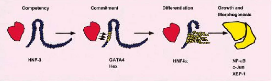
Hepatogenesis can be considered in distinct developmental stages:
competence, commitment, differentiation and morphogenesis. Although these stages reflect key aspects of hepatic development it is worth bearing in mind that they are closely related, interdependent, and often overlapping processes.
From Endoderm to Liver; Determination, Commitment, and Differentiation
This process begins on embryonal day (ED) 8.5 in the mouse with proliferation of undifferentiated endodermal cells of the ventral foregut and their migration into the septum transversum, where they come into contact with mesenchymal cells (Figure 1.4).
At this point, they are already specified to enter the liver lineage (determination) and form the hepatic diverticulum (Zaret, 2001). Mouse foregut-derived cells begin to express AFP at ED9.0 and then albumin at ∼ED9.5, followed by placental alkaline phosphatase and intermediate filament proteins, cytokeratins 14, 8 and 18 (Cascio and Zaret, 1991). Fibroblast growth factors (FGFs), particularly FGFs 1 and 2, can cause cardiac mesenchyme in inducing albumin gene expression (Jung et al., 1999). The morphology of the cells then changes to that of the hepatoblast (an early progenitor or stem/progenitor cell), expressing δ-glutamyl transpeptidase, α-antitrypsin, glutathione-S-transferase and fetal isoforms of aldolase, lactic dehyrogenase and pyruvate kinase in addition to AFP and albumin (Fausto, 1990; Brill et al., 1995; Thorgeirsson, 1996). The cells proliferate rapidly between ED12 and ED16 and subsequently diverge along two distinct lineages, the hepatocyte and cholangiocyte, beginning just prior to ED16. This time period has been referred to as a differentiation window in hepatic development (Marceau et al., 1992). In cell culture prior to ED16, fetal liver epithelial cells (early progenitor cells) appear to have the ability to change their phenotypic gene expression pattern from hepatocytic to ductular or reverse, depending on the environmental (cell culture) conditions (Marceau et al., 1992; Blouin et al., 1995). However, after ED16, the cells are committed to progress along one or the other of these lineages and no longer
retain their bipotential properties, although they continue to proliferate (i.e. “committed” or late progenitor cells). However, the irreversible nature of these changes no longer holds, as even phenotypically fully mature cells isolated from the adult liver appear to switch their phenotype in culture between hepatocytic and ductal, depending on experimental conditions (Michalopoulos et al., 2001).
Figure 1.4 Schematic diagram of fetal liver development in the mouse (Shafritz and Dabeva, 2002)
After commitment, hepatocyte progenitor cells express antigens HBD-1, H-2, transferrin receptor, c-CAM and HES6 in addition to the proteins mentioned above and biliary epithelial cell progenitors also express CK7 and 19, OC-2, OV-6 and BD-1 (Shiojiri et al., BD-199BD-1; Fausto, BD-1990; Thorgeirsson, BD-1996; Marceau et al., BD-1992; Blouin et al., 1995). As organogenesis proceeds, intrahepatic bile ducts are formed in the vicinity of large portal vein branches, beginning on ED17. This is evidenced by formation of ductular structures containing CK19 positive epithelial cells that have the appearance of “strings of pearls”. The basic lobular structure is then formed, although the hepatic parenchymal plates or cords do not become fully mature until several weeks after birth.
Transcriptional Regulation of Liver Development
Although the different steps involved in liver development are well described from the hepatic specification of ventral endoderm to the generation of hepatoblasts and the differentiation of mature hepatocytes, the control mechanisms underlying these steps are still poorly understood. Recent experiments with primary tissue explants of foregut endoderm have suggested the influence of positive and negative extracellular signals, respectively provided by cardiac and dorsal mesoderm, during early hepatic specification (Gualdi et al., 1996). Gene disruption in mice has demonstrated those factors as diverse as c-jun (Hilberg et al., 1993), RelA (Beg et al., 1995), and both hepatocyte growth factor/scatter factor (HGF/SF; (Schmidt et al., 1995) and its receptor Met (Bladt et al., 1995). It is generally accepted that particular combinations of members of four families of liver-enriched transcription factors (LEFT), including hepatocyte nuclear factor (HNF)3, HNF4, HNF1, and CCAAT/enhancer binding protein (C/EBP) control critical steps in hepatic differentiation, and that a hierarchy of expression of these transcription factors exists (Cereghini, 1996)(Figure 1.4)
Figure 1.5 Key stages in liver development (Duncan, 2000)
The HNF3 family of transcription factors has important roles in determining hepatic competency of endoderm. The initial expression of hnf-3 occurs before the onset of hepatic development with hnf-3β preceding to that of hnf-3α in mouse. HNF3 proteins share a highly conserved novel DNA binding domain and, along with the Drosophila forkhead protein, are the founding members of the winged helix family of transcription factors. HNF3 remodels the configuration of chromatin around transcriptional regulatory regions containing hnf-3 binding sites. It can interact with its DNA binding element within the context of chromatin. Upon
binding, HNF3 acts to disrupt linker histone binding and repositions nucleosomes around the regulatory region. This enhances the accessibility of other transcription factors such as GATA4 which acts cooperatively with HNF3 to regulate gene expression (Cirillo and Zaret, 1999) and Hex (hematopoietically expressed homeobox) that is also essential for the earliest stages of liver development (Martinez Barbera et al., 2000) then commitment of endoderm to a hepatic fate has started by the expression of AFP and Albumin.
Once the pre-hepatic cells become committed to the liver-cell fate, the process of differentiation starts. HNF4α, which is required for complete differentiation of hepatocyte, binds DNA strictly as a homodimer. Expression of HNF4α closely correlates with the expression of the transcription factor HNF1α (Bulla, 1997). HNF4α and 1α combination has a key role in the expression of diverse sets of genes that control multiple aspects of metabolism, detoxification, and serum factor secretion in normal liver physiology (Spath and Weiss, 1997).
1-3.4 Identification of Stem-Like Cells in Animal and Mouse Models
In response to the loss in the hepatic mass after partial hepatosectomy, hepatocytes readily proliferate to reconstitute the liver. However, when proliferation of hepatocytes was inhibited, a novel cell population proliferates in the liver; these cells are collectively termed “oval cells”. It is suggested that the oval cell compartment contains putative hepatic stem cells and/or their partially committed progeny (Sell and Pierce, 1994;Fausto, 1994).
When the hepatic cells were labeled in vivo using retroviral vectors to follow their fate during early stages of chemical induced hepatocarcinogenesis in rats, it has been demonstrated that preneoplastic foci can originate from mature hepatocytes as similar as what dedifferentiation theory tells about the origin of HCC. However, workers still can not exclude the possibility that oval cells may contribute to the generation of foci either directly or by differentiation of oval cells to hepatocytes that will subsequently dedifferentiate and give rise to foci (Gournay et al., 2002)