journal homepage:www.elsevier.com/locate/bioorg
Synthesis of novel methyl jasmonate derivatives and evaluation of their
biological activity in various cancer cell lines
Bilgesu Onur Sucu
a,b, Ozgecan Savlug Ipek
b,c, Sukran Ozdatli Kurtulus
b, Busra Emine Yazici
b,d,
Nihal Karakas
b,d, Mustafa Guzel
b,e,⁎aIstanbul Medipol University, Vocational School of Health Services, Pharmacy Services, Kavacik Campus, Kavacik-Beykoz, Istanbul 34810, Turkey bIstanbul Medipol University, Regenerative and Restorative Medicine Research Center (REMER), Kavacik Campus, Kavacik-Beykoz, Istanbul 34810, Turkey cYildiz Technical University, Graduate School of Natural and Applied Sciences, Department of Chemistry, Besiktas, Istanbul 34349, Turkey
dIstanbul Medipol University, School of Medicine, Department of Medical Biology, Kavacik Campus, Kavacik-Beykoz, Istanbul 34810, Turkey
eIstanbul Medipol University, International School of Medicine, Department of Medical Pharmacology, Kavacik Campus, Kavacik-Beykoz, Istanbul 34810, Turkey
A R T I C L E I N F O Keywords: Cancer therapy Aerobic glycolysis Warburg effect Hexokinase-II inhibition Methyl jasmonate
Novel drug discovery and development
A B S T R A C T
Warburg hypothesized that the energy consumption of cancer cells is different than the normal cells. When compared to normal conditions, cancer cells do not undergo tricarboxylic acid (TCA) cycle therefore resulting in more lactate in the cells. Glycolysis pathway is a way of cancer cells to provide energy. The first step in glycolysis is the phosphorylation of glucose to glucose-6-phosphate. This reaction is catalyzed by the hexokinase-II enzyme (HK-II) which is known to be overexpressed in tumor cells. The feeding of cancer cells can be prevented by inhibiting the hexokinase-II enzyme in the first step of aerobic glycolysis. In literature, Methyl Jasmonate (MJ) is known as a Hexokinase-II inhibitor since it disposes VDAC and HK-II interaction on mitochondrial membrane. In our study, we aimed to increase the activity by synthesizing the novel MJ analogues with appropriate mod-ifications. Here we report Hexokinase-2 enzyme and cell viability study results in different cancer cells. Based on the three different cancer cell lines we investigated, our novel MJ analogues proved to be more potent than the original molecule. Thus this research may provide more efficacious/novel HK-II inhibitors and may shed light to develop new anti-cancer agents.
1. Introduction
Glycolysis, which is utilized by organisms that perform cellular re-spiration, is the first step in the breakdown of glucose to obtain energy (ATP) for the cellular metabolism. In addition to this, glycolysis does not occur only in the presence of oxygen, a lot of anaerobic organisms also have glycolytic metabolism of glucose. This pathway has almost 10 steps that give adenosine triphosphate (ATP), NADH, pyruvate mole-cules, etc. These intermediates are necessary for synthesis of other cellular constituents [1]. Cancer and normal cells depart from each other with regard to morphology and metabolic conduct. Normal cells mainly obtain their source of energy by oxidative phosphorylation in mitochondria. Otto Warburg proposed that cancer cells prefer a high rate of glycolytic pathway even in the presence of abundant oxygen for their energy production rather than the tricarboxylic acid (TCA) cycle. This metabolic state involving increased glucose uptake leads to local acidification through enhanced lactate production. This hypothesis is
verified with many studies in literature[2]. The first step of glycolytic pathway is phosphorylation of glucose to glucose-6-phosphate (G-6-P) via phosphate transfer from ATP. This reaction is catalyzed with Hex-okinase (HK) enzyme[3]. There are four HK isozymes categorized as HK-I, II, III and IV. These isozymes found in different regions in the mammalian tissues. HK-I generally occupied in most tissues; on a vast scale in brain, kidney, and red blood cells (RBCs). Hexokinase II is high abundance in the cardiac and skeletal muscle in a healthy tissue. In all tissues HK-III isozyme is evenly distrubitued, despite of less existing percentage amount when compared to other isozymes. Hexokinase-IV, which is also known as glucokinase (GK), is predominantly located in liver, pancreas and intestine. All of these HK isozymes are important in cellular metabolism and play crucial role in major cellular events[4]. According to literature, the expression level of HK-II in cancer cells is much higher than in normal cells[3]. It has been postulated that Mi-tochondrial Outer Membrane (MOM) via interaction with the voltage-dependent anion channel (VDAC) is balanced by HK-II to prevent
https://doi.org/10.1016/j.bioorg.2019.103146
Received 24 April 2019; Received in revised form 19 July 2019; Accepted 22 July 2019
⁎Corresponding author at: Istanbul Medipol University, Regenerative and Restorative Medicine Research Center (REMER), Kavacik Campus, Kavacik-Beykoz,
Istanbul 34810, Turkey.
E-mail address:[email protected](M. Guzel).
Available online 26 July 2019
0045-2068/ © 2019 Elsevier Inc. All rights reserved.
apoptosis. In tumors the HK-II gene is elevated and HK-II RNA level is overexpressed [5]. This isozyme is a dominant in insulin-responsive tissues such as heart, skeletal muscle, adipose tissue and various types of cancers, such as ovarian, gastric, breast, cervical carcinoma. More-over, HK-II is upregulated in many types of tumors by enhanced aerobic glycolysis in tumor cells[6]. Notably, these important affects of HK-II show that inhibition of this enzyme can be an effective and attractive target for anticancer drug development.
Based on this information we thought that potent and efficacious new molecules that target the selective inhibition of this isozyme can provide good potential candidates as anti-cancer agents. In literature, currently four clinical and pre-clinical candidates have been reported as small molecule HK-II inhibitors which are Methyl Jasmonate (MJ), 3-Bromopyruvate (3-BP), 2-deoxyglucose (2-DG), and Lonidamine (Fig. 1).
Recently, there has been many reports that MJ exhibits reasonably good anti-cancer activity in vitro and in vivo experiments. The previous studies demonstrated that MJ induces suppression of cellular pro-liferation and death in diverse human and mouse cancer cell lines, in-volving breast, prostate, melanoma, lymphoblastic leukemia, and lymphoma cells [7]. Moreover important anti-inflammatory activity was also reported for MJ analogues that showed increased activity than natural anti-inflammatory prostaglandins. These results have triggered much research interest in jasmonates as class of versatile bioactive molecules[8]. In the light of these findings and based on literature search we have designed handful of novel MJ analogues that has never been synthesized before and investigated their anti-cancer activity in various cancer cell lines. Thus, in the present study we report that the synthesis, enzyme activities and cell viabilities of novel methyl jasmo-nate analogues.
2. Results and discussion 2.1. Synthetic chemistry
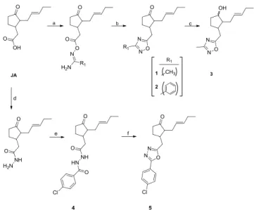
Initially we started modifiying the ester portion of MJ by installing druggable handles such as oxodiazole moieties. Therefore, we started from jasmonic acid (JA) from which was obtained initial building block pseudo ester with DCC coupling strategy followed by basic condensa-tion provided novel 1,2,4- oxadiazoles 1 and 2. In order to obtain 1,3,4-oxadiazoles we started with hydrazide formation from JA, followed by Schotten-Baumann reaction with 4-chlorobenzoyl chloride and POCl3
condensation yielded us desired molecule 5 (Scheme 1).
Based on the initial biological findings and cell based assay results (Table 1), among one of the synthesized more active oxadiazole (1) (IC50= 0.40 μM) molecule was subjected to reduction procedure to
obtain compound 3 (IC50= 0.27 μM) which demonstrated much more
activity.
After obtaining positive results we then decided to synthesize var-ious amide analogues (6 and 7) as well as modifying it to methyl car-bamate (9). Amides were routinely obtained via HBTU coupling pro-cedure as shown Scheme 2. Compound 9 was synthesized from an isocyanate intermediate (8) using Curtius rearrangement procedure. Overall these amides showed reasonable enzyme inhibition activity, however cell based results did not translate the same activity.
We then turned our attention to reversed esters and fused
molecules. Therefore, after reduction of the ester to its corresponding primary alcohol (10), two heteroatom containing aromotic esters (13 and 14) were obtained via CDI coupling procedure. In order to modify the ketone portion of MJ, we formylated the -α-carbon of the ketone followed by phenyl hydrazine addition afforded the fused phenyl-pyr-azole 12 (Scheme 3). The preliminary enzyme assay results of these novel analogues when compared to initial series (compounds 1–3) surprisingly did not provide better activity, however there may need to be more reversed ester analogues synthesized in order to see the clear evidence in inferiority and observe solid structural activity relationship (SAR).(SeeScheme 4.)
Our strategy to modify the ketone portion continued with reductive amination followed by synthesizing novel urea analogues 17 and 18. We first converted MJ to its corresponding amines via reductive amination procedure using Na(OAc)3BH to obtain the desired secondary amines
which were transformed to urea analogues with isocyanates (17 and 18). Our observation for the urea analogues is that the elongation of the MJ on ketone side did not seem to increase activity (IC50= 38.14 and
14.0 µM respectively) since the molecular weight increased dramatically. Fig. 1. Small molecule Hexokinase-II inhibitors known in literature.
Scheme 1. Synthesis of Methyl Jasmonate Analogues 1-5a. aReagents and
conditions: (a) For 1: DCC, acetamide oxime, DMF, rt. For 2: DCC, benzamide oxime, acetone, rt. (b) For 1: DCC, DMF, 115 °C. For 2: KOH, DMSO, rt. (c) NaBH4, MeOH (d) hydrazine monohydrate, 80 °C, 3 h. (e) 4-chlorobenzoyl
chloride, ACN/H2O, 0 °C. (f) POCl3, toluene, 110 °C, 16 h.
Scheme 2. Synthesis of Methyl Jasmonate Analogues 6-9a. aReagents and
conditions: (a) HBTU, TEA, (2-aminoethyl)morpholine (for 6), D-(+)-Glucosamine hydrochloride (for 7), (b) DMAP, DPPA, TEA, toluene, 110 °C. (c) NaOMe, MeOH, rt.
This can be hypothesized that increasing the bulk in ketone portion of MJ somehow changes the binding mode of these molecules with HK-II en-yzme. We are still investigating the binding mode of these novel analo-gues and will be reported in due course after verification.
We then pursued much challenging modifications on MJ which is shown onScheme 5. Our aim was to displace the alkyl side chain of MJ with an appropriate substituents or aromotic structures. Starting with 2-cyclopenten-1-one, α -bromination, Suzuki coupling and Michael ad-dition reactions provided compound 21 with overall good yield. Bio-logical activity of this molecule did not make much difference when compared to other modifications, which may suggest that the side chain
The inhibitory enzyme activities against hexokinase II (HK-II) ex-pressed as IC50values (Table 1).
Based on the IC50values of HK-II enzyme inhibition, compounds that
demonstrates less than 1 μM IC50 activity are listed as
3 > 2 > 1 > 7 > 6. The order of inhibitory activity was established taking into account inorganic phosphate release (seeTable 1). Methyl jasmonate used as reference standard. Preliminary enzyme inhibition studies revealed that we have identified handful of novel MJ analogues much more potent than MJ itself.
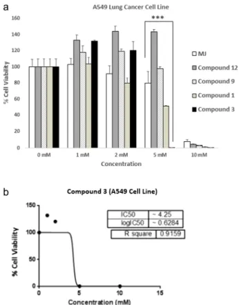
To evaluate the effecs of these novel analogues on the viability of A549 (lung cancer) cell line, cells were treated with different novel compounds and among all tested, compound-3 derivatives were found extremely potent when compared to commercial HK-II inhibitor Methyl Jasmonate (MJ) (Fig. 2a; p < 0.0001). Additionally, compound 3 showed cytotoxic effects on A549 cell line with significant decrease in cell viability to 0.36% (IC50values: 4.250 mM) (seeFig. 2b).
The cells were treated with increased doses (1–10 mM) of novel compounds and Methyl Jasmonate (MJ) as a reference standard. Control wells were treated with equal concentrations of DMSO solvent. (a) After 24 h of treatment, cells were applied to viability assay. According to measurements, cell viability was decreased to 0.36% in 5 mM compound 3 treated cells. Plots indicate relative IC50values of
compound 3. (b) compound 3 treated A549 cell line. The results were expressed as the mean ± SD from three independent experiments (p*** < 0.0001).
A549 cells were treated with all different novel HK-II inhibitors and their effects on cell viability expressed as IC50values, are summarized
inTable 2.
Based on the IC50values, compounds 1 and 3 demonstrated
rea-sonably good activity when compared to MJ. The order of cytotoxic activity was established considering the percentage of viable cells. Methyl jasmonate used as a reference standard.
According to cell viability assays, measurable cytotoxic activity of novel HK-II inhibitors on lung cancer cell line were recorded. Then we further investigated the cytotoxicity of various HK-II inhibitors on SKOV-3 ovarian cancer cell line. Our findings showed that among these novel inhibitors, compound 3 was the most potent HK-II inhibitor as compared to other compounds synthesized in our laboratory. Highly significant cell death percentages were recorded in SKOV-3 as well as A549 cell lines upon treatment with 5 mM compound 3 derivatives for Scheme 3. Synthesis of Methyl Jasmonate Analogues 10–14a.aReagents and
conditions: (a) TMS, NaI, TEA, ACN 40 °C, 1.5 h. (b) NaOMe, ethyl formate, toluene, rt. (c),phenyl hydrazine, MeOH. (d): NaH, DMF, CDI-activated car-boxylic acid 0 °C → rt.
Scheme 4. Synthesis of Methyl Jasmonate Analogues 15–18a.aReagents and
conditions: (a) Amine, ACN, 2 h. (b) Na(OAc)3BH, overnight. (c)
2-metox-yphenyl isocyanate, DCM, rt. (d) 1,4-diisocyanate, DCM, rt.
Scheme 5. Synthesis of Methyl Jasmonate Analogues 19–21a.aReagents and
conditions: (a) OXONE®, DCM, HBr, TEA, rt, 4 h. (b) Phenyl boronic acid, Na2CO3, Pd(PPh3)4, toluene/H2O, 100 °C, 24 h. (c) Dimethyl malonate, NaH dry
THF, 0 °C.
Table 1
The result of enzyme activity.
Compounds IC50(µM) MJ 7.47 Compound 1 0.40 Compound 2 0.29 Compound 3 0.27 Compound 5 1.86 Compound 6 1.01 Compound 7 0.99 Compound 9 2.15 Compound 12 2.53 Compound 13 5.70 Compound 14 16.8 Compound 17 38.1 Compound 18 14.0 Compound 21 27.1
The results were expressed as the mean of duplicates ± SD.
24 h (Fig. 3a; p < 0.0001). Additionally, IC50values of compound 3
treated SKOV-3 cell line was calculated as 1.772 mM. This data in-dicates compound 3 is more potent than MJ and any other analogues on SKOV-3 ovarian cancer cell line (Fig. 3b).
The cells were treated with increased doses (1–10 mM) of novel HK-II inhibitors and Methyl Jasmonate (MJ) as a reference standard. Control wells were treated with equal concentrations of DMSO solvent. (a) After 24 h of treatment, cells were applied to viability assay. According to measurements, cell viability was decreased to 0.28% in 5 mM compound 3 treated cells. Plots indicate relative IC50values of (b)
compound 3 treated with SKOV-3 cell line. The results were expressed as the mean ± SD from triple replicates (p*** < 0.0001).
SKOV-3 cells treated with the different HK-II inhibitors and their cytotoxic effects expressed as IC50values, were summarized inTable 3.
According to the IC50values, compound 3 proved to more potent on
SKOV-3 cell line after 24 h exposure. The order of cytotoxic activity was established considering the percentage of viable cells. Methyl jasmo-nate used as a reference standard. The cell viability results evidently
show that there might be some cell specificity of these novel com-pounds against different cancer cell lines.
Furthermore, effects of HK-II inhibition on HK-II protein expression levels and biochemical verification of cell death were studied by wes-tern blot analysis.
According to our results, HK-II expression was increased when treated with novel HK-II inhibitor compound 3 (Fig. 4a). This is most likely due to a cellular defense to compansate HK-II activity by in-creasing its protein expression levels. Since the expression level is not linked to inhibitory effect of HK-II inhibitors, the cell death is inevitable once the cells recieve the inhibitory signals.
Poly-ADP-ribose polymerase (PARP) cleavage is one of the best biochemical marker of cell death and PARP is cleaved at the very downstream of apoptotic cascade, thus indicating the last stages of cell death at which DNA damage occurs[9,10]. Therefore, we further stu-died compound 3 triggered cell death at the protein level and showed the presence of cleaved PARP as well as decresed levels of its precursor Fig. 2. Cytotoxic activity of novel MJ analogues on A549 lung cancer cell line.
Table 2
The IC50values of novel molecules in A549 cell line.
The Results of A549 cell line
Compounds IC50Values MJ 6.383 mM Compound 1 4.564 mM Compound 3 4.25 mM Compound 5 9.778 mM Compound 9 7.115 mM Compound 12 9.011 mM Compound 13 10.88 mM Compound 14 9.878 mM Compound 17 ND
The results were expressed as the mean of duplicates ± SD.
Fig. 3. Cytotoxic activity of novel HK-II inhibitors on SKOV-3 ovarian cancer cell line.
Table 3
The IC50values of novel molecules in SKOV-3 cell line.
The Results of SKOV-3 cell line
Compounds IC50Values MJ 4.17 mM Compound 1 6.077 mM Compound 2 ND Compound 3 1.772 mM Compound 5 ND Compound 6 ND Compound 9 5.758 mM Compound 12 5.82 mM Compound13 ND Compound 14 7.68 mM Compound 17 ND
The results were expressed as the mean of duplicates ± SD.
(PARP). Beside that, we did not detect cleaved PARP in control cells treated with equal concentrations of DMSO as expected (Fig. 4b).
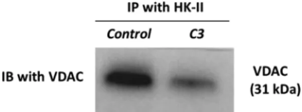
Based on the literature, HK-II shows its activity in two ways: either catalyzing the reaction of 6-glucose phosphate formation from glucose in glycolysis or binding VDAC on mitochondria[12–15]. Inhibition of both actions results in cell death. HK-II inhibitors such as 2DG prevent the catalytic activity of the enyzme while MJ acts through the inter-ruption of its mitochondrial interaction[16]. Therefore, MJ analogues produced in our laboratory as HK-II inhibitors are predicted to de-monstrate the same mechanism of action with MJ. For this aim, we investigated the inhibitory effect of Compound 3 (C3) on HK-II and VDAC interaction via immunoprecipitation of HK-II followed by wes-tern blotting of VDAC (seeFig. 5).
Based on our results, we identified more potent HK-II inhibitor (ex: C3) than MJ itself[11]. We also determined that the most potent MJ analogue C3 acts by disposing VDAC and HK-II interaction on mi-tochondrial membrane; therefore induces cell death. Even though MJ by itself is not reported for its enzymatic inhibitory role on HK-II, our enzyme inhibition assay suggests that the novel MJ analogues can be potential candidates for HK-II inhibition. In order to fully undertsand the mechanism of action on inhibiton of HK-II catalytic activity more investigations need to be conducted to clarify their behaviour. These studies suggest that newly syntesized MJ analogues may have future potential as anti-cancer agents. Therefore, we will continue to pursue additional in vitro experiments and in vivo efficacy studies of these novel analogues which will be reported on due course.
3. Experimental section 3.1. General
Anhydrous solvents and all reagents were purchased from Sigma-Aldrich, Merck and Alfa Aesar. Proton nuclear magnetic resonance (1H
NMR) spectra were recorded at Varian NMR (600, 500, 150 and 160 MHz, respectively). Chemical shifts were reported in ppm down-field from tetramethylsilane (TMS) for proton and carbon. For1H NMR
spectra, chemical shifts are reported in parts per million (ppm) and are reported relative to residual non-deuterated solvent signals. Coupling constants are reported in hertz (Hz). The following abbreviations (or a combination, thereof) are used to describe splitting patterns: s, singlet; d, doublet; t, triplet; q, quartet; quint, quintet; m, multiplet; comp, overlapping multiplets of non-magnetically equivalent protons; br, broad. The mass spectra were obtained using a Shimadzu LC-MS 2040C equipped by Electrospray Source (ESI) operating in both positive and negative ions. The solvents used in MS measures were methanol (Chromasolv grade), purchased from Sigma-Aldrich and mQ water 18 MΩ, obtained from Millipore's Simplicity system. For the ionization, 5 mM ammonium acetate in H2O was used in LC-MS. Analytical
thin-layer chromatography (TLC) was carried out on Merck silica gel F-254 plates. Coloumn chromatography purifications were performed on Fig. 4. HK-II protein expression and PARP cleavage levels of Compound 3 (C3)
treated A549 lung cancer cell line. The cells were treated with Compound 3 (IC50= 4.25 mM) and control medium (DMSO solvent control) for 24 h to
de-tect expression of HK-II and cell death proteins. Protein expression levels were determined by ImageJ and normalized to β-actin levels. (a) Treatment of A549 cell line with II inhibitor compound 3 resulted in significantly increased HK-II expressions as compared to control treatment, (b) Compound 3 treated cells showed decreased PARP and increased c-PARP levels significantly as compared to control. The results were expressed as the mean ± SD from three in-dependent experiments. (p#< 0.01; p* < 0.005; and p** < 0.0005).
Fig. 5. HK-II bound VDAC expression in mitochondrial protein extracts of C3 and DMSO (control) treated A549 lung cancer cell line. The cells were treated with C3 (IC50= 4.25 mM) and control medium (DMSO solvent control) for 24 h
and mitochondrial proteins were extracted. To detect HK-II activity on VDAC binding, HK-II was immunoprecipitated (IP) and HK-II bound VDAC expressions were detected by immunoblotting (IB). A549 cells treated with C3; MJ analogue showed decreased expression of HK-II bound VDAC.
Merck Silica gel 60 (0.063–0.200 mm ASTM) as the stationary phase. TLC plates were visualized with UV light an also stained with ninhy-drin, anisaldehyde or permanganat.
3.2. Chemical synthesis
Synthesis of jasmonic acid; To methyl jasmonate (224.3 mg, 1 mmol) in 5 mL THF and 5 mL water was added LiOH·H2O (84 mg, 2 mmol).
The reaction mixture was stirred at rt for 1 h and then diluted with 30 mL water. The aqueous layer was washed by hexanes (30 mL), acidified by 1 N HCl and extracted with EtOAc. Evaporation of the solvent after dried over MgSO4gave the product as light yellow oil[8].
Compound 1: (3-((3-methyl-1,2,4-oxadiazol-5-yl)methyl)-2-(pent-2-en-1-yl)cyclopentanone; (a) 1 mL of a 0.5 M jasmonic acid solution (0.5 mmol) in DMF was added to reaction flask, followed by addition of 0.55 mL of 1.0 M solution (0.55 mmol) of DCC in DMF. After mixing for 30 min, 1.0 mL of a 0.55 M acetamide oxime solution (0.55 mmol) in DMF was added and the resulting solution was mixed for 4 h at 25 °C. (b) A further 0.55 mL of 1.0 M DCC (0.55 mmol) in DMF was added and the reaction mixtures were heated to 115 °C for 6 h to effect cyclode-hydration. After cooling to room temperature, 7 mL of DCM was added to each vessel, followed by 3 mL of water. After 10 min of agitation, DCM layer washed with 1 × 3 mL H20, 1 × 3 mL 1 N HCI, 1 × 3 mL sat.
NaHCO3, and 1 × 3 mL brine. The organic layers were dried with
MgSO4and filtrated. Purified by column chromatography (3:2 EA:Hxn)
[17]. Yellow oil (56%).1H NMR (500 MHz, CDCl 3) δ 5.49–5.42 (m, 1H), 5.28–5.21 (m, 1H), 3.22 (dd, J = 15.2, 4.5 Hz, 1H), 2.87 (dd, J = 15.3, 9.1 Hz, 1H), 2.43–2.38 (m, 3H), 2.38 (s, 3H), 2.37–2.33 (m, 1H), 2.21–2.15 (m, 1H), 2.15–2.08 (m, 1H), 2.08–2.00 (m, 2H), 2.00–1.94 (m, 1H), 1.59–1.50 (m, 1H), 0.94 (t, J = 7.5 Hz, 3H).13C NMR
(125 MHz, CDCl3) δ 218.09 (C]O), 177.69 (CqAr), 167.17 (CqAr),
134.43 (CH), 124.62 (CH), 53.88 (CH), 39.30(CH), 37.61 (CH2), 30.94
(CH2), 26.94 (CH2), 25.52 (CH2), 20.60 (CH2), 14.09 (CH3), 11.54
(CH3). LCMS m/z: [M+H], found 248.32. C14H20N2O2 requires 249.15.
Compound 2: 2-(pent-2-en-1-yl)-3-((3-phenyl-1,2,4-oxadiazol-5-yl) methyl)cyclopentanone; (a) To a mixture of the jasmonic acid (1 mmol) in acetone (4 mL) was added DCC (1.1 mmol). The reaction mixture was stirred at room temperature for 30 min, then the benzamide oxime (1 mmol) was added. The resulting mixture was stirred at room tem-perature for 12 h. Acetone was evaporated at reduced pressure and to the residue was added water (20 mL). The mixture was concentrated to give a residue. (b) To a solution of O-acylbenzamidoxime 1 (1 mmol) in DMSO (2–3 mL) was added KOH (1 mmol). The reaction mixture was stirred at room temperature for 10– 20 min (TLC or precipitation of the product). The reaction mixture was diluted with 30 mL of cold water. The resulting was washed with water (30 mL). Purified by coloumn chromatography with 2:3 (EA:Hxn) solvent mixture [18]. White oil (60%).1H NMR (500 MHz, CDCl 3) δ 8.02–7.99 (m, 2H), 7.45–7.39 (m, 3H), 5.45–5.38 (m, 1H), 5.25–5.18 (m, 1H), 3.26 (dd, J = 15.2, 4.7 Hz, 1H), 2.92 (dd, J = 15.2, 9.0 Hz, 1H), 2.47–2.39 (m, 1H), 2.38–2.34 (m, J = 3.4 Hz, 2H), 2.34–2.30 (m, 1H), 2.22–2.15 (m, 1H), 2.07 (ddd, J = 18.9, 11.3, 8.9 Hz, 1H), 2.03–1.95 (m, 3H), 1.58–1.52 (m, 1H), 0.89 (t, J = 7.5 Hz, 3H).13C NMR (126 MHz, CDCl 3) δ 218.15 (C]O),
177.98 (CqAr), 168.40 (CqAr), 134.49 (CH), 131.24 (CHAr), 128.88 (CHArX2), 127.43 (CHArX2), 126.74 (CqAr), 124.66 (CH), 53.94 (CH), 39.40 (CH), 37.65 (CH2), 31.13 (CH2), 27.01 (CH2), 25.62 (CH2), 20.65
(CH2), 14.11 (CH3). LCMS m/z: [M+H], found 310.39. C19H22N2O2
requires 311.05.
Compound 3: 3-((3-methyl-1,2,4-oxadiazol-5-yl)methyl)-2-(pent-2-en-1-yl)cyclopentanol; Added sodium borohydride (1.5 eq) to a solution of 1 (1 eq) in methanol (10 mL) at 0 °C. The reaction mixture was stirred for 3 h. Quenched the reaction mixture with of 1 M HCl. The mixture was diluted with ethyl acetate. Removed the organic layer, the aqueous layer. The organic layers were dried with MgSO4and filtrated. Purified
by coloumn chromatography.1H NMR (500 MHz, CDCl 3) δ 5.50–5.41 (m, 1H), 5.41–5.32 (m, 1H), 3.96–3.89 (m, 1H), 3.05 (dd, J = 15.3, 5.3 Hz, 1H), 2.85 (dd, J = 15.3, 8.7 Hz, 1H), 2.36 (s, J = 8.4 Hz, 3H), 2.15–2.09 (m, 2H), 2.08–1.99 (m, 3H), 1.89–1.78 (m, 2H), 1.67–1.52 (m, 3H), 0.95 (t, J = 7.5 Hz, 3H).13C NMR (126 MHz, CDCl 3) δ 178.84 (Car), 166.87 (Car), 133.74 (CH), 126.26 (CH), 78.26 (CH), 53.51 (CH), 41.32(CH), 33.28 (CH2), 31.74 (CH2), 30.21 (CH2), 28.85 (CH2), 20.62 (CH2), 14.19 (CH3), 11.51 (CH3). LCMS m/z: [M+H], found 251. C14H22N2O2requires 250.34. Compound 5: 3-((5-(4-chlorophenyl)-1,3,4-oxadiazol-2-yl)methyl)-2-(pent-2-en-1-yl)cyclopentanone; 2 mL of hydrazine monohydrate was placed in a 2 necked flask. Upon addition of jasmonic acid (1 mmol) dropwise, the system was stirred at 80 °C. After 3 h the reaction was terminated by LC-MS control. To avoid excess hydrazine, toluene was added to evaporate the solvent in the rotary evaporator. LCMS m/z: [M +H]: 225. 2-(3-oxo-2-(pent-2-en-1-yl)cyclopentyl)acetohydrazide (1 mmol) was dissolved in 5 mL of water and cooled to 0 °C. Then 4-chlorobenzoyl chloride (3 mmol) was carefully added dropwise. Acetonitrile was added to the reaction mixture, which was solid while standing, and allowed to stir for 2 h. After 2 h, the solvent was evapo-rated on a rotary evaporator. A white solid was obtained. The reaction was terminated by LC-MS. LCMS m/z: [M+H]: 363. 4 was dissolved in toluene. An excess amount of phosphorus (V) oxychloride was added and the mixture was stirred at 110 °C under reflux for 16 h. The reaction was terminated by LC-MS control. The reaction mixture was cooled and neutralized by adding dropwise to the NaHCO3solution. Extraction was
carried out with ethyl acetate and the organic phase was dried over MgSO4and then the solvent was evaporated in a rotary evaporator. It
was purified by column chromatography with a 10: 1.5 EA: Hxn solu-tion mixture[19].1H NMR (500 MHz, CDCl 3) δ 7.97 (d, J = 8.5 Hz, 2H), 7.49 (d, J = 8.5 Hz, 2H), 5.52–5.42 (m, 1H), 5.32–5.23 (m, 1H), 3.31 (dd, J = 15.4, 4.6 Hz, 1H), 2.96 (dd, J = 15.4, 9.0 Hz, 1H), 2.52–2.35 (m, 3H), 2.30–2.21 (m, 1H), 2.15 (dd, J = 19.2, 10.6 Hz, 1H), 2.10–1.99 (m, 3H), 1.71–1.56 (m, 2H), 0.95 (t, J = 7.5 Hz, 3H). 13C NMR (126 MHz, CDCl 3) δ 218.07 (C]O), 165.18 (Car), 164.14 (Car), 138.02(Car), 134.40 (CH), 129.47 (CH), 128.04 (CH), 124.68 (CH), 122.29 (Car), 53.97 (CH), 39.23 (CH), 37.61 (CH2), 30.06 (CH2), 27.04 (CH2), 25.63 (CH2), 20.63 (CH2), 14.12 (CH). LCMS m/z: [M+H] found 345, C19H21ClN2O2requires 344.84. Compound 6: N-(2-morpholinoethyl)-2-(3-oxo-2-(pent-2-en-1-yl)cyclo-pentyl)acetamide; To a mixture of compound jasmonic acid in EtOAc and triethylamine (TEA) (2 eq), was added N,N,N′,N′-Tetramethyl-O-(1H-benzotriazol-1-yl)uronium hexafluorophosphate (HBTU) (1 eq). After stirring for 1 h at room temperature, 4-(2-aminoethyl)morpholine (2 eq) was added and the reaction mixture was stirred for 12 h. The mixture was washed with water, dried, and concentrated to give a re-sidue which was purified by coloumn chromatography with 10:3 (EA:Hxn) solvent mixture[7]. Yellow oil (83%).1H NMR (500 MHz,
CDCl3) δ 5.98 (b, 1H), 5.42–5.35 (m, 1H), 5.26–5.19 (m, 1H), 3.64 (t, 4H), 3.34–3.28 (m, 2H), 2.52 (dd, J = 14.2, 4.6 Hz, 1H), 2.42 (t, J = 6.0 Hz, 2H), 2.41–2.38 (m, 4H), 2.32–2.25 (m, 4H), 2.20–2.13 (m, 1H), 2.11–2.05 (m, 1H), 2.04–1.96 (m, 3H), 1.86–1.81 (m, 1H), 1.50–1.40 (m, 1H), 0.90 (t, J = 7.5 Hz, 3H). 13C NMR (125 MHz, CDCl3) δ 171.14 (C]O), 133.92 (CH), 125.25 (CH), 66.85 (CH2x2), 57.11 (CH2), 54.14 (CH), 53.35 (CH2x2), 41.25 (CH2), 38.64 (CH), 37.73 (CH2), 35.51 (CH2), 27.19 (CH2), 25.64 (CH2), 20.62 (CH2), 14.16 (CH3). LCMS m/z: [M+H], found 322.44. C18H30N2O3requires 323.10. Compound 7: 2-(3-oxo-2-(pent-2-en-1-yl)cyclopentyl)-N-(2,4,5-trihy-droxy-6-(hydroxymethyl)tetrahydro-2H-pyran-3-yl)acetamide; To a mix-ture of compound jasmonic acid in EtOAc and TEA (2 eq), was added HBTU (1 eq). After stirring for 1 h at room temperature, D-(+)-Glucosamine hydrochloride (2 eq) in 2 mL water was added and the reaction mixture was stirred for 12 h. The mixture was washed with water, dried, and concentrated to give a residue. Purified by coloumn chromatography with 10:2 (DCM:MeOH) solvent mixture[7]. Yellow oil (70%).1H NMR (500 MHz, CD
3OD) δ 5.47–5.40 (m, 1H), 5.36–5.27
38.20 (CH), 37.23 (CH2), 26.38 (CH2), 24.81 (CH2), 20.13 (CH2),
13.12(CH3). LCMS m/z: [M+H], found 371.43. C18H29NO7 requires
372.10.
Compound 9: Methyl ((3-oxo-2-(pent-2-en-1-yl)cyclopentyl)methyl) carbamate; A dry flask containing jasmonic acid (1 mmol), 4-(Dimethylamino) pyridine (1.1 mmol) and TEA (1.1 mmol) in toluene was stirred under nitrogen and heated to 110 °C. The diphenylpho-sphoryl azide (DPPA) (1.1 mmol) was then added dropwise at 110 °C and left under reflux for 16 h. The reaction was terminated by LC-MS control. After toluene was evaporated, it was extracted with ethyl acetate, washed with brine. The organic phase was dried over Na2SO4,
then the solvent was evaporated. It was used without any purification. LCMS m/z: [M+H]: 208. To a mechanically stirred suspension of NaOMe in MeOH at room temperature was added 8 (1 mmol) in MeOH. After 6 h later, the solvent was evaporated and acidified with con-centrated HCl to a pH of 1. Then extracted with H2O and ether. The
aqueous layer was separated and the combined organic layers were dried with MgSO4, concentrated to yellow oil. Purified by coloumn
chromatography with 10:1.5 (EtOAc:HXN) solvent mixture. Yellow oil (45%).1H NMR (500 MHz, CDCl 3) δ 5.49–5.42 (m, 1H), 5.27–5.18 (m, 1H), 4.86 (s, 1H), 3.66 (s, 3H), 3.46–3.35 (m, 1H), 3.29–3.16 (m, 1H), 2.46–2.28 (m, 3H), 2.14–2.01 (m, 5H), 1.92–1.83 (m, 1H), 1.57–1.45 (m, 1H), 0.95 (t, J = 7.5 Hz, 3H).13C NMR (126 MHz, CDCl 3) δ 219.13 (C]O), 157.24 (C]O), 134.17 (CH), 125.07 (CH), 52.26 (CH3), 52.17 (CH), 44.25 (CH2), 41.82, 37.53 (CH2), 25.74 (CH2), 24.93 (CH2), 20.57 (CH2), 14.12 (CH3). LCMS m/z: [M+H] found 240.20, C13H21NO3requires 239.31. Compound 10: 3-(2-hydroxyethyl)-2-(pent-2-en-1-yl)cyclopentanone; Chlorotrimethylsilane (TMS) (1.25 eq) was added to a solution of so-dium iodide (1.25 eq) in dry acetonitrile (5 mL). A solution of the me-thyl jasmonate (1 mmol) and trieme-thylamine (0.3 mL) was added, and the resulting mixture stirred at 40 °C for 1.5 h. The product was extracted with light petroleum, and evaporated to give the silyl ether as an yellow oil. The silyl ether was dissolved in dry THF and added dropwise to a suspension of lithium aluminium hydride (4 eq) in THF at 0 °C. The mixture was stirred at room temperature for 2 h. Work-up with water and purified by coloumn chromatography. Light yellow oil (55%).1H
NMR (500 MHz, CDCl3) δ 5.46–5.39 (m, 1H), 5.29–5.22 (m, 1H), 3.80–3.69 (m, 2H), 2.38–2.34 (m, 2H), 2.34–2.31 (m, 1H), 2.22–2.16 (m, 1H), 2.10–1.95 (m, J = 10.9 Hz, 5H), 1.88–1.80 (m, 1H), 1.56–1.51 (m, 1H), 1.46–1.41 (m, 1H), 0.95 (t, J = 7.5 Hz, 3H). 13C NMR (126 MHz, CDCl3) δ 133.79 (CH), 125.33 (CH), 60.95 (CH2), 54.91 (CH), 38.09 (CH), 38.04 (CH2), 37.67 (CH2), 27.27 (CH2), 25.53 (CH2), 20.64 (CH2), 14.19 (CH3). LCMS m/z: [M+H], found 196.29. C12H20O2 requires 197.20. Compound 12: 2-(6-(pent-2-en-1-yl)-2-phenyl-2,4,5,6-tetra-hydrocyclopenta[c]pyrazol-5-yl)ethanol; To a mechanically stirred sus-pension of NaOMe in toluene at room temperature was added 10 in toluene followed by ethyl formate added dropwise. The mixture was stirred for 3–4 h and then acidified with concentrated HCl to a pH of 1. Then extracted with H2O and ether. The aqueous layer was separated
and the combined organic layers were dried with MgSO4, concentrated
to yellow oil[20]. To a stirred solution of 11 (1 eq) in MeOH (10 mL) was added phenyl hydrazine (1.1 eq), and the mixture was stirred for 1.5 h at room temperature. Solvent was removed under reduced pres-sure, and the residue was dissolved in CH2Cl2(300 mL), washed with
H2O, dried with Na2O4, and concentrated to oil. Purified by coloumn
chromatography with 1:1 (EA:Hxn) solvent mixture [21]. Silica Gel Chromatography (SGC) afforded the title compound described with NMR spectroscopy.1H NMR (500 MHz, CDCl 3) δ 7.57 (d, J = 8.0 Hz, 2H), 7.42 (t, J = 7.8 Hz, 2H), 7.36 (s, J = 4.2 Hz, 1H), 7.26 (t, 1H), 126.20 (CH), 124.95 (CH), 120.55 (CH), 61.05 (CH2), 47.56 (CH), 45.08 (CH), 39.57 (CH2), 30.03 (CH2), 28.43 (CH2), 20.50 (CH2), 14.10 (CH3). LCMS m/z: [M+H], found 297.55, C19H24N2O requires 296.41.
Compound 13: 2-(3-oxo-2-(pent-2-en-1-yl)cyclopentyl)ethyl 3-methyli-soxazole-5-carboxylate; 10 (1 mmol) was dissolved in DMF and cooled to 0 °C. NaH(1.25 mmol) was added under nitrogen to the mixture for alkoxide formation. Simultaneously, 3-methylisoxazole-5-carboxylic acid (1 mmol) was dissolved in DMF and carbonyldiimidazole (1.2 mmol) was added and stirred for activation of the acid for 30 min. After 30 min, CDI-activated 3-methylisoxazole-5-carboxylic acid was added dropwise with stirring the alkoxide added under nitrogen. The reaction was allowed to warm to room temperature and stirred. The mixture was stirred for 5 h and then acidified with concentrated HCl to a pH of 1. Then extracted ethyl acetate and H2O. The organic phase was
dried over MgSO4 and then the solvent was evaporated in a rotary
evaporator. Purified by column chromatography with a 1: 2 mixture of EA: Hxn solution. Light yellow oil (70%).1H NMR (500 MHz, CDCl
3) δ
6.75 (d, J = 2.9 Hz, 1H), 5.44–5.37 (m, 1H), 5.24–5.17 (m, 1H), 4.42 (t, J = 6.7 Hz, 2H), 2.36–2.32 (m, 6H), 2.24–2.16 (m, 2H), 2.11–1.97 (m, 5H), 1.89–1.79 (m, 2H), 0.93–0.89 (m, 3H).13C NMR (126 MHz, CDCl
3)
δ 219.18 (C]O), 160.40 (Car), 159.95 (C]O), 156.79 (Car), 133.91 (CH), 124.96 (CH), 110.10 (CH), 64.18 (CH2), 54.63 (CH), 38.30 (CH),
37.84 (CH2), 33.33 (CH2), 27.07 (CH2), 25.39 (CH2), 20.54 (CH2),
14.10 (CH3), 11.41 (CH3). LCMS m/z: [M+H] found 306.10,
C17H23NO4requires 305.37.
Compound 14: 2-(3-oxo-2-(pent-2-en-1-yl)cyclopentyl)ethyl thiazole-4-carboxylate; 10 (1 mmol) was dissolved in DMF and cooled to 0 °C. NaH (1.25 mmol) was added under nitrogen to the mixture for alkoxide formation. Simultaneously, 3-methylisoxazole-5-carboxylic acid (1 mmol) was dissolved in DMF and carbonyldiimidazole (1.2 mmol) was added and stirred for activation of the acid for 30 min. After 30 min, CDI-activated 3-methylisoxazole-5-carboxylic acid was added dropwise with stirring the alkoxide added under nitrogen. The reaction was allowed to warm to room temperature and stirred. The mixture was stirred for 5 h and then acidified with concentrated HCl to a pH of 1. Then extracted ethyl acetate and H2O. The organic phase was dried
over MgSO4 and then the solvent was evaporated in a rotary
eva-porator. Purified by column chromatography with a 2: 1 mixture of EA: Hxn solution. Yellow oil (60%).1H NMR (500 MHz, CDCl
3) δ 8.86 (d,
J = 2.1 Hz, 1H), 8.23 (d, J = 2.1 Hz, 1H), 5.46–5.37 (m, 1H), 5.30–5.20 (m, 1H), 4.50–4.44 (m, 2H), 2.41–2.32 (m, 3H), 2.32–2.20 (m, 2H), 2.14–2.08 (m, 1H), 2.08–1.99 (m, 3H), 1.92–1.84 (m, 1H), 1.80–1.71 (m, 1H), 1.56–1.45 (m, 1H), 0.92 (t, J = 7.5 Hz, 3H). 13C NMR
(126 MHz, CDCl3) δ 219.47 (C]O), 161.21 (C]O), 153.49 (CHar),
147.95 (Car), 133.89 (CHar), 127.31 (CH), 125.04 (CH), 63.66 (CH2),
54.75 (CH), 38.39 (CH), 37.92 (CH2), 33.61 (CH2), 27.17 (CH2), 25.42
(CH2), 20.57 (CH2), 14.14 (CH3). LCMS m/z: [M+H] found 308.50.
C16H21NO3S requires 307.41.
Compound 17: Methyl 2-(3-(1-benzyl-3-(2-methoxyphenyl)ureido)-2-(pent-2-en-1-yl)cyclopentyl)acetate; Methy jasmonate (1 eq) ve benzyla-mine (1 eq) 5–10 mL in acetonitrile was added to reaction flask. After mixing for 1–2 h, Na(OAc)3BH (1.5 eq) was added and the resulting
solution was stirred over night. To compound of 7 in DCM was added 2-metoxyphenyl isocyanate (1 eq). The reaction mixture was controlled by LC-MS and TLC. Purified by coloumn chromatography with a 1:2 mixture of EA: Hxn solution. Light yellow oil (70%).1H NMR (500 MHz,
CDCl3) δ 8.15–8.12 (m, 1H), 7.39–7.26 (m, 5H), 7.05 (s, 1H), 6.93–6.86 (m, 2H), 6.75–6.71 (m, 1H), 5.45–5.32 (m, 2H), 4.84–4.75 (m, 1H), 4.69 (d, J = 17.6 Hz, 1H), 4.50 (d, J = 17.3 Hz, 1H), 3.65 (s, J = 4.5 Hz, 3H), 3.55 (s, 2H), 2.64 (dd, J = 15.1, 4.3 Hz, 1H), 2.27–2.20 (m, 1H), 2.20–1.93 (m, 8H), 1.86–1.72 (m, 1H), 1.29–1.17
(m, 1H), 0.95 (t, J = 7.5 Hz, 3H).13C NMR (126 MHz, CDCl3) δ 173.21
(C]O), 155.80 (C]O), 147.68 (Car), 138.08 (Car), 132.78 (CH), 129.13 (Car), 128.80 (CH), 127.39 (CH), 127.27 (CH), 126.36 (CH), 121.82 (CH), 121.07 (CH), 119.01 (CH), 109.82 (CH), 58.87, 55.49, 51.40, 49.36 (CH2), 47.04, 41.21, 40.45 (CH2), 30.18 (CH2), 29.14
(CH2), 28.07 (CH2), 20.76 (CH2), 14.19 (CH3). LCMS m/z: [M+H]
found 465.15. C28H36N2O4requires 464.60.
Compound 18: Dimethyl 2,2′-((1,14-dimorpholino-4,11-dioxo- 3,5,10,12-tetraazatetradecane-3,12-diyl)bis(2-(pent-2-en-1-yl)cyclo-pentane-3,1-diyl))diacetate; Methy jasmonate (2 eq) ve 4-(2-aminoethyl) morpholine (2 eq) 5–10 mL in acetonitrile was added to reaction flask. After mixing for 1–2 h, Na(OAc)3BH (3 eq) was added and the resulting
solution was stirred over night. To compound of 8 in DCM was added 1,4-diisocyanate (1 eq). The reaction mixture was controlled by LC-MS and TLC. Purified by coloumn chromatography with a 1: 2 mixture of EA: Hxn solution. Light yellow oil (65%).1H NMR (500 MHz, CDCl
3) δ
5.38–5.21 (m, 4H), 3.71–3.64 (m, 8H), 3.62 (d, J = 5.1 Hz, 6H), 3.44–3.05 (m, 9H), 2.61–2.39 (m, 14H), 2.19–1.81 (m, 18H), 1.67–1.48 (m, 6H), 1.46–1.07 (m, 5H), 0.94–0.89 (m, 6H).13C NMR (126 MHz,
CDCl3) δ 173.38 (C]O), 173.21 (C]O), 159.86 (C]O), 159.78 (C]
O), 132.96 (CH), 132.41 (CH), 127.61 (CH), 126.56 (CH), 66.90 (CH2), 60.65, 58.49 (CH), 54.14 (CH), 54.08 (CH), 51.34 (CH2), 46.92, 46.48, 41.29, 40.52, 40.47 (CH2), 39.55(CH2), 38.55, 30.14 (CH2), 29.35 (CH2), 28.86 (CH2), 28.54 (CH2), 28.43 (CH2), 28.41(CH2), 28.39 (CH2), 28.36 (CH2), 28.04 (CH2), 27.14 (CH2), 20.67 (CH2), 20.56 (CH2), 14.20 (CH3), 14.17(CH3). LCMS m/z [M+H] found 818.10. C44H76N6O8requires 817.11.
Compound 21: Dimethyl (3-oxo-phenylcyclopentyl)malonate; 2-Cyclopentenone (1 mmol) and OXONE® (1.2 mmol) were dissolved in dry DCM in a 2 necked flask. 2 N HBr (2.2 mmol) was added and the solvent turned orange. TEA (2 mL) was added carefully after stirring at room temperature for 4 h. After stirring overnight at room temperature, the reaction mixture was neutralized by pH control, extracted with ethyl acetate, and the organic phase was dried over MgSO4and the
solvent was evaporated in a rotary evaporator [22]. Brown solid (30%).Phenyl boronic acid (1.5 mmol), Na2CO3(3 mmol) was added to
a stirred mixture under nitrogen by adding a 1/1 10 mL toluene/water mixture. 19 was added via dissolution in toluene. Finally, moles of 2% Pd (PPh3)4were added and heated at 100 °C under nitrogen. After 24 h,
it was cooled to room temperature. Extraction was carried out with ethyl acetate and the organic phase was dried over MgSO4and then the
solvent was evaporated in a rotary evaporator. Light yellow oil (30%).
1H NMR (500 MHz, DMSO) δ 8.10 (t, J = 2.8 Hz, 1H), 7.69 (d,
J = 7.5 Hz, 2H), 7.36 (t, J = 7.5 Hz, 2H), 7.30 (t, J = 7.3 Hz, 1H), 2.51–2.46 (m, 4H). 13C NMR (126 MHz, DMSO) δ 206.39 (C]O),
160.01 (CAr), 140.65 (Cq), 130.80 (Cq), 127.38 (CHArx2), 127.18 (CH), 125.80 (CHArx2), 34.57 (CH2), 25.00 (CH2). LC-MS m/z: [M+H]:
159. Dimethyl malonate (1.5 mmol) in a 2 necked flask was placed under nitrogen and dissolved in 10 mL dry THF and cooled to 0 °C. After 30 min NaH (3 mmol) was added rapidly under nitrogen and the reac-tion continued at 0 °C. After 1 h 20 (1 mmol) was added to the reacreac-tion medium. The reaction was allowed to warm to room temperature and allowed to stir for 4 h. The reaction was terminated by TLC and LC-MS control. Extraction was carried out with 1 N HCl and DCM. The organic phase was dried over MgSO4and then the solvent was evaporated in a
rotary evaporator. The column was purified by column chromatography with a 4:1 mixture of EA: Hxn solution. Colorless oil (70%).1H NMR
(500 MHz, CDCl3) δ 7.33 (t, J = 7.5 Hz, 2H), 7.25 (t, J = 7.4 Hz, 1H),
7.09 (d, J = 7.2 Hz, 2H), 3.69 (s, J = 9.7 Hz, 3H), 3.49 (d, J = 7.0 Hz, 1H), 3.35 (s, 3H), 3.30 (d, J = 12.2 Hz, 1H), 3.09–2.97 (m, 1H), 2.61–2.51 (m, 1H), 2.50–2.34 (m, 2H), 1.86–1.72 (m, 1H). 13C NMR
(126 MHz, CDCl3) δ 215.74 (C]O), 168.16 (C]O), 168.07 (C]O),
136.77 (Car), 129.03(CHarx2), 128.65 (CHarx2), 127.38 (CHar), 59.60 (CH), 54.23 (CH), 52.47 (CH3), 52.18 (CH3), 43.90 (CH), 37.88 (CH2),
25.06 (CH2). LCMS m/z: [M+H] found 291.20. C16H18O5 requires
290.31.
3.3. Biological activity studies
3.3.1. Hexokinase - II enzyme activity assay
The Universal Enzyme Assay Kit (R & D, EA004) was used to de-termine the hexokinase II enzyme inhibition for novel molecules. This kit quantitatively separates inorganic phosphate as it converts ADP to AMP via coupling phosphatase found in its structure. The liberated inorganic phosphate complexes with the malachite green phosphate detection reagents. The resulting complex ratio is proportional to the formation of AMP from ADP by phosphatase. For this reason, the rate of inorganic phosphate production reflects the kinetics of the kinase re-action. Enzyme activity assay was applied according to manufacturer’s instructions. Briefly, a substrate mixture was prepared by using 0.5 mM ATP and 25 mM glucose. The human hexokinase-2 enzyme (rhHK2) was prepared to be 7.5 ng/μL and the coupling phosphatase 4 enzyme to be 10 μg/mL. Respectively, 20 μL of buffer, 20 μL of rhHK2, 2 μL of sample and 10 μL of cCoupling phosphatase 4 were added to the well and in-cubated for 10 min. And then, 30 μL of malachite green reagent A, 100 μL of ultrapure water, 30 μL of malachite green reagent B was added and incubated for 20 min. After incubation, the absorbance was read at 620 nm and the results were calculated according to the fol-lowing equation.
=
Specific Activity(pmol/min/µg)
Adjusted phosphate release*(nmol) x (1000 pmol/nmol) Incubation time (min) x amount of enzyme (µg) x coupling rate** * Derived from the linear curve of the phosphate standard and ad-justed to the control.
** The coupling ratio in specified conditions is 0.475. 3.3.2. Cell viability assay
The human lung carcinoma cell line A549 (ATCC # CCL-185) and the human ovarian cancer cell line SKOV-3 (ATCC # HTB-77) used in the experiments were obtained from ATCC (USA). A549 cells were cultured in RPMI (Gibco) medium containing 10% FBS (Gibco), 1% Penicillin/Streptomycin (Gibco) and 1%L-Glutamatine. The cells were incubated at 37 °C in a 5% CO2 incubator. While SKOV-3 cells were cultured in McCoy’s 5a Modified Medium (Gibco) at the same condi-tions. After achieving confluence, 1.5 x10⁴cells/well seeded in 96-well
black plates and treated with novel molecules for 24 h at six different concentrations (0.1–10 mM), and each concentration was prepared as triplicates. The cell viability assay Cell-Titer Glo (Promega, Madison, WI, USA) was performed according to manufacturer’s instructions. MJ used as control and DMSO in all of the concentrations was 2.5%. Three independent experiments were performed. The luminescence signal was read on the SpectraMax i3x Multi-Mode Detection Platform (Moleculer Devices, USA) to determine the percentage of viable cells. IC50values
were determined by Graphpad Prism 7. 3.3.3. Mitochondrial protein extraction
A549 cancer cells were seeded in 6-well plates at a density of 2 × 10⁵cells/well and incubated in 5% CO
2incubator. Next day, the
cells were treated with compound 3 with its IC50dose for A549 cell line.
After 24 h, the medium was removed and centrifuged. The cells were obtained from the wells in RIPA (Thermo Fischer) buffer with protease inhibitor (Roche, cOmplete™, Mini, EDTA-free Protease Inhibitor Cocktail). The cells were collected as lysate and incubated at +4 °C on the shaker for 20 min. Next, the lysate was centrifuged and the super-natant was collected. The supersuper-natant was then centrifuged at 5000 rpm for 10 min. After centrifuging, the supernatant was collected and centrifuged at 13,000 rpm for 30 min. The supernatant was re-moved and the pellet was suspended in RIPA buffer. Protein amount was determined for each sample by using nanodrop spectrophotometric protein concentration calculator (IMPLEN P330).
the tubes were centrifuged at 300 rpm for 2 min and the supernatant was collected. 1 µg of Hexokinase-2 antibody (Cell Signaling, Hexokinase II, C64G5) was added into the supernatant and the samples were kept on the shaker at +4 °C for about 1 h. Afterwards, 75 µL of beads were added to each sample and incubated overnight at +4 °C on the shaker. The next day the samples were centrifuged at 3000 rpm for 2 min and the supernatant was discarded. Laemmli buffer with 10% β-mercaptoethanol (Biorad) was added into the pellets and the samples were incubated for 5–10 min at 100 °C. Then western blot procedure was applied as explained below.
3.3.5. Western Blot
For total protein collection as whole cell lysate, 2 × 10⁵cells were
seeded into each well of 6 well plates. Then the cells were incubated at 37 °C in 5% CO2for 24 h. The culture medium was discarded and cells
were treated with proper amount of C3 according to IC50values of each
cell line. After 24 h of treatment, protein lysates were collected using Ripa lysis buffer with protease inhibitor and incubated at 4 °C for 30 min on shaker. Then pelleted and protein samples in supernatant were collected seperately. Total protein concentration measured by nanodrop spectrophotometric protein concentration calculator (IMPLEN P330) and SDS treated samples were prepared by using Laemmli buffer (Biorad). Equal amounts of protein (25 μg) were elec-trophoresed by loading into 4–12% polyacrylamide gel and the proteins were separated by their molecular weight. And then proteins were transferred to polyvinylidene difluoride (PVDF) membranes by using the Trans-Blot TurboTransfer System (BioRad). After that, blocking of membranes was performed by using 5% non-fat milk dissolved in TBST (137 mM NaCl, 20 mM Tris, 0.1% Tween-20) for 1 h at room tem-perature. Membranes were incubated overnight with primary mono-clonal antibodies at 4 °C at on a shaker. Antibodies used in the ex-periments were as follows: monoclonal rabbit Hexokinase-2 antibody (12885S, cell signaling) (1:750); monoclonal mouse β-actin antibody (CST #3700s; Cell Signaling) (1:1000); and anti- poly-ADP-ribose polymerase (PARP) (CST #9542) (1:2000). The following day, the membranes were washed 3 times for 5 min with TBST and then in-cubated with HRP-conjugated secondary antibodies (anti-mouse CST#7076S; anti-rabbit CST#7074S) (anti-rabbit 1: 2500; anti-mouse 1: 2500) which dissolved in TBST containing 5% non-fat milk powder for 1 h at room temperature. Membranes were washed 3 times with TBST for 5 min. Membrane images were obtained using ECL HRP sub-strate (thermo fisher, Pierce) by ChemiDoc MP System (Bio-Rad). Protein levels were analyzed using the ImageJ program and band in-tensities were normalized according to β-actin blots.
3.3.6. Statistics
Data were analyzed by ANOVA Tukey test with SPSS 19.0 when comparing two groups. Data were expressed with the mean of duplicates ± SD and the differences were determined as p#< 0.01;
p* < 0.005; p** < 0.0005 and p*** < 0.0001. 4. Conclusion
To our knowledge, the novel MJ analogues inhibit HK-II activity through VDAC detachment from mitochondria. Further studies of these molecules will provide us their possible mechanism of action on the conversion of glucose to glucose 6-phosphate in glycolysis pathway. Our next set of MJ analogues might shed light on developing new po-tent anti-cancer agents. This will guide us in preparing more and multi potent, efficacious and soluble drug candidates. In conclusion, we have
This project (215S890) is funded by TUBITAK. We kindly appreciate for their support. Authors OSI, and BEY, received funding from TUBITAK as a stipend for this project (TUBİTAK project no 215S890). We thank Yasemin Yozgat for her invaluable assistance in biological assays, Melike Aybala Guzel for proofreading our manuscript, and Ozan Topcu for his technical support in IP experiments in this study. Appendix A. Supplementary material
Supplementary data to this article can be found online athttps:// doi.org/10.1016/j.bioorg.2019.103146.
References
[1] J.E. Wilson, Isozymes of mammalian hexokinase: structure, subcellular localization and metabolic function, J. Exp. Biol. 206 (2003) 2049–2057.
[2] V. Bhardwaj, N. Rizvi, M.B. Lai, J.K. Lai, A. Bhushan, Glycolytic enzyme inhibitors affect pancreatic cancer survival by modulating its signaling and energetics, Anticancer Res. 30 (2010) 743–750.
[3] F. Baoa, K. Yangb, C. Wub, S. Gaoa, P. Wanga, L. Chena, H. Lia, New natural in-hibitors of hexokinase 2 (HK2): steroids from Ganoderma sinense, Fitoterapia 125 (2018) 123–129.
[4] Z. Chen, H. Zhang, W. Lu, P. Huang, Role of mitochondria-associated hexokinase II in cancer cell death induced by 3-bromopyruvate, Biochim. Biophys. Acta 1787 (2009) 553–560.
[5] H. Lin, J. Zeng, J.I. Luengo, et al., Discovery of a novel 2,6-disubstituted glucosa-mine series of potent and selective hexokinase 2 inhibitors, ACS Med. Chem. Lett. 7 (3) (2015) 217–222.
[6] D. Xu, J. Jin, H. Yu, Z. Zhao, D. Ma, C. Zhang, H. Jiang, Chrysin inhibited tumor glycolysis and induced apoptosis in hepatocellular carcinoma by targeting hex-okinase-2, J. Exp. Clin. Cancer Res. 36 (2017) 44.
[7] H.T. Dang, H.J. Lee, E.S. Yoo, J. Hong, B. Bao, J.S. Choi, J.H. Jung, New jasmonate analogues as potential anti-inflammatory agents, Bioorg. Med. Chem. 16 (2008) 10228–10235.
[8] S. Liu, W.H. Wang, Y.L. Dang, Y. Fu, R. Sang, Rational design and efficient synthesis of a fluorescent-labeled jasmonate, Tetrahedron Lett. 53 (2012) 4235–4239. [9] I. Kitazumi, M. Tsukahara, Regulation of DNA fragmentation: the role of caspases
and phosphorylation, FEBS J. 278 (3) (2011) 427–441.
[10] M. Tewari, L. Quan, K. O’Rourke, S. Desnoyers, Z. Zeng, D.R. Beidler, G.G. Poirier, G.S. Salvesen, V.M. Dixit, Yama/ CPP32b, a mammalian homolog of ced-3, is a CrmA-inhibitable protease that cleaves the death substrate poly(ADP-ribose) poly-merase, Cell 81 (1995) 801–809.
[11] M. Guzel, B.O. Sucu, O. Savlug, S. Ozdatli, Methyl jasmonate derivatives as possible drug candidates for use in treatment of cancer, World patent PCT/TR2018/050522 (2018) 09 25.
[12] N. Goldin, L. Arzoine, A. Heyfets, A. Israelson, Z. Zaslavsky, T. Bravman, V. Bronner, A. Notcovich, V. Shoshan-Barmatz, E. Flescher, Methyl jasmonate binds to and detaches mitochondria-bound hexokinase, Oncogene 27 (2008) 4636–4643. [13] S.P. Mathupala, Y.H. Ko, P.L. Pedersen, Hexokinase II: cancer’s double-edged sword acting as both facilitator and gatekeeper of malignancywhen bound to mitochon-dria, Oncogene 25 (2006) 4777–4786.
[14] L. Galluzzi, O. Kepp, N. Tajeddine, G. Kroemer, Disruption of the hexokinase–VDAC complex for tumor therapy, Oncogene 27 (2008) 4633–4635.
[15] I.M. Cesari, E. Carvalho, M.F. Rodrigues, B.S. Mendonça, N.D. Amôedo, F.D. Rumjanek, Methyl jasmonate: putative mechanisms of action on cancer cells cycle metabolism, and apoptosis, Int. J. Cell Biol. 2014 (2014) 572097. [16] D. Zhang, J. Li, F. Wang, J. Hu, S. Wang, Y. Sun, Deoxy-D-glucose targeting of
glucose metabolism in cancer cells as a potential therapy, Cancer Lett. 355 (2014) 176–183.
[17] T. Deegan, T. Nitz, D. Cebzanov, D. Pufko, J. ve Porco, Parallel synthesis of 1,2,4-oxadiazoles using CDI activation, Bioorgan. Med. Chem. Lett. 9 (1999) 209–212. [18] S. Baykov, T. Sharonova, A. Osipyan, S. Rozhkov, A. Shetnev, A. ve Smirnov, A
convenient and mild method for 1,2,4-oxadiazole preparation: cyclodehydration of O-acylamidoximes in the superbase system MOH/DMSO, Tetrahedron Lett. 57 (2016) 2898–2900.
[19] John Mccall et al., Pyrazolopyrimidinone compounds for the inhibition of PASK and their preparation, 2014066795 (2014) PCT Int. Appl.
[20] H.C. Gibbard, C.J. Moody, C.W. ve Rees, 3aH-lndenes, Part 5.' Preparation and reactions of 3-methoxy- and 3-trimethylsiloxy-3a-substituted -3aH- indenes, J. Chem. Soc. Perkin Trans. I (1985) 723.
[21] D.D. LeCloux, C.J. Tokar, M. Osawa, R.P. Houser, M.C. Keyes, W.B. ve Tolman, Optically active and a-symmetric tris (pyrazolyl) hydr oborate and tris (pyrazolyl) phosphine oxide ligands: synthesis and structural characterization, Organometallics 13 (1994) 2855–2866.
[22] K.W. Kim, I.H. Park, A convenient halogenation of a, b-unsaturated carbonyl compounds with OXONE® and hydrohalic acid (HBr, HCl), Synthesis 16 (2004) 2641–2644.