1092
http://journals.tubitak.gov.tr/medical/
Turkish Journal of Medical Sciences Turk J Med Sci
(2018) 48: 1092-1095 © TÜBİTAK
doi:10.3906/sag-1707-173
Patient symptoms and magnetic resonance imaging correlation
in temporomandibular joint internal derangement
Osman AKDAĞ, Gökçe YILDIRAN*, Mehtap KARAMEŞE
Department of Plastic, Reconstructive, and Aesthetic Surgery, Faculty of Medicine, Selçuk University, Konya, Turkey
* Correspondence: [email protected]
1. Introduction
Temporomandibular joint (TMJ) disorders commonly present with clinical symptoms such as pain, tenderness, joint sounds, deviating mandibular function, and headache or earache (1,2). Temporomandibular internal derangement refers to a disc displacement with reduction or without reduction, which describes the joint disc’s abnormal localization (1).
Pain is a very irritating symptom and usually the main symptom among these patients, and it may evoke for the clinician a temporomandibular internal derangement. On the other hand, each patient may describe an etiological agent and how the pain started. Commonly the reasons that cause pain include bruxism, depression, dental therapy, and hard foods (3).
Magnetic resonance imaging (MRI) is the most commonly used and the most accurate imaging method for evaluating the internal derangement of TMJ (4). However, it is known that sometimes patient symptoms or complaints may not compatible with MRI results. In this study, the relationship between MRI and patient symptoms has been revealed.
2. Materials and methods 2.1. Patients
One hundred and four patients who were admitted with unilateral pain in the TMJ region were evaluated between March 2013 and October 2015 retrospectively with MRI and TMJ examination charts in our plastic surgery outpatient clinics.
Subjects with hypermobile condyles, perforated discs, and TMJ masses were excluded from the study. Patients whose dominant symptom was unilateral TMJ pain and who had MRI performed were included.
Written informed consent was obtained from all patients while filling out the examination charts. The study was designed in accordance with the Helsinki Declaration. 2.2. TMJ examination cards
We routinely use patient examination cards in our outpatient clinics for TMJ patients, which evaluate the patient’s age, sex, current pain status and level, duration of symptoms, painful joint laterality, bruxism presence, depressed mood presence, and how the pain began.
The cards include a visual analog scale (VAS) that evaluates the current pain from 0 to 10 (0 = no pain; 10 = worst pain imaginable)
Background/aim: Temporomandibular joint (TMJ) internal derangements consist of reduced and nonreduced disc dislocations and
are often diagnosed by magnetic resonance imaging (MRI). The main symptom of these derangements is TMJ pain. This study aimed to evaluate whether there is a correlation between TMJ pain and disc dislocation.
Materials and methods: One hundred and four patients experiencing pain were evaluated retrospectively with the examination charts
that we use routinely in our clinics and MRI. Patients were separated into two main groups as MRI(+) and MRI(-) groups according to their internal derangement findings in MRI. Mean VAS levels were compared between these two groups.
Results: There were no significant differences between the MRI(+) and MRI(-) groups and no differences between the reduced disc
dislocation group and nonreduced disc dislocation group.
Conclusion: Although pain is maybe the most irritating symptom for TMJ patients, it cannot take the diagnostic place of MRI and MRI
is still the gold standard method to make the exact diagnose of internal derangement.
Key words: Internal derangement, magnetic resonance imaging, pain, temporomandibular joint
Received: 26.07.2017 Accepted/Published Online: 30.11.2017 Final Version: 12.12.2018
1093 AKDAĞ et al. / Turk J Med Sci
2.3. MRI
All MRI findings were evaluated by the same radiologist by using 1.5-T MRI equipment. Patients with normal MRI findings formed the MRI(-) group. Patients with MRI findings related to internal derangement (either reduced or nonreduced disc dislocation) formed the MRI(+) group.
The MRI(+) group was divided into two groups: “reduced disc dislocation group” (RDD) and “nonreduced disc dislocation group” (NDD).
2.4. Evaluation
All patients were evaluated and all procedures (examination cards, VAS, MRI) were applied before any possible intervention surgery.
VAS scores were compared between the MRI(+) and MRI(-) groups in order to evaluate if disc dislocation was related to pain status. VAS scores between the RDD and NDD groups were also compared in order to evaluate if the pain level was related to reducibility.
For specifying the effects of etiological agents on the MRI findings, MRI(+) and MRI(-) patients were evaluated according to the initial reason for pain starting, like bruxism, hard foods, dental therapy, etc. Patients were asked directly about depressive mood, bruxism, and reasons for pain starting.
All data were collected and statistically evaluated with SPSS 16.0 and t-tests.
3. Results
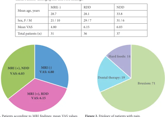
Eighty-one female and 23 male patients with a mean age of 30.3 years (range: 15–71) were evaluated. Thirty-one patients (29.8%) were specified by the radiologist as totally normal. There were 73 patients (70.1%) in the MRI(+) group who had either reduced or nonreduced disc dislocation. Thirty-six of the 73 MRI(+) patients had disc displacement with reduction (RDD) and 37 patients had disc displacement without reduction (NDD) (Table).
The mean VAS value of the MRI(-) group was 4.80 and that of the MRI(+) group was 6.09. There was not a statistically significant difference for VAS scores between these groups.
The mean VAS value of the RDD group was 6.15 and that of the NDD group was 6.03 (Figure 1). There was not a statistically significant correlation between VAS and reducibility.
Etiologically, the most common cause of pain was bruxism (68.2%), followed by dental therapy (18.2%) and hard foods (13.4%). The most common pain cause of both the MRI(-) and MRI (+) groups was bruxism (24% in the negative group and 44.2% in the positive group) (Figure 2).
Table. Patients’ demographics and MRI findings.
Mean age, years MRI(-) RDD NDD
28.7 28.1 33.8 Sex, F / M 21 / 10 29 / 7 31 / 6 Mean VAS 4.80 6.15 6.03 Total patients (n) 31 36 37 MRI (-) VAS: 4.80 MRI (+), RDD VAS: 6.15 MRI (+), NDD VAS: 6.03 Bruxism: 71 Dental therapy: 19 Hard foods: 14
Figure 1. Patients according to MRI findings: mean VAS values
1094
AKDAĞ et al. / Turk J Med Sci Sixty out of 104 patients (57.6%) indicated that they
had a depressive mood and all of those 60 patients were female (74%).
4. Discussion
TMJ disorder prevalence is more than 40% in the general population and the most irritating symptom of these patients is pain (5,6).
Although it is known that MRI has limited availability and long scan time, MRI is still the most accurate and gold-standard diagnosis method for temporomandibular internal derangements, because of evaluating the disc’s exact localization while the mouth is opened or closed (7). However, there are limited studies evaluating the correlation between MRI and patient pain, so it was studied to evaluate if pain could be a good indicator in these disorders. Murakami et al. studied the relationship of TMJ pain and joint effusion in MRI; however, they showed that there was no correlation between joint effusion and pain (8). Their study included 19 patients and they proposed that this correlation should be studied with a larger series. We evaluated 104 patients and also evaluated the internal derangement correlation rather than joint effusion on MRI, which is much more closely related to joint pain.
Patients who suffer from TMJ may present with many clinical symptoms (1,2). However, in this study, we excluded patients admitted with other complaints from pain and patients whose MRI findings were anything other than any type of temporomandibular internal derangement in order to evaluate the MRI findings only with pain.
Evaluating only pain in patients who were admitted to outpatient clinics, it was tried to evaluate whether pain can be a good indicator in these disorders. Patients with only unilateral pain were included in this study. Grading the pain and scoring the VAS might be less reliable in cases of bilateral pain, because while evaluating the pain contralateral joint pain may be a confounding factor. These assessments increase subjective efficacy during VAS scoring. Also, myofascial pain, psychiatric pain, and extraarticular pain are more likely to be bilateral. In order to be fair, only unilateral patients were included in the study.
In our study, 81 of 104 patients (77.8%) were female, and this is compatible with the literature. Warren et al. claimed that women are much more likely to feel TMJ pain and more tend to allodynia (9). Despite the fact that women patients who participated in this study are more likely to have depressive moods (74% of female patients), we associated the high ratio of female/male patients with the pronociceptive TMJ effect of estradiol as shown by Wu et al. whereby pain is such a subjective criterion that it varies even according to sex (10).
Vieira-Queiroz et al. determined that patients who have positive MRI findings for an internal derangement are more likely to have pain (7). They established that patients with pain are more likely to have internal derangement findings in TMJ MRI. This was compatible with our study. In our study, 73 of 104 patients with pain (70.1%) had positive MRI results (either reduced or nonreduced). This ratio indicates that patients who suffer from pain are apparently more likely to have an internal derangement. However, nearly 1/3 of patients with pain had normal MRI findings and this is not a negligible ratio. Pain is not related only to internal derangement in the TMJ region but also to personal factors and it may be related to disorders that cannot be detected by MRI (11).
VAS scoring is used for making pain a measurable subject (12). We wondered if there was a relationship between pain and MRI and we used a VAS in order to measure pain levels. In this study, there was not a statistically significant difference between the VAS values and MRI positivity. We interpreted this as follows: although patients with are much more likely to have positive MRI findings, the severity of pain as evaluated with the VAS is not correlated with MRI findings and patients with more pain may not be more likely to have positive MRI findings. We attribute this to the subjectivity of pain.
De Melo et al. evaluated patients under 21 years old and found that there was a significant correlation between nonreduced disc dislocation and pain (2). In our study, all patients had pain so we did not evaluate whether the nonreduced group was more likely to suffer from pain. However, we evaluated and compared the reduced and nonreduced disc dislocation patients’ pain severity according to their VAS scores. We could not find significant difference between reduced and nonreduced disc dislocation patients’ VAS values, from which we concluded that pain does not show the reducibility. The RDD group had more pain, surprisingly, even if it was not statistically significant. This made us think that a patient who has more pain may not be more likely to have a nonreduced disc dislocation rather than a reduced dislocation, because he or she may perceive more pain because of the clicking sound and deviating mandible.
Many papers in the literature claim that TMJ disorders are correlated with etiology (13). Etiologically, the most common cause of pain was bruxism in our study, which is compatible with the literature. The most common cause of pain for both the MRI(-) and MRI(+) group was bruxism. However, we believe that perceived bruxism is less than the actual rate. The perceived bruxism rate may be increased with some more questions about headache, night sleep quality, daytime sleepiness, temple pain upon waking, etc. in the questionnaire. However, we asked questions about bruxism directly to patients and their spouses because
1095 AKDAĞ et al. / Turk J Med Sci
those questions, which may increase the found rate of bruxism, also may indicate other disorders than TMJ. In order to prevent bias, we did not ask those questions at the expense of not determining the correct rate of perceived bruxism.
Even if the patients with TMJ disorders presented mostly with pain, nearly one-third of these patients had no findings in their MRI results. Thus, the TMJ pain might
not be related only to TMJ internal derangement. Patients who have complaints from TMJ should be evaluated based on patient history, physical examination, VAS, and MRI.
In conclusion, MRI is still the most accurate and gold-standard method for TMJ patients with pain. However, there is not a correlation between pain level and MRI findings. In TMJ assessment, it may not possible to expect worse MRI findings in patients with more pain.
References
1. Rudisch A, Innerhofer K, Bertram S, Emshoff R. Magnetic resonance imaging findings of internal derangement and effusion in patients with unilateral temporomandibular joint pain. Oral Surg Oral Med Oral Pathol Oral Radiol Endod 2001; 92: 566-571.
2. de Melo DP, Sousa Melo SL, de Andrade Freitas Oliveira LS, Ramos-Perez FM, Campos PS. Evaluation of temporomandibular joint disk displacement and its correlation with pain and osseous abnormalities in symptomatic young patients with magnetic resonance imaging. Oral Surg Oral Med Oral Pathol Oral Radiol 2015; 119: 107-112.
3. Emodi-Perlman A, Yoffe T, Rosenberg N, Eli I, Alter Z, Winocur E. Prevalence of psychologic, dental, and temporomandibular signs and symptoms among chronic eating disorders patients: a comparative control study. J Orofac Pain 2008; 22: 201-208. 4. Tasaki MM, Westesson PL, Raubertas RF. Observer variation
in interpretation of magnetic resonance images of the temporomandibular joint. Oral Surg Oral Med Oral Pathol 1993; 76: 231-234.
5. Ryalat S, Baqain ZH, Amin WM, Sawair F, Samara O, Badran DH. Prevalence of temporomandibular joint disorders among students of the University of Jordan. J Clin Med Res 2009; 1: 158-164.
6. Chantaracherd P, John MT, Hodges JS, Schiffman EL. Temporomandibular joint disorders’ impact on pain, function, and disability. J Dent Res 2015; 94: 79-86.
7. Vieira-Queiroz I, Gomes Torres MG, de Oliveira-Santos C, Flores Campos PS, Crusoé-Rebello IM. Biometric parameters of the temporomandibular joint and association with disc displacement and pain: a magnetic resonance imaging study. Int J Oral Maxillofac Surg 2013; 42: 765-770.
8. Murakami K, Nishida M, Bessho K, Iizuka T, Tsuda Y, Konishi J. MRI evidence of high signal intensity and temporomandibular arthralgia and relating pain. Does the high signal correlate to the pain? Br J Oral Maxillofac Surg. 1996; 34: 220-224. 9. Warren MP, Fried JL. Temporomandibular disorders and
hormones in women. Cells Tissues Organs 2001; 169: 187-192. 10. Wu YW, Bi YP, Kou XX, Xu W, Ma LQ, Wang KW, Gan YH,
Ma XC. 17-Beta-estradiol enhanced allodynia of inflammatory temporomandibular joint through upregulation of hippocampal TRPV1 in ovariectomized rats. J Neurosci 2010; 30: 8710-8719.
11. Fujiwara M, Honda K, Hasegawa Y, Hasegawa M, Urade M. Comparison of joint pain in patients diagnosed with and without articular disc displacement without reduction based on the Research Diagnostic Criteria for Temporomandibular Disorders. Oral Surg Oral Med Oral Pathol Oral Radiol 2013; 116: 9-15.
12. Kliger M, Stahl S, Haddad M, Suzan E, Adler R, Eisenberg E. Measuring the intensity of chronic pain: are the visual analogue scale and the verbal rating scale interchangeable? Pain Pract 2015; 15: 538-547.
13. Sharma S, Gupta DS, Pal US, Jurel SK. Etiological factors of temporomandibular joint disorders. Natl J Maxillofac Surg 2011; 2: 116-119.