The Osteochondrosis of Superior Pole Of Patella: A Case
Report
Patellanın Üst Kutup Osteonekrozu: Olgu Sunumu
İsmail Ağır, Emin Özkul
Bismil Devlet Hastanesi Ortopedi ve Travmatoloji Kliniği
The osteochondroses are a heterogeneous group of injuries to the epiphyses and apophyses of children or adolescents. The breakdown of endochondral ossification and bone fragmentation are seen radiologically. The aetiology of osteochondroses remains uncertain. Catch-up growth or strenuous sports activities have been thought to be the cause of this benign disease. In condition of patellar osteochondrosis, rarely the primary ossification centre is affected known as Köhler’s disease. More frequently, the secondary ossification centre, usually at the distal pole known as Sinding- Larsen-Johansson. Superior pole osteochondrosis of patella has been reported in the literature on only a handful of occasions. We presented the clinic and radiologic findings of an eleven year-old boy with superior pole osteochondrosis patella.
Key Words: Patella, Osteochondrosis, Fragmentation, Superior
Osteokondrozlar çocuk ve adolesan çağda çeșitli bölgelerdeki apofizleri, epifizleri ve epifizoid kemikleri etkileyen bir hastalık grubudur. Endokondral ossifikasyon bölgelerinde çökme ve kemik fragmantasyonları görülür. Osteokondrozlarda etiyoloji kesin olarak bilinmemektedir. Hızlı büyümenin ve ağır spor aktivitelerinin bu iyi huylu hastalığa sebep olduğu düșünülmek-tedir. Patellanın primer ossifikasyon merkezi nadir etkilenir ve Köhler hastalığı olarak bilinir. Daha sık olarak patellanın sekonder ossifikasyon merkezleri özellikle alt kutbu etkilenir ve Sinding- Larsen-Johansson hastalığı olarak bilinir. Literatürde patella üst kutup osteonekrozu nadir bildirilmiștir. Bu çalıșmada patella üst kutup osteonekrozu görülen 11 yașındaki hastanın klinik ve radyolojik bulgularını sunduk.
Anahtar Sözcükler: Patella, Ostekondroz, Fragmantasyon, Üst kutup
Osteochondroses are self-limiting
breakdown of endochondral
ossification characterised
radiologically by bone fragmentation (1). The aetiology of osteochondroses remains uncertain and rather speculative. Catch-up growth or strenuous sports activities have been thought to be the cause of this benign disease in children between 5 and 9 years of age (1-5). Because of traction exerted through the ligament, the osteochondritic changes may occur at either the upper or the lower attacments of the ligamentum patellae as known Sinding
Larsen-Johanson(SLJ) and
Osgood-Schlatter’s(OS) disease (6).
Superior pole osteochondrosis of patella has been reported in the literature on only a handful of occasions (7-9). We presented the clinic and radiologic findings of an eleven year-old boy
with superior pole osteochondrosis patella.
CASE
The patient is an eleven year-old boy who initially presented with a right knee effusion, right knee pain which were continued for two weeks. He reported no prior history of trauma to his knee. Their parents reported no history of upper respiratory tract infection and no fever for last 2 months which was asked for to exclude acute rheumatoid fever disease and other infectious and systemic diseases. Physical examination revealed a mild right knee effusion and minimal pain at the superior border of patella but there were no other remarkable findings such as erythema and local heat increases. There was no limition of range of motion of affected knee. Received: 14.04.2011 • Accepted: 24.04.2012
Corresponding Author İsmail Ağır
Bismil Devlet Hastanesi Ortopedi ve Travmatoloji Kliniği E-posta: [email protected]
GSM: 0412 415 23 10
Ankara Üniversitesi Tıp Fakültesi Mecmuası 2012, 65 (2) DOI: 10.1501/Tıpfak_000000823
CERRAHİ TIP BİLİMLERİ/SURGICAL SCIENCES
Ankara Üniversitesi Tıp Fakültesi Mecmuası 2012, 65 (2)
The Osteochondrosis of Superior Pole of Patela: A case report 130
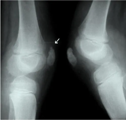
Figure 1: Fragmentation at superior pole of patella. There was no complaints at left knee
of patient. At the conventional radiography there was fragmentation at superior pole of patella (Figure 1) At the laboratory findings; sedimentation was 15 mm/h (normal range 12-24 mm/h) , C-reactive pro-tein (CRP) was 3 mg/L (normal range 0-6 mg/L).
We prescribed non-steroidal anti-inflammatory drug (NSAID) to patient with activity restriction. The symtoms was regressed with NSAID. At 6 month of follow-up clinical
symtoms was improved but
radiological findings was continued.
DISCUSSION
Osteochondroses represent a
heterogeneous group of self-limiting disturbances of endochondral ossification of the epiphyses and apophyses of children or adolescents (1,5,6). In condition of patellar osteochondrosis, rarely the primary ossification centre is affected and seen more often between the ages of 5 and 9 known as Köhler’s disease (1-4). More frequently, the secondary ossification centre, usually at the distal pole, is involved between the ages of 9 and 11 known as SLJ (1,2). Superior pole osteochondrosis of patella has been reported in the literature on only a handful of occasions was no name (6-8). Why osteochondrosis so rarely develops
at the superior pole of the patella remains unknown. The broader insertion of the quadriceps in this area probably distributes traction forces more evenly (7). Superior pole osteochondrosis of patella can be distinguished from bipartite changes by both radiographically and clinical symptomatology. The condition is found more often in young males when reviewing the case reports published ( 6-8).
The largest case series, by Batten and Menelaus, presented the condition in six boys (6). The young men ranged in ages from 10 to 11 years, and all
were active. Radiographic findings of the proximal pole of the patella in that series were similar to that seen in SLJ disease. Besides that four of the patients had radiographic evidence of SLJ or OS disease either in the same knee or the opposite knee. Based on these findings, the authors suggested that similar processes take place in all three conditions.
Grogan and colleagues (9) reported on seven cases of proximal pole fragmentation which they believed was an avulsion of direct trauma. However in our case there was no history of direct trauma. In the series of grogan, despite the direct trauma, the proximal pole was still the least common form of patellar avulsion. The only cases with histological
documentation was reported by Tyler W and McCarthy EF (7) in 2002. They found that the histological
apperence like other side
osteochandrosis. The histological findings according to study of Tyler and McCarthy support that the osteochandrosis of superior pole is also a chronic traction apophysitis. Although the catch-up growth or
strenuous sports activities are thougt as causes of osteochondrosis,
according to keats almost
asymptomatic cases can be seen and concluded that the irregular ossification, fragmentation and sclerosis of the patella could be a normal developmental variation( 10). Therefore the question remains whether sports activities really are the cause of the disease rather than the key factor for the osteochondrosis to become symptomatic. As a result, the authors acknowledge that further research about this finding is warranted.
Ankara Üniversitesi Tıp Fakültesi Mecmuası 2012, 65 (2)
İsmail Ağır, Emin Özkul 131
REFERENCES
1. Corten K, Vandenneucker H, Molenaers G, et al. Bilateral patellar Köhler's disease in an eleven-year-old child with growth retardation: a case report. Acta Orthop
Belg. 2009 ; 75:273-6.
2. Franceschi F, Barnaba SA, Rojas M et al. Multiple osteochondroses f bilateral knee joints: a case report. Knee Surg Sports Traumatol Arthrosc 2007; 15: 431-435. 3. Kohler A. Uber eine haufige, bisher
anscheinend unbekannte Erkrankung einzelner kindlicher Knochen. Munch Med Wochenschr 1908; 55: 1923-1926.
4. Moffat BW. Kohler’s disease of the patella. J Bone Joint Urg 1929; 11-A: 579-581. 5. Traverso A, Baldari A, Catalani F. The
coexistence f Osgood-Schlatter’s disease with Sinding-Larsen- ohansson’s disease. Case report in an adolescent soccer layer. J Sports Med Phys Fitness 1990; 30: 331-333.
6. Batten J. Menelaus M.B. Fragmentation of the proximal pole of the patella: Another manifestation of juvenile traction osteochondritis? J Bone Joint Surg Br 1985; 67: 249-251.
7. Tyler W. McCarthy E.F. Osteochondrosis of the superior pole of the patella: Two cases with histologic correlation. Iowa Orthop J 2002; 22: 86-89.
8. Linscheid R.L. Dahlin D.C. Unusual lesions of the patella. J Bone Joint Surg Am 1966; 48: 1359-1366.
9.Grogan D. P. Carey T.P. Leffers D. et al. Avulsion fractures of the patella. J Pediatr Orthop 1990; 10: 721-730.
10. Keats TE. An Atlas of Normal Roentgen Variants that may imulate Disease. 1996, 6th ed. Mosby, St. Louis, pp 584- 91.