The Relationship Between
Schizophrenia and Cavum
Septum Pellucidum:
A Case Study
Kadir Ascibasi
1, Orkun Aydin
1,
Duygu Kuzu
2, Artuner Deveci
31Assist. Dr., Celal Bayar University, Faculty of Medicine,
Department of Psychiatry, Manisa - Turkey
2Psychologist, Istanbul Arel University, Department of
Psychology, Istanbul - Turkey
3Assist. Prof. Dr., Celal Bayar University, Faculty of
Medicine, Department of Pschiatry, Manisa - Turkey
ABSTRACT
The relationship between schizophrenia and cavum septum pellucidum: a case study
Septum pellucidum that forming the medial wall of the lateral ventricles, a thin layer is formed by two laminae. Cavum septum pellucidum (CSP), a space between the two leaflets of the septi pellucidi, is thought to be a neurodevelopmental anomaly associated with schizophrenia. In normal fetal development, the formation of septum pellucidum occurs at the same time with the development of the neighboring limbic system structures such as hippocampus, amygdala, corpus callosum, septal nucleus. Therefore, the anatomical differences that are detected in the septum pellucidum may reflect the embryonal development disorders of the adjacent anatomical structures. Several magnetic resonance imaging studies have reported a higher prevalence of enlarged CSP in patients with schizophrenia than in normal subjects. In the literature of psychiatric disorders, CSP is most associated with schizophrenia. Wide CSP may support the neurodevelopmental etiology of schizophrenia. In this report we present a schizophrenia case. And we tried to discuss the patient’s clinical appearance and neurocognitive disability.
Key words: Cavum septum pellucidum, magnetic resonance imaging, schizophrenia ÖZET
Şizofreni ile kavum septum pellusidum arasındaki ilişki: Bir olgu sunumu
Lateral ventriküllerin medial duvarını oluşturan septum pellucidum iki laminadan oluşan ince bir tabakadır. Kavum septum pellucidum (KSP), septum pellucidumun iki yaprağı arasındaki bir boşluktur. KSP’un şizofreni ile ilişkili nörogelişimsel bir anomali olduğu düşünülmektedir. Normal fetal gelişimde septum pellucidumun oluşumu hipokampus, amigdala, korpus kallosum, septal nükleus gibi komşu limbik yapılarla eş zamanlı olmaktadır. Bu yüzden septum pellucidumda saptanan anatomik farklılıklar, komşu anatomik yapıların embriyonal gelişim bozukluklarını yansıtabilir. Birkaç manyetik rezonans görüntüleme çalışmasında, şizofreni hastalarında normal kişilere göre daha yüksek prevalansta geniş KSP saptanmıştır. Psikiyatrik bozukluklarla ilgili literatüre baktığımızda KSP’nin en çok şizofreni ile bağlantılı olduğunu görebiliriz. Geniş KSP şizofreninin etyolojik nedenlerinden biri olarak gösterilen nörogelişimsel modeli desteklemektedir. Biz bu olgu sunumunda, manyetik rezonans görüntüleme yöntemi ve nörobilişsel yeti yitimini tartıştık.
Anahtar kelimeler: Kavum septum pellucidum, manyetik rezonans görüntüleme, şizofreni
Address reprint requests to / Yazışma adresi: Assist. Dr. Kadir Ascibasi,
Celal Bayar University, Faculty of Medicine, Department of Pschiatry, Manisa - Turkey Phone / Telefon: +90-236-236-0330/3855 E-mail address / Elektronik posta adresi: [email protected]
Date of receipt / Geliş tarihi: March 26, 2013 / 26 Mart 2013 Date of acceptance / Kabul tarihi: September 9, 2013 / 9 Eylül 2013
INTRODUCTION
E
ven though schizophrenia is a disorder of whichetiology has not been enlightened yet, it is accepted that environmental and biological (genetic, physiological, biochemical and developmental) factors played a significant role. Neurodevelopmental model is one of the most prominent hypotheses in the schizophrenia etiology. It was showed with postmortem examination and brain imaging techniques that important number of schizophrenia patients have
developmental defects like corpus collosum agenesia (CCA), arachnoid vesicles and other abnormalities (1). For example, CCA is a rarely seen abnormality. Out of the 19.000 autopsies only 1 case, out of 40.000 autopsies 2 cases were reported (2). Although prevalence of CCA shows variability in schizophrenia population, in the series which were done with 140 schizophrenia patients by Swayze et.al. (3) 1.4% of prevalence was reported. Septum pellucidum is a thin layer which is formed by two laminas and takes place at medial barrier of the lateral ventricles’. When laminas combine, cavity which
is named “cavum septum pellucidum” (CSP) is formed (4). CSP is seen in all premature infants and fetuses. But, during the intrauterine 3-6 month period, rear half leaves of laminae’s join together (5). The front part of laminae’s join together in 3-6 month period after birth (4). The formation process of the septum pellucidum coincides with the development of neighbours corpus callosum, hippocampus, amygdala and limbic structures such as septal nucleus. Therefore, anatomical differences which detected inseptum pellucidum can reflect the embryonic development disorders of the adjacent anatomical structures (4,6). During fetal development, the growth of the hippocampus and corpus callosum cause the adhesion of septum pellucidum leaves by pushing from back to front. It is assumed that if a dysgenesis happens in these adjacent structures, it can cause a disruption of the usual adhesion process of septum pellucidum (4,7,8).
Known proportion of CSP in adults change in wide range between 0.1% to 85% (7). The reason for such a wide range is developments in imaging studies of the brain. These developments lead to detection of small sized patologies by making more imaging and using thinner sections (7). While being shorter than 6mm in the width CSP is agredded to be the “variant of the normal”, being 6mm and over is admitted that it can be reletad to development of child and adult psychiatric disorders (7,10,11).
In literature it is found that CSP is mostly related to schizophrenia among psychiatric disorders (12,13). Also, the reason why schizophrenia patients have higher proportion of “wide CSP” is related to anatomical finding which sustains neurodevelopmental model for schizophrenia etiology (7). In this case study, schizophrenia patient who was diagnosed with “wide CSP” is tried to be discussed in association with clinical aspect and neurocognitive ability loss.
CASE
39 years old, elementary school graduated, never married male patient was working as a farmer. According to patient his complaint was feeling himself bad, while according to his relatives and friends he was moody,
introvert, he had inability to communicate and he was talking to himself. Because of patient’s complaints were getting worse he was brought to policlinic and after first evaluation he was decided to be hospitalized in our clinic. After patient had finished elementary school successfully he wanted to continue his education but because of his family’s request he started to work as a farmer instead of continuing his school. During this time, there was no any social relational problem both with his parents and his friends. Before he had gone to military service he was engaged and decided to marry. At the age of 20 he went to military service and after completed the service he returned home at the age 21. Psychological complaints have started at that time as introversion, inability to set a relationship, going away from people and talking less and less gradually. Because of his symptoms, he was left by his fiancé. During his psychiatry consultation he was prescribed a medicine of which name he did not remember. He used this medicine approximately 6 months and had a little benefit. He did not have any psychiatric admission and he did not take any treatment during the following 17 years and his complaints increased significantly during this time period. In the last year addition to those complaints he started to talk to himself. He were spending much of his day as working in the farm and sitting at home alone.
Except the psychiatric history there was not any important finding in patient’s background. There was no cigarette, alcohol and substance consumption. In family history, there was not any psychiatric medical history.
In the psychiatric examination, his self-care was sufficient, general appearance and age was coherent with sociocultural citation, speech quantity was lessened, content of the speech was poor and he was talking to himself and laughing. It is detected that his conscious was clear, but orientation was distorted, he had inability to concentrate and to sustain attention and his memory was poor. Restricted affect, visual and auditory hallucination was present.
Association of thought process was scattered, persecution and reference delusions were present in thought content. Judgement, abstract thinking and
evaluation of realization were insufficient. Patient had psychomotor retardation, anhedonia, anergy, lack of motivation and avolution. Social functioning was markedly poor. Patient was having trouble with doing daily works and getting help from his parents. And also his insight about his disorder was poor. There was not any pathology in physical and neurological examination. After psychiatric examination he was diagnosed with schizophrenia and 2mg/day risperidon treatment was prescribed and increased step by step to 6mg/day. He used treatment for 1.5 month but had no benefit from the treatment. It was reported in several research (24,25) that paliperidone increase social and occupational functioning and provide progress in some neurocognitive areas. For this reason treatment was changed as palliperidone 6mg/day and increased step by step to 12mg/day.
Patient did not respond to treatment. During the period of hospitalization scores of Scale for the Assesment of Negative Symptoms (SANS) and Scale for the Assesment of Positive Symptoms (SAPS) were 104 and 61 respectively.
Hemogram, biochemical, thyroid functions, B12 vitamine, folic asid, level of ferritine, serology, EEG, ECG, cranial MRI was asked. In his serology results
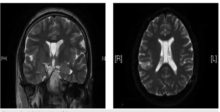
Hepatitis A was detected. A gliotic point which was millimeter-sized and near the anterior horn of lateral ventricle at the left frontal region and; a CSP variation which was 12x22x46 mm in size at the level of the lateral ventricles were observed on the patient’s MRI (Figure 1). Neurology and neurosurgery consultations were wanted. It was stated that there was no need for an intervention according to present findings.
DISCUSSION
It is well known that septum pellucidum fusion is related to faster development of hippocampus, corpus collosum and other midline structures associated to schizophrenia (4,5,14). That’s why the presence of CSP may be an indicator of early developmental disorders in the areas mentioned before for schizophrenia patients. Even though it is not seen as possible, any defect in localized structures like CSP may play an important role in the etiology of schizophrenia. On the other hand the abnormalities of CSP are not special to schizophrenia and other psychotic disorders because those findings can be seen in developmental disorders like Fetal Alcohol Syndrome, Sotos and Apert syndromes (15-17). Though, among the disorders like
panic disorder the width and the prevalence are in the normal borders. On the other hand, in the first episode psychoses with mood symptoms (11); bipolar mood disorders and schizotypal personality disorders (18) high prevalence of CSP was reported. Based on this, some authors claim that a portion of psychosis which are associated with schizophrenia and bipolar mood disorder, are related with development of midline structures. And they think that schizotypal personality disorder may be a mild form of schizophrenia spectrum disorder (11).
It has not found a direct relationship between schizophrenia and presence or size of CSP so far. Wide CSP is related with symptoms like severe thought defects and excessive risk of suicide are seen as having relationship between intellectual functioning, verbal learning and losing some areas such as memory in schizophrenia patients (13,19). Because CSP is in close relation with limbic system it has been mentioned that it might be an early indicator for the detection of neuropathological changes like retardation, seizures, micro/macrocephaly in early period (20).
A research on schizophrenia patients with normal and wide CSP, reported that wide CSP patients have higher clinical symptom severity than normal width CSP patients and this group have worse neuropsychological test success (13). In our case according to Nopoulos (21) criteria’s because of the width of CSP over than 6mm (it must be seen 1,5mm space and at least 4 coronal sections) it can be accepted as a “wide CSP”. Thus, in our case low scores in neuropsychological tests can be related to this situation. Schizophrenia patients who have wide CSP have higher SANS scores than normal width CSP patients (13,22). In our case, patient had score of 104 from SANS.
Moreover, there have been two studies which evaluated the cognitive functions and CSP until this time. These researches indicated that individuals who have wide CSP are worse than normal width CSP in verbal learning and memory functions. Septum pellucidum provides important connections between hypothalamus, hippocampus, amygdala, habenula and reticular formation (9,13,22,23). In our case total recalling point in verbal memory test was 15 and there was severe
retardation in long and short term memory. And also he was unsuccessful in visual memory test. In a research it was found that there is a significant correlation between the size of wide CSP’s and scores of IQ. This relationship was not found in normal size CSP and in control group. It was just found in patients who have wide CSP (23). In our case mild mental retardation (IQ=63) was detected in intelligence assesment.
Also, when patient is evaluated for, in schizophrenia deficit syndrome patient should have primary negative symptoms. Those negative effects should not be related with medicine side effects, anxiety, skepticism, hallucination, delusion, mental retardation and depression. Those symptoms should be permanent characteristics of remission period. Negative symptom in deficit syndrome shows dominant, primary and continuous characteristics. In deficit syndrome at least two features among the restricted affect, decreased emotion, poor talking, losing interest, loosing sense, decreased social impulse should be present and clinically significant in last 12 months and they should continue even at clinically stable times. When our case was evaluated for restricted affect, facial expression, descriptive gestures and changes in the voice tone was little. Evaluating for decreased emotion it was clear that expression of pleasure and averse was poor. While amount of speech is decreased, transmission of information by himself is seen more rather than answering question. Interest in environment and events was clearly decreased. Launching goal directed events and sustaining was decreased. There was no social interaction will and attempt.
Our case is compatible with the literature both for the severity of symptoms or the results of the neuropsychological tests. Being resistant to treatment is significant for patient’s clinical follow up and it is needed to focus on this matter. Even though the relationship between CSP and schizophrenia with severe clinical picture is being investigated, there is not any research on comparison of treatment responses. In future studies, this topic is significant in order to evaluate both clinician’s evaluation of the effects for treatment of psychiatric disorders in terms of neurodevelopmental pathologies and patient’s functionality.
REFERENCES
1. Hallak JE, Crippa JA, Pinto JP, Machado de Sousa JP, Trzesniak C, Dursun SM, McGuire P, Deakin JF, Zuardi AW. Total agenesis of the corpus callosum in a patient with childhood-onset schizophrenia. Arq Neuropsiquiatr 2007; 65:1216-1219. 2. David AS, Wacharasindhu A, Lishman WA. Severe psychiatric
disturbances and abnormalities of the corpus callosum. J Neurol Neurosurg Psychiatry 1993; 56:85-93.
3. Swayze VW, Andreasen NC, Ehrhardt JC, Yuh WT, Alliger RJ, Cohen GA. Developmental abnormalities of the corpus callosum in schizophrenia. Arch Neurol 1990; 47:805-808.
4. Sarwar M. The septum pellucidum: normaland abnormal. AJNR Am J Neuroradiol 1989; 10:989-1005.
5. Shaw CM, Alvord EC. Cavum septi pellucidi et vergae: their normal and pathological states. Brain 1969; 92:213-223. 6. Kim KJ, Peterson BS. Cavum septi pellucidi in Tourette syndrome.
Biol Psychiatry 2003; 54:76-85.
7. Nopoulos P, Swayze V, Flaum M, Ehrhardt JC, Yuh WT, Andreasen NC. Cavum septi pellucidi in normals and patients with schizophrenia as detected by magnetic resonance imaging. Biol Psychiatry 1997; 41:1102-1108.
8. Kwon JS, Shenton ME, Hirayasu Y, Salisbury DF, Fischer IA, Dickey CC, Yurgelun-Todd D, Tohen M, Kikinis R, Jolesz FA, McCarley RW. MRI study of cavum septi pellucidi in schizophrenia, affective disorder, and schizotypal personality disorder. Am J Psychiatry 1998; 155:509-515.
9. Bodensteiner JB, Schaefer GB, Craft JM. Cavum septi pellucidi and cavum vergae in normal and developmentally delayed populations. J Child Neurol 1998; 13:120-121.
10. Hagino H, Suzuki M, Kurokawa K, Mori K, Nohara S, Takahashi T, Yamashita I, Yotsutsuji T, Kurachi M, Seto H. Magnetic resonance imaging study of the cavum septi pellucidi in patients with schizophrenia. Am J Psychiatry 2001; 158:1717-1719. 11. Kasai K, McCarley RW, Salisbury DF, Onitsuka T, Demeo S,
Yurgelun-Todd D, Kikinis R, Jolesz JA, Shenton ME. Cavum septi pellucidi in first-episode schizophrenia and first-episode affective psychosis: an MRI study. Schizophr Res 2004; 71:65-76. 12. DeLisi LE, Hoff AL, Kushner M, Degreef G. Increased prevalence
of cavum septum pellucidum in schizophrenia. Psychiatry Res 1993; 50:193-199.
13. Flashman LA, Roth RM, Pixley HS, Cleavinger HB, McAllister TW, Vidaver R, Saykin AJ. Cavum septum pellucidum in schizophrenia: clinical and neuropsychological correlates. Psychiatry Res 2007; 154:147-155.
14. Shenton ME, Dickey CC, Frumin M, McCarley RW. A review of MRI findings in schizophrenia. Schizophr Res 2001; 49:1-52. 15. Coulter CL, Leech RW, Schaefer GB, Scheithauer BW, Brumback
RA. Midline cerebral dysgenesis, dysfunction of the hypothalamic-pituitary axis, and fetal alcohol effects. Arch Neurol 1993; 50:771-775.
16. Chen CP, Lin SP, Chang TY, Chiu NC, Shih SL, Lin CJ, Wang W, Hsu HC. Perinatal imaging findings of inherited Sotos syndrome. Prenat Diagn 2002; 22:887-892.
17. Yacubian-Fernandes A, Palhares A, Giglio A, Gabarra RC, Zanini S, Portela L, Plese JP. Apert syndrome: analysis of associated brain malformations and conformational changes determined by surgical treatment. J Neuroradiol 2004; 31:116-122.
18. Dickey CC, McCarley RW, Xu ML, Seidman LJ, Voglmaier MM, Niznikiewicz MA, Connor E, Shenton ME. MRI abnormalities of the hippocampus and cavum septi pellucidi in females with schizotypal personality disorder. Schizophr Res 2007; 89:49-58. 19. de Souza Crippa JA, Zuardi AW, Busatto GF, Sanches RF, Santos
AC, Araújo D, Amaro E, Hallak JE, Ng V, McGuire PK. Cavum septum pellucidum and adhesio interthalamica in schizophrenia: an MRI study. Eur Psychiatry 2006; 21:291-299.
20. Schaefer GB, Bodensteiner JB, Thompson JN Jr. Subtle anomalies of the septum pellucidum and neurodevelopmental deficits. Dev Med Child Neurol 1994; 36:554-559.
21. Nopoulos PC, Giedd JN, Andreasen NC, Rapoport JL. Frequency and severity of enlarged cavum septi pellucidi in childhood-onset schizophrenia. Am J Psychiatry 1998; 155:1074-1079.
22. Bruyn G. Agenesis septi pellucidi, cavum septi pellucidi, cavum vergae, and cavum veli interpositi: In Vinken PJ, Bruyn GW (editors). Handbook of Clinical Neurology, Vol. 30. Amsterdam: Elsevier 1977; 299-366.
23. Nopoulos P, Krie A, Andreasen NC. Enlarged cavum septi pellucidi in patients with schizophrenia: clinical and cognitive correlates. J Neuropsychiatry Clin Neurosci 2000; 12:344-349. 24. Kim SW, Chung YC, Lee YH, Lee JH, Kim SY, Bae KY, Kim JM, Shin
IS, Yoon JS. Paliperidone ER versus risperidone for neurocognitive function in patients with schizophrenia: a randomized, open-label, controlled trial. Int Clin Psychopharmacol 2012; 27:267-274.
25. Aydemir O. Functioning in schizophrenia and paliperidone: a review of studies with personal and social performance scale. Bulletin of Clinical Psychopharmacology 2009; 19 (Suppl.2):335-340.