窑Clinical Research窑
Evaluation of choroidal thickness using spectral-domain
optical coherence tomography in patients with severe
obstructive sleep apnea syndrome: a comparative study
1Department of Ophthalmology, School of Medicine, BaskentUniversity, Konya 42080, Turkey
2Department of Pulmonary Medicine, School of Medicine, Baskent University, Konya 42080, Turkey
Correspondence to: Aylin Karalezli. Baskent Uygulama ve Arastirma Hastanesi, Goz Hastaliklari, Hocacihan Mah, Saray Cad, No.1, Selcuklu, Konya 42080, Turkey. [email protected]
Received: 2013-12-05 Accepted: 2014-04-09
Abstract
·
AIM: To assess choroidal thickness in patients with severe obstructive sleep apnea syndrome (OSAS) and compare them with healthy controls, using spectral domain optical coherence tomography (OCT).·
METHODS: In this observational, cross-sectional study, choroidal thicknesses of 23 newly severe OSAS patients and 23 body mass index- age- and sex-matched healthy subjects were measured using a high speed, high -resolution frequency domain -OCT device (姿=840 nm, 26 000 A -scans/s, 5 滋m axial resolution). All patients underwent a complete ophthalmic examination before the measurements. OCT measurements were taken at the same time of day (9:00 a.m.), in order to minimize the effects of diurnal variation.·
RESULTS: There was a statistically significant difference in median choroidal thickness between the OSAS patients (201 滋m; range 145 -237 滋m) and the controls (324 滋m; range 296 -383 滋m; <0.001). There were significant differences at all measurement points ( <0.001 for all). The apnea -hypopnea index (AHI) values were more than 30 in all OSAS patients and the mean AHI was 48.57 依6.54. The interexaminer intraclass correlation coefficient (ICC) for the mean choroidal thickness was 0.938 (95% CI, 0.908-0.985) and ICC was greater than 0.90 for all measurement points.·
CONCLUSION: The decreased choroidal thickness of patients with severe OSAS might be related to the the autonomic disregulation associated with this disease.Further studies are needed to evaluate the etiopathologic relationship between choroidal thickness and OSAS.
·
KEYWORDS: choroidal thickness; optical coherence tomography; obstructive sleep apnea syndromeDOI:10.3980/j.issn.2222-3959.2014.06.22
Karalezli A, Eroglu FC, Kivanc T, Dogan R. Evaluation of choroidal thickness using spectral-domain optical coherence tomography in patients with severe obstructive sleep apnea syndrome: a comparative
study. 2014;7(6):1030-1034
INTRODUCTION
O
bstructive sleep apnea syndrome (OSAS) is defined as repeated episodes of upper airway obstruction during sleep, leading to periods of hypoxemia and hypercapnia [1]. It is characterized by snoring, excessive daytime sleepness, chronic fatigue, and decreased cognitive abilities. The prevelance of OSAS is 2%-4% in the middle age population. Risk factors include obesity, male gender, thick neck, consumption of alcohol, and abnormal upper respiratory tract. Diagnosis of OSAS is made by overnight polisomnography (PSG)[2,3].OSAS-related hypoxia, hypercapnia, and decreased oxygen saturation have been shown to generate systemic and pulmonary hypertension, atherosclerosis, coronary artery disease, and increased intracranial pressure [4,5]. Recent evidence suggests that inflammatory processes, oxidative stress, endothelial dysfunction and vascular remodelling may play roles in the pathogenesis of vascular complications in OSAS [6,7]. OSAS can also be responsible for autonomic dysfunction with high sympathetic tone and elevated muscle sympathetic nerve activity[7].
There have been several reports that OSAS is also associated with ophthalmic disordes, including glaucoma, floppy eyelid syndrome, nonarteritic anterior ischemic optic neuropathy (NAION), optic disc edema, retinal vein occlusions, and central serous chorioretinopathy (CSCR)[8-14].
栽藻造押8629原愿圆圆源缘员苑圆 8629-82210956 耘皂葬蚤造押ijopress岳员远猿援糟燥皂
(OCT) was introduced as an effective tool for evaluating choroidal thickness [15]. New-generation spectral domain OCT devices with enhanced scanning speed provide the opportunity to achieve high-resolution images and more accurate measurements. Thus, it has become possible to evaluate ocular tissues located at deeper levels than that of the retina, which was not possible with time domain OCT[16-18]. In recent studies, a decreased retinal nerve fiber layer thickness and increased optic nerve head area-volume parameters measured with OCT have been reported in patients with OSAS [19,20]. But, there have not been any reports to investigate choroidal thickness in patients with OSAS. Therefore, we intended to evaluate the choroidal thickness of patients with severe OSAS and to compare them with that of healthy controls.
SUBJECTS AND METHODS
Twenty-three newly diagnosed OSAS patients who have apnea-hypopnea index (AHI) >30 (regarded as severe OSAS) (20 males and 3 females) and 23 control subjects (20 males and 3 females) were consecutively enrolled in this case-control prospective study. Patients suspected of clinically of having OSAS examinated and underwent standart overnight PSG in the "Sleep Unit" of the Pulmonary Medicine department of our university. OSAS was diagnosed and graded according to the AHI values. Diagnosis of OSAS was made when the AHI was over 5. When the AHI was between 5/h and 15/h, OSAS was regarded as "mild", between 16/h and 30/h as "moderate", and when it was over than 30/h, it was regarded as "severe". After polysomnographic study, patients with an AHI 逸30 were considered as severe OSAS and AHI <5 were included as control subjects that were matched 1:1 with OSAS patients for body mass index (BMI), sex, and age.
The study protocol was approved by the local ethics committee of Baskent University School of Medicine. The research adhered to the tenets of the Declaration of Helsinki, and a detailed written informed consent form was obtained prior to each individual's participation in the study. Patients with a history of ocular surgery, ocular trauma, anterior or posterior segment disease, glaucoma, heavy smoking, alcohol abuse, bronchial asthma, interstitial lung diseases, cerebrovascular disease, systemic hypertension, diabetes mellitus were excluded from the study. Patients who were using any systemic or ocular drug were also excluded. Blood pressure values were between 110/80 mm Hg and 130/90 mm Hg in all patients. Moreover, in order to obtain clear images and to minimize the effect of axial length on choroidal thickness, patients with best corrected visual acuity worse than 20/20 and a refractive error higher than 1 D were excluded from the study[21].
All patients underwent a detailed ophthalmic examination,
including visual acuity testing, refraction assessment, biomicroscopy, intraocular pressure measurement with non-contact tonometry, fundus examination, and choroidal thickness measurements by OCT. One eye per patient was randomly selected for OCT measurement. Randomization was done using a random number generator.
Choroidal thickness measurements were performed by the same experienced technician using a high-speed and high-resolution spectral-domain OCT device (姿=840 nm, 26000 A-scans/s, and 5 滋m axial resolution), and results were analyzed using Optovue RTVue software version 3.5 (Optovue Inc., Fremont, CA, USA). The scan pattern was the retina cross line, consisting of 2 orthogonally oriented 6-mm lines that contained 1024 A-scans. By automatically inverting the image, the chorioretinal interface became adjacent to the zero delay. The retina cross-line scan had 32 frames in average, 16 per direction, without tracking[22].
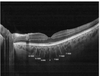
Choroidal thickness was measured perpendicularly from the outer edge of the retinal pigment epithelium to the choroid-sclera boundary at the fovea and at 7 more points located at fovea, at 500 滋m nasal to the fovea, 1000 滋m nasal to the fovea, 1500 滋m nasal to the fovea; 500 滋m temporal to the fovea, 1000 滋m temporal to the fovea, and 1500 滋m temporal to the fovea (Figure 1). Choroidal thickness measurements were performed by two masked physicians (Karalezli A and Eroglu FC). All basal OCT scans were performed at the same time of the day (in the morning) to avoid diurnal fluctuations [23]. The average of the two measurements was taken; the differences between readings of the masked physicians were found to be within 10滋m of the
Figure 1 Choroidal thickness measurements of a patient T-1500: Choroidal thickness at 1500 滋m temporal to the fovea; T-1000: Choroidal thickness at 1000 滋m temporal to the fovea; T-500: Choroidal thickness at 500 滋m temporal to the fovea; F: Choroidal thickness at fovea; N-500: Choroidal thickness at 500滋m nasal to the fovea; N-1000: Choroidal thickness at 1000滋m nasal to the fovea, N-1500: Choroidal thickness at 1500 滋m nasal to the fovea.
mean. The inter examiner reproducibility of the choroidal thickness measurements was assessed by measuring the intraclass correlation coefficient (ICC).
Statistical Analysis Statistical analysis was performed using the statistical package SPSS v15.0 (SPSS Inc., Chicago, USA, 2006). Data were analyzed by Mann-Whitney test. The categorical variables between the groups were analyzed using the 2test and to quantify the reproducibility of manual re-measurements of the choroidal thickness in cases of alignment errors, the ICC was calculated. The value <0.05 was considered statistically significant
RESULTS
The mean age of the OSAS patients was 58.6依9.7y (range 42-84y) and that of the controls was 58.2依7.1y (range 43-82y). There was no statistically significant difference in age between the groups ( =0.765). The AHI values were more than 30 in all OSAS patients and the mean AHI was 48.57依6.54 (regarding as several OSAS).
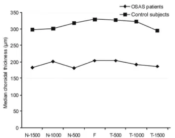
The median choroidal thickness was measured as 201 滋m (range 145-237 滋m) in OSAS patients and 324 滋m (range 296-383滋m) in the control subjects. The median choroidal thickness was statistically significant between groups ( <0 . 001 ) . Moreover, as shown in Figure 2, the choroidal thicknesses of the patients with OSAS were all thinner than those of the controls in all eyes and at all seven measurement points.
The interexaminer ICC for the choroidal thickness was 0.938 (95%CI, 0.908-0.985) and ICC was greater than 0.90 for all measurement points. Figure 3 shows the comparison of the median chroidal thichness measurements by two different observers in OSAS and control groups.
DISCUSSION
There are many reports in the literature that show the association between OSAS and many ocular disorders[8-14]. Intermittent airway obstruction in OSAS causes hypoxia and decrease main saturation of oxygen. Recurrent hypoxia and reperfusion episodes can lead to oxidative stress and inflammation, and also damage vascular endothelium and decreased responsiveness to vasodilatator agents, such as nitric oxide (NO). Moreover, drop in oxygen saturation leads to activation of the adrenergic system, high sympathetic tone and elevated muscle sympathetic nerve activity[7]. All of these conditions lead to vascular dysregulation, autonomic dysfunction and altered blood flow, because of imbalanced of vasodilatation and vasoconstriction [7,24]. This vascular phenomenon might compromise optic nerve perfusion and oxygenation, and several authors have mentioned a high prevalence of NAION and glaucoma in OSAS patients[11,12]. Retinal vein occlusions have been associated with OSAS as they may be result of a slowdown of blood circulation secondary to hypoxemia [13]. OSAS also has been recognized to be a risk factor for CSCR, because of elevated levels of
circulating catecholamines in patients with OSAS[5] .
The ocular blood flow can be assessed using color duplex imaging, laser speckle method, or Doppler flowmetry[25-27]. However, because the choroid receives aproximately 95% of all ocular blood flow, changes in its structure would help to evaluate choroidal, thus ocular blood flow[25-27].
Therefore, a precise clinical understanding of choroidal morphology should be important for understanding of the pathogenesis of many retinal and choroidal disease[28]. Certain Figure 2 Choroidal thickness measurements in OSAS patients and control subjects N-1500: Choroidal thickness at 1500 滋m nasal to the fovea; N-1000: Choroidal thickness at 1000滋m nasal to the fovea; N-500: Choroidal thickness at 500滋m nasal to the fovea; F: Choroidal thickness at fovea; T-500: Choroidal thickness at 500 滋m temporal to the fovea; T-1000: Choroidal thickness at 1000 滋m temporal to the fovea; T-1500: Choroidal thickness at 1500 滋m temporal to the fovea.
Figure 3 Comparison of the choroidal thickness measurements of the OSAS patients and control subjects between two different observers N-1500: Choroidal thickness at 1500 滋m nasal to the fovea; N-1000: Choroidal thickness at 1000滋m nasal to the fovea; N-500: Choroidal thickness at 500滋m nasal to the fovea; F: Choroidal thickness at fovea; T-500: Choroidal thickness at 500滋m temporal to the fovea; T-1000: Choroidal thickness at 1000 滋m temporal to the fovea; T-1500: Choroidal thickness at 1500 滋m temporal to the fovea.
栽藻造押8629原愿圆圆源缘员苑圆 8629-82210956 耘皂葬蚤造押ijopress岳员远猿援糟燥皂
characteristics of the choroid have been challenging to study histologically. To date, spectral domain OCT featuring high resolution and scanning speeds has allowed the choriod to be accesed [22]. Spaide [29] described a "enhanced deep imaging" (EDI) technique to optimize the parameters of OCT and this tecnique allows to visualise the full thickness of the choroid.
In the recent clinical studies, choroidal thickness was reported significantly decreased in patients with pathologic myopia, age-related macular degeneration, glaucoma and diabetic retinopathy or increased in patients with Voght-Koyanagi-Harada disease and CSCR [30-34]. In these studies, the authors speculated that abnormal choroidal thickness may be associated with choroidal circulation. Sogawa [35] reported that there were no correlation between the choroidal thickness and choroidal blood flow in the healthy young subjects. But, in two different studies, the authors mentioned that sildenafil citrate increases choroidal thickness because of a vasodilatory effect of sildenafil citrate on the choroidal circulation[36,37].
There are several studies related to choroidal blood flow in the patients with OSAS [38,39]. Tonini [38]reported that choroidal vascular response to hypoxia or hyperoxia does not alter in the patients with OSAS. Khayi [39]also reported that choroidal vascular changes in blood pressure were similar to healthy subjects. This result was explained with an autoregulation mechanism of the choroid. Contrary to retinal and optic nerve head vasculature, choroid vessels are subjected to autonomic regulation. But, ability to authoregulate of the choroid may be altered in smokers, age-related maculopathy, CSCR and glaucoma[40-43].
In our study, we showed that the median choroidal thickness was statistically thinner in the eye of patients with severe OSAS than that of the controls. We think that hypoxia and reperfusion episodes are responsible for pathophysiology of the OSAS, and these episodes cause oxidative stress, inflammation, damage to vascular endothelium and decreased responsiveness of the vessels to nitric oxide. Insufficient vasodilatation of vessels may lead to decrease in choroidal blood flow. We speculated that impaired autoregulation of the choroid in severe OSAS may be responsible for thinner choroidal thickness than that of controls.
There is a shortcoming regarding our study. A larger cohort would have allowed more meaningful analysis on the relation between the severity of OSAS and choroidal thickness; however, this can be achieved in a further study based on the preliminary findings of this study. Moreover, the design of the study with age, BMI and sex-matched control group and high ICCs between the two operators (ICC>0.90, for all measurement points) make the results more valuable.
In conclusion, patients with severe OSAS tend to have thinner choroids than healthy individuals, which in turn,
might be a result of the autonomic disregulation associated with this disease. Further studies with larger subgroups of this disease, including mild-modarete OSAS, are necessary to come to a more definite conclusion.
ACKNOWLEDGEMENTS
Conflicts of Interest: Karalezli A, None; Eroglu FC, None;Kivanc T, None; Dogan R, None.
REFERENCES
1 Kiekens S, Veva De Groot, Coeckelbergh T, Tassignon MJ, van de Heyning P, Wilfried De Backer, Verbraecken J. Continuous positive airway pressure therapy in associated with an increase in intraocular pressure in obstructive sleep apnea syndrome. 2008;49 (3): 934-940
2 Punjabi NM. The epidemiology of adult obstructive sleep apnea. 2008;5(2):136-143
3 Guilleminault C. Clinical features and evaluation of obstructive sleep apnea. In: Karger MH, Roth T, Dement WC, editors. Principles and practice of sleep medicine. London: Sauders; 1994;165-170
4 Caples SM, Garcia-Touchard A, Somers VK. Sleep-disordered breathing and cardiovascular risk. 2007;30(3):291-303
5 Marrone O, Riccobono L, Salvaggio A, Mirabella A, Bonanno A, Bonsignore MR. Catecholamines and blood pressure in obstructive sleep apnea syndrome. 1993;103(3):722-727
6 Arnaud C, Dematteis M, Pepin JL, Baguet JP, L佴vy P. Obstructive sleep apnea, immuno-inflamation, and atherosclerosis.
2009;31(1):13-125
7 Jelic S, Padeletti M, Kawut SM, Higgins C, Canfield SM, Onat D, Colombo PC, Basner RC, Factor P, LeJemtel TH. Inflammation, oxidative stress, and repair capacity of the vascular endothelium in obstructive sleep apnea. 2008;117(17):2270-2278
8 Mojon DS, Hess CW, Goldblum D, Boehnke M, Koerner F, Gugger M, Bassetti C, Mathis J. Normal tension glaucoma is associated with sleep apnea syndrome. 2002;216(3):180-184
9 Bendel RE, Kaplan J, Heckman M, Fredrickson PA, Lin SC. Prevelance of glaucoma in patients with obstructive sleep apnoea- a cross-sectional case-series. 2008;22(9):1105-1109
10 Karger RA, White WA, Park WC, Rosales AG, McLaren JW, Olson EJ, Woog JJ. Prevalence of floppy eyelid syndrome in obstructive sleep apnea syndrome. 2006;113(9):1669-1674
11 Mojon DS, Mathis J, Zulauf M, Koerner F, Hess CW. Optic neuropathy associated with sleep apnea syndrome. 1998;105 (5): 874-877
12 Bucci FA Jr, Krohel GB. Optic nerve swelling secondary to the obstructive sleep apnea syndrome. 1986;105(4):428-430 13 Glacet-Bernard A, Leroux les Jardins G, Lasry S, Coscas G, Soubrane G, Souied E, Housset B. Obstructive sleep apnea among patients with retinal vein occlusion . 2010;128(12):1533-1538 14 Jain AK, Kaines A, Schwartz S. Bilateral central serous chorioretinopathy resolving rapidly with treatment for obstructive sleep apnea. 2010;248(7):1037-1039 15 Maul EA, Friedman DS, Chang DS, Boland MV, Ramulu PY, Jampel HD, Quigley HA. Choroidal thickness measured by spectral domain optical coherence tomography: factors affecting thickness in glaucoma patients.
2011;118(8):1571-1579
16 Manjunath V, Taha M, Fujimoto JG, Duker JS. Choroidal thickness in normal eyes measured using Cirrus-HD optical coherence tomography.
17 Drexler W, Fujimoto JG. State-of-the art retinal optical choerence tomography. 2008;27(1):45-88
18 Sander B, Larsen M, Thrane L, Hougaard JL, Jongersen TM. Enhanced optical coherence tomography imaging by multiple scan averaging.
2005;89(2):207-212
19 Lin PW, Friedman M, Lin HC, Chang HW, Pulver TM, Chin CH. Decreased retinal nerve fiber layer thickness in patients with obstructive sleep apnea/hypopnea syndrome. 2011; 249(4):585-593
20 Casas P, Ascaso FJ, Vicente E, Tejero-Garc佴s G, Adiego MI, Crist佼bal JA. Retinal and optic nerve evaluation by optical coherence tomography in adults with obstructive sleep apnea hypopnea syndome (OSAHS).
2013;251(6):1625-1634
21 Fujiwara T, Imamura Y, Margolis R, Slakter JS, Spaide RF. Enhanced depth imaging optical coherence tomography of the choroid in highly myopic eyes. 2009;148(3):445-450
22 Branchini L, Regatieri CV, Flores-Moreno I, Baumann B, Fujimoto JG, Duker JS. Reproducibility of choroidal thickness measurements across the three spectral domain optical coherence tomography systems.
2012;119(1):119-123
23 Tan CS, Ouyang Y, Ruiz H, Sadda SR. Diurnal variation of choroidal thickness in normal, healthy subjects measured by spectral domain optical coherence tomography. 2012;53(1):261-266 24 Ip MS, Tse HF, Lam B, Tsang KW, Lam WK. Endothelial function in obstructive sleep apnea and respond to treatment.
2004;169(3):348-353
25 Sugiyama T, Araie M, Riva CE, Schmetterer L, Orgul S. Use of laser speckle flowgraphy in ocular blood flow research. 2010; 88(7):723-729
26 Portman N, Gugleta K, Kochkorov A, Polunina A, Flammer J, Orgul S. Choroidal blood flow response to isometric exercise in glaucoma patients and patients with ocular hypertension. 2011;52 (10):7068-7073
27 Bill A. Circulation in the eye. In: Renkin EM, Michel CC, eds. Handbook in physiology. Microcirculation, Part 2. The American Physiological Society, Bethesda, MD; 1984:1001-1035
28 Polska E, Polak K, Luksch A, Fuchsjager-Mayrl G, Petternel V, Findl O, Schmetterer L. Twelve hour reproducibility of choroidal blood flow parameters in healthy subjects. 2004;88(4):533-537 29 Spaide RF, Koizumi H, Pozzoni MC. Enhanced depth imaging spectral domain optical coherence tomography. 2008;146 (4): 496-500
30 Chung SE, Kang SW, Lee JH, Kim YT. Choroidal thickness in polipoidal choroidal vasculopathy and exudative age-related macular degeneration. 2011;118(5);840-845
31 Kubota T, Jonas JB, Naumann GO. Decreased choroidal thickness in
eyes with secondary age ange glaucoma. An aetiological factor for deep retinal changes in glaucoma? 1993;77(7):430-432 32 Esmaeelpour M, Povazay B, Hermann B, Hofer B, Kajic V, Hale SL, North RV, Drexler W, Sheen NJ. Mapping choroidal and retinal thickness variation in type 2 diabetes using three-dimensional 1060-nm optical coherence tomography. Sci 2011;52(8):5311-5316 33 Fong AH, Li KK, Wong D. Choroidal evaluation using enhanced depth imaging spectral-domain optical coherence tomography in Vogt-Koyanagi-Harada disease. 2011;31(3):502-509
34 Immura Y, Fujiwara T, Margolis R, Spaide RF. Enhanced depth imaging optical coherence tomography of the choroid in central serous chorioretinopathy. 2009;29(10):1469-1473
35 Sogawa K, Nagaoka T, Takahashi, A, Tanano I, Tani T, Ishibazawa A, Yoshida A. Relationship between choroidal thickness and choroidal circulation in healthy young subjects. 2011;153 (6): 1129-1132.e1
36 Vance SK, Imamura Y, Freund KB. The effects of sildenafil citrate on choroidal thickness as determined by enhanced depth imaging optical coherence tomography. 2011;31(2):332-335
37 Kim DY, Silverman RH, Chan RV, Khanifar AA, Rondeau M, Lloyd H, Schlegel P, Coleman DJ. Measurement of choroidal perfusion and thickness following systemic sildenafil (Viagra誖 ). 2013;91 (2): 183-188
38 Tonini M, Khayi H, Pepin JL, Renard E, Baguet JP, L佴vy P, Romanet JP, Geiser MH, Chiquet C. Choroidal blood-flow responses to hyperoxia and hypercapnia in men with obstructive sleep apnea. 2010;33 (6): 811-818
39 Khayi H, Pepin JL, Geiser MH, Tonini M, Tamisier R, Renard E, Baguet JP, Levy P, Romanet JP, Chiquet C. Choroidal blood flow regulation after posture change or isometric exercise in man with obstructive sleep apnea syndrome. 2011;52(13):9489-9496
40 Wimpissinger B, Resch H, Berisha F, Weigert G, Schmetterer L, Polak K. Response of choroidal blood flow to carbogen breathing in smokers and non-smokers. 2004;88(6):776-781
41 Pournaras CJ, Logean E, Riva CE, Petrig BL, Chamot SR, Coscas G, Soubrane G. Regulation of subfoveal choroidal blood flow in age-related macular degeneration. 2006;47(4):1581-1586 42 Tittl M, Maar N, Polska E, Weigert G, Stur M, Schmetterer L. Choroidal hemodynamic changes during isometrix exercise in patients with inactive central serous chorioretinopathy. 2005;46 (12): 4717-4721
43 Emre M, Orgul S, Gugleta K, Flammer J. Ocular blood flow alteration in glaucoma is related to systemic vascular dysregulation.