CONJUGATED POLYMER NANOPARTICLES FOR
BIOMEDICAL APPLICATIONS INCLUDING BIOIMAGING
AND DRUG DELIVERY
A THESIS
SUBMITTED TO THE DEPARTMENT OF CHEMISTRY
AND THE GRADUATE SCHOOL OF ENGINEERING AND SCIENCE OF BİLKENT UNIVERSITY
IN PARTIAL FULFILLMENT OF THE REQUIREMENTS
FOR THE DEGREE OF
MASTER OF SCIENCE
By ÖZLEM ÜNAL
I certify that I have read this thesis and that in my opinion it is fully adequate, in scope and in quality, as a thesis for the degree of Master of Science.
………. Assoc. Prof. Dr. Dönüş TUNCEL (Advisor)
I certify that I have read this thesis and that in my opinion it is fully adequate, in scope and in quality, as a thesis for the degree of Master of Science.
………. Prof. Dr. Engin Umut AKKAYA
I certify that I have read this thesis and that in my opinion it is fully adequate, in scope and in quality, as a thesis for the degree of Master of Science.
………. Assist. Prof. Dr. Özlen KONU
Approved for the Graduate School of Engineering and Science:
………. Prof. Dr. Levent ONURAL
iii
ABSTRACT
CONJUGATED POLYMER NANOPARTICLES FOR BIOMEDICAL APPLICATIONS INCLUDING BIOIMAGING AND DRUG DELIVERY
Özlem Ünal
M.S. in Department of Chemistry Supervisor: Assoc. Prof. Dr. Dönüş TUNCEL
July, 2013
In this study, the ability of fluorene-based conjugated polymer nanoparticles in the delivery of anticancer therapeutics and bioimaging was investigated through in vitro cytotoxicity assessments and fluorescence imaging. In order to prepare the nanoparticles, green light emitting polymer,poly[(9,9-bis{6-dimethylaminohexyl}fluorenyl-2,7-diyl)-co-(1,4-benzo-{2,1,3}-thiodiazole)]
(PDAFBT),was synthesized via Suzuki coupling reaction and characterized by nuclear magnetic resonance (NMR), electrospray ionization mass spectroscopy (ESI-MS), matrix assisted laser desorption ionization mass spectroscopy (MALDI), fourier transform infrared spectroscopy (FT-IR), UV-visible absorption spectroscopy (UV-Vis) and fluorescence spectroscopies. PDAFBT nanoparticles were synthesized through reprecipitation method which is based on the injection of the polymer solution in a good solvent into a poor solvent for the polymer to enhance the collapse of polymer chains in the form of spherical nanoparticles. Anti-cancer drug, camptothecin (CPT), was entrapped into the PDAFBT nanoparticles via hydrophobic interaction during the nanoparticle formation. Size, surface charge, morphology and optical characterizations of blank and CPT loaded nanoparticles were investigated by dynamic light scattering (DLS), scanning electron microscopy (SEM), transmission electron microscopy (TEM), UV-Vis and fluorescence spectrocopies. In order to investigate the properties of nanoparticles as drug carrier, drug encapsulation efficiency (EE) and drug loading efficiency (LE) were determined and EE was found to be as 92% and 75% for the 1:35 and 1:25 polymer to drug ratios, respectively, while LE of PDAFBT nanoparticles was determined as 2.67±0.3 %. Preliminary drug release profile which needs to be strengthen statistically, of PDAFBT nanoparticles was analyzed and showed that almost 100% of the loaded CPT was
iv
released steadily during first 48h time period indicating that the release of CPT is not a burst process. Stability of blank and CPT loaded PDAFBT nanoparticles was examined in different biological media including cell culture medium, bovine serum albumine (BSA) andhuman serum. The precipitation in nanoparticle dispersion was observed due to adsorption of proteins onto the surface of polymers.
In vitro dose dependent cytotoxicity of blank and CPT loaded PDAFBT
nanoparticles was assessed through 3-[4,5-dimethylthiazol-2-yl]-2,5-diphenyl tetrazoliumbromide (MTT) toxicology assay on human carcinoma breast cancer cell lines, namely,MDA-MB-231, MDA-MB-157 and MCF-7 cells. The blank PDAFBT nanoparticlesdo not exhibit significant toxicity to MDA-MB-231 and MDA-MB-157 cells up to 40 μM, however, a linear decrease on the viability of MCF-7 cells was detected ata concentration higher than 2.5 μM and nearly 40% of the cells were dead at 40 μM.The half maximal inhibitory concentration (IC50 ) values of free CPT and CPT loaded PDAFBT NPs were calculated to be 4.50μM and 1.35μM for MDA-MB-157 cell line, respectively, and 1.97μM and 1.05μM for MDA-MB-231 cells, respectively. These results indicate clearly that the CT-loaded nanoparticles are more efficient than free CPT in destroying the cancer cells. Fluorescent microscope images show also the efficient internalization of blank and CPT-loaded nanoparticles by MDA-MB-157 and MDA-MB-213 cell lines. Preliminarily in vivo studies have been performed on embryonic zebra fish and C. Elegans animal models using these nanoparticles and the efficient uptake of nanoparticles by the tissues have been observed by fluorescent microscopy. This results indicate that CPNs are promising for imaging and delivery vehicles for time and dose dependent treatments.
v
ÖZET
KONJUGE POLİMER NANOPARÇACIKLARIN İLAÇ TAŞINIMI VE BİYOLOJİK GÖRÜNTÜLEME İÇEREN BİYOMEDİKAL
UYGULAMALARI Özlem Ünal
Kimya Bölümü Yüksek Lisans Tezi Tez Yöneticisi : Doç. Dr. Dönüş TUNCEL
Temmuz, 2013
Bu çalışmada, floren tabanlı konjuge polimer nanoparçacıkların antikanser terapatik taşınımı ve biolojik görüntüleme verimi, canlı dışı toksik inceleme ve floresan görüntüleme yöntemleriyle incelenmiştir. Nanoparçacıkların hazırlanmasında yeşil ışıyan poli[(9,9-bis{6-dimetilaminohekzil}florenil-2,7-dil)-ko-(1,4-benzo-{2,1,3}-tiyadiazol)](PDAFBT) polimeri kullanılmıştır. PDAFBT polimeri, Suzuki kenetleme reaksiyonu ile sentezlenmiş, karekterizasyonu nükleer manyetik rezonans spektroskopileri ( H1-NMR, C13-NMR), elektrosprey ionizasyon kütle spektroskopisi (ESI-MS), matris destekli lazer salınımlı kütle spektroskopisi (MALDI), fourier transformu kızılötesi spektroskopisi (FT-IR), UV-Vis absorbans ve floresans spektroskopileri ile yapılmışmıştır. PDAFBT nanoparçacıkları, çözücünün hidrofilikliğinin azaltılmasına dayanan ve polimer zincirlerinin çökerek küresel forma dönüşmesine dayanan geriçöktürme yöntemi ile hazırlanmıştır. Anti-kanser terapatiği olarak kullanılan kamptotesin, PDAFBT nanoparçacıklarının içerisine nanoparçacık oluşumu sırasında etkin olan hydrofobik etkileşimle yüklenmiştir. Nanoparçacıkların boyut, yüzey yükü, şekilsel ve ışıksal karekterizasyonları dinamik ışık saçılımı (DLS), geçirimli elektron (TEM), taramalı elektron mikroskopileri (SEM), UV-Vis absorbans ve fluoresans spektroskopileri ile yapılmıştır. Nanoparçacıkların ilaç taşıyıcı olarak özelliklerinin incelenmesi amacıyla, ilaç kapsülleme (EE) ve ilaç taşıma (LE) verimleri analiz edilmiş ve ilaç kapsülleme verimi 1:35 ilaç-polimer oranında % 92, 1:25 ilaç-polimer oranında % 75 olarak bulunurken, ilaç taşıma verimi ise % 2.67±0.3 olarak belirlenmiştir. Öncül bir çalışma olan ve sonuçları istatiksel olarak güçlendirilecek PDAFBT
vi
nanoparçacıkların ilaç salınım profili analiz edilmiş ve ilaç miktarının yaklaşık % 100 ünün 48 saat içinde salındığı gözlenmiştir.
Boş ve kamptotesin yüklü PDAFBT nanoparçacıkların hücre kültürü ortamı, bovin serum albumin (BSA) ve insan serumu içeren farklı biyolojik ortamlardaki stabilitesi test edilmiş, insan serumu ve bovin serum albumin ortamında proteinlerin polimer yüzeyine tutunması nedeniyle nanoparçacık solusyonunda çökme gözlenmiştir. Boş ve ilaç yüklü nanoparçacıkların canlı dışı (in vitro) doza bağlı toksik incelenmesi, tetraazolyum boyasının redüklenmesine dayanan MDA-MB-231, MDA-MB-157, MCF-7 insan göğüs kanseri hücre hatları üzerinde MTT toksikoloji testi ile yapılmıştır. Sadece kamptotesin ve kamptotesin yüklü PDAFBT nanoparçacıklar için % 50 inhibe edici konsantrasyon (IC50) değerleri MDA-MB-157 hücreleri için 4.50 μM ,1.35 μM ve MDA-MB-231 hücleri için 1.97μM ve 1.05μM’dır ve polimer kapsuller sayesinde ilacın hücrerele etkin alınımı gösterir. PDAFBT ve PPFBT nanoparçacıkların yaşayan canlı modelleri olan zebra balığı ve C. Elgans üzerindeki yapılan görüntülemeleri nanoparçacıkların dokulara etkin bir şekilde nüfuz ettiğini desteklemektedir ayrıca zamana ve doze bağlı uygulamalar için umut vericidir.
Anahtar Kelimeler: Konjuge polimer nanoparçacıklar, ilaç taşıma, biyolojik görüntüleme
vii
Dedicated to the memory of
Burak Alisir
viii
ACKNOWLEDGEMENT
First of all, I would like to express my sincere appreciation to my research supervisor Assoc. Prof. Dönüş Tuncel for her guidance, support, trust and encouragement for not only my reseach project, but also for my academic plans. Her positive attitude towards my studies have helped me to ahead, despite all the academic challenges and huddles I faced.
I would also thank examining committee members, Prof. Engin Umut Akkaya and Assist. Prof. Özlen Konu, for their time and valuable suggestions on my thesis.
I am deeply grateful to Prof. Özlen Konu and her PhD student Ermira Jahja for their worthy colaboration and and significant support to biological part of the study. Moreover I would like to thank to Prof. Arzu Atalay and Selen Güçlü due to their trainings for C. Elegans experiments.
I would like to extend special thanks to Vusale İbrahimova for her guidance in the laboratory and her valuable friendship.
I also would like to express my appreciation to all of those with whom I have had the pleasure to work within the past three years. I am sincerely grateful to my old and new colleagues Meltem Aygüler, Müge Artar, Şeyma Ekiz, Eda Kocak, Özlem Gezici, Muazzam Idris, Josheed PK., Hamidou Keita, Esra Soner and Sinem Gürbüz.
I have to express my thanks to Alp Özgün for donation of his blood for the experiments with “human serum” in my research and sharing his ideas.
I would like to thank to my close friends, Gözde Barım, Merve Doğaç, Tuğçe Durgut, Tuba Yaşar, Duygu Demircioğlu, Melis Tunalı, Menekşe Koca, Seda Selçuk, Merve Taner, Merve Şahinsoy, Gülşen Uğrar, Mert Demirdelen, Yiğit Altay, Aykut Aydın, and Yiğit Can Yüceyurt for their worthy fellowship and significant contrubition to my life.
Lastly, I wish to sincerely thank to my mother, father and brother for their encouragement, trust and motivation. It is very important for me to feel their love and suppot during my life.
ix
CONTENTS
CHAPTERS
CHAPTER 1 ... 1 1. INTRODUCTION ... 1 CHAPTER 2 ... 2 2. BACKROUND ... 2 2.1. Conjugated Polymers ... 22.1.1 Fluorene Based Conjugated Polymers ... 5
2.1.2.Synthesis methods of conjugated polymers ... 6
2.2.Conjugated Polymer Nanoparticles ... 10
2.2.1 Preparation Methods of Conjugated Polymer Nanoparticles ... 11
2.2.2 Photophysical Properties and Optoelectronic Applications of CPNs .... 12
2.2.3 Conjugated Polymer Nanoparticles in Nanomedicine ... 14
CHAPTER 3 ... 23
3.EXPERIMENT RESULTS ... 24
3.1. General ... 24
3.2 Syntheses of Monomers and Polymer ... 25
3.2.1. Synthesisof 2,7-dibromo-9,9-bis-(6-bromo-hexyl)-9H-fluorene (M1) . 25 3.2.2. The Synthesis of {6-[2,7-Dibromo-9-(6-dimethylamino-hexyl)-9H-fluoren-9-yl]-hexyl}-dimethyl-amine ... 26
3.2.3. Synthesis of Poly[(9,9-bis{6-dimethylaminohexyl}fluorenyl-2,7-diyl)-co-(1,4-benzo-{2,1,3}-thiodiazole)] (PDAFBT) ... 27
3.2.4.Synthesis of PDAFBT Nanoparticles... 28
3.2.5. Synthesis of Drug Loaded PDAFBT Nanoparticles and Determination of Drug Loading Capacity of PDAFBT Nanoparticles ... 28
x
3.2.6. Drug Release Study ... 29
3.2.7. Sample Preparation from PDAFBT Nanoparticles for TEM imaging . 29 3.2.8. Preparation of PDAFBT Nanoparticle Solutions in Bovine Serum Albumin and Human Serum Media ... 30
3.3 Biological Assays ... 30
3.3.1. 3-[4,5-dimethylthiazol-2-yl]-2,5-diphenyl tetrazoliumbromide (MTT) In Vitro Toxicology Assay for Culture Cells Treated with Blank and Drug Loaded Nanoparticles... 30
3.3.2. 5-Bromo-2-deoxyuridine (BrdU) Cell Labeling and Proliferations Assay for Culture Cells Treated with Blank and Drug Loaded Nanoparticles ... 30
3.3.3. Bradford Protein Assay to Determine the Quantitative Amount of Human Plasma Proteins Adsorbed on the Surface of Nanoparticles ... 31
3.3.4 PDAFBT Nanoparticle Staining Protocol on Embryonic Zebra Fish .... 31
3.3.5 PPFBT Nanoparticle Staining protocol on C. Elegans ... 32
CHAPTER 4 ... 33
4. EVALUATION ... 33
4.1. Synthesis and Characterization of Monomers ... 34
4.1.1.Synthesis and Characterization of 2,7-dibromo-9,9-bis-(6-bromo-hexyl)-9H-fluorene (M1) ... 34
4.1.2.The Synthesis and Characterization of The Synthesis of {6-[2,7- Dibromo-9-(6-dimethylamino-hexyl)-9H-fluoren-9-yl]-hexyl}-dimethyl-amine (M2) ... 35
4.1.3. Synthesis and Characterization of Poly[9,9-bis{6-dimethylaminohexyl} fluorenyl-2,7-diyl)-co-(1,4-benzo-{2,1,3}-thiodiazole)] (PDAFBT) ... 38
4.2. Synthesis and Characterization of Blank and Drug Loaded Water Dispersible Conjugated Polymer Nanoparticles (CPNs) ... 43
xi
4.2.1. Synthesis and Characterization of poly[9,9-bis{6-dimethylaminohexyl} fluorenyl-2,7-diyl)-co-(1,4-benzo-{2,1,3}-thiodiazole)] (PDAFBT)
Nanoparticles... 43
4.2.2 Synthesis and Characterization of Drug Loaded poly[9,9-bis{6-dimethyl aminohexyl}fluorenyl-2,7-diyl)-co-(1,4-benzo-{2,1,3}-thiodiazole)](PDAFBT) Nanoparticles ... 47
4.2.3 Determination of the Drug Encapsulation Efficiency of PDAFBT Nanoparticles... 47
4.2.4. Characterization of Drug Loaded PDAFBT Nanoparticles ... 51
4.2.5. Determination Drug Release Profile of CPT Loaded PDAFBT Nanoparticles... 53
4.3. Biological Applications of Blank and Drug Loaded PDAFBT Nanoparticles ... 57
4.3.2. Determination of In-vitro Cyctoxicity of Blank and CPT Loaded PDAFBT Nanoparticles ... 61
4.3.3. In Vitro and In Vivo Screening of blank PDAFBT and PPFBT Nanoparticles... 67
4.3.3.1.Identification of PDAFBT Treated Cells by BrdU Labeling ... 67
4.3.3.2 In Vivo Screening of PDAFBT nanoparticles in Zebrafish Embryos as Model Organism ... 68
4.3.3.3.In Vivo Screening and Toxicity Assesment of PPFBT nanoparticles in C. Elegans as a Model Organism ... 70
CHAPTER 5 ... 73
5. CONCLUSION ... 73
BIBLIOGRAPHY ... 74
xii
LIST OF FIGURES
Figure 2.1. Conjugated and isolated π bonds in 1,3,6heptene.. ... 2
Figure 2.2. General band picture of an insulator, a semiconductor,a metal and conductivity of conjugated polymers compared to those of materials. ... 3
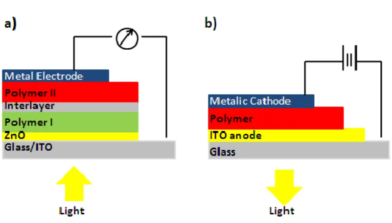
Figure 2.3. Ilustration of radiative and nonradiative transitions between the electronic states in a molecule after absorption of a photon by Jablonski diagram.12 . 4 Figure 2.4. Schematic representation of (a) photovoltaic cell (b) light emitting diode based on conjugated polymer. ... 5
Figure 2.5. Different emission colors based on backbone structure of co-polyfluorenes. ... 6
Figure 2.6. Basic representation of metal catalyzed C-C bond formation. ... 8
Figure 2.7. Reaction Mechanism of Heckcross-couplings. ... 8
Figure 2.8. Reaction Mechanism of Suzuki Cross Coupling ... 9
Figure 2. 9. Reaction Mechanism of Stille Cross Coupling. ... 10
Figure 2.10. Miniemulsion method for the preparation of polymer nanoparticles. .. 11
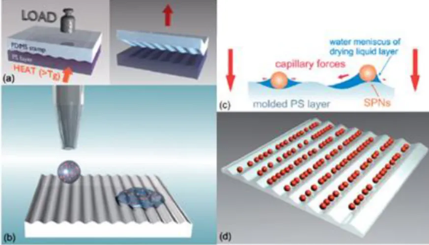
Figure 2. 11. Reprecipitation method for the preparation of polymer nanoparticles. 12 Figure2.12.Schematic representation showing fabrication process of semiconducting polymer nanoparticles for optoelectronic applications. ... 13
Figure 2.13. Differential contrast and fluorescence images of macrophage cells labeled with PFO PFBT polymer dots. ... 15
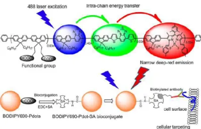
Figure 2.14. Schematic illustration of narrow emissive CPN bioconjugates for specific cellular targeting. ... 17
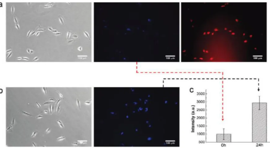
Figure 2.15. Light and fluorescence miscroscopy images of A549 cell after incubation with PFO/PG-DOX ... 18
Figure 2.16. Shematic representation of complex formation between poly(phenylene ethylene) CPNs and siRNA for actine B gene delivery. ... 19
Figure 2.17. Epifluorescence and epifluorescence/ DIC merged images of wild type C. Elegans A) untreated young adults, worms fed with fluorescent nanodiamonds for B) 2 h and C)12 h. ... 22
xiii
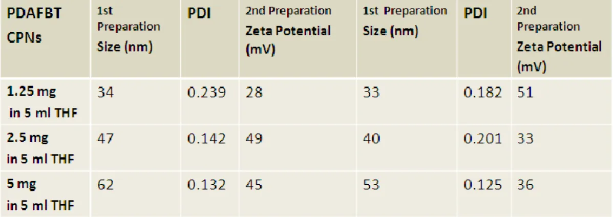
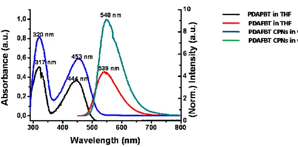
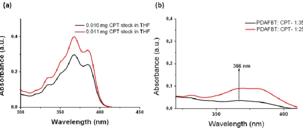
Figure 4.1.Schematic illustration of thesis work. ... 33 Figure 4.2. (a) 1H-NMR and (b)13C-NMR spectra of 2,7-dibromo-9,9-bis-(6-bromo-hexyl)-9H-fluorene (M1). ... 35 Figure4.3. (a)1H-NMR and (b)13C-NMR spectra of {6-[2,7-Dibromo-9-(6-dimethylamino-hexyl)-9H-fluoren-9-yl]-hexyl}-dimethyl-amine (M2)...37 Figure 4.4. TOF-MS spectrum of M2. ... 38 Figure4.5. (a)1H-NMR and (b)13C-NMR spectra of poly[9,9-bis{6-dimethylaminohexyl}fluorenyl-2,7-diyl)-co-(1,4-benzo-{2,1,3}-thiodiazole)] ... 40 Figure 4.6. (a) ESI-MS of PDAFBT (b) MALDI-MS of PDAFBT (c) MALDI-MS of PDAFBT in the wide spectrum. ... 41 Figure 4.7. FT-IR spectrum of poly[9,9-bis{6-dimethylaminohexyl}fluorenyl-2,7-diyl)-co-(1,4-benzo-{2,1,3}-thiodiazole)] ... 42 Figure 4.8. UV-Vis absorption and fluorescence emission spectra of poly[9,9-bis{6-dimethylaminohexyl}fluorenyl-2,7-diyl)-co-(1,4-benzo-{2,1,3}-thiodiazole)] ... 42 Figure 4.9. Cartoon representation of the preparation of PDAFBT nanoparticles. .. 43 Figure4.10. (a)Number avaragesize distrubution by histogram from DLS masurement and (b) zeta potential measurement of PDAFBT nanoparticles. ... 44 Figure 4.11. UV-Vis absorption and fluorescence emission spectra of PDAFBT in THF and dispersion of PDAFBT nanoparticles in water. ... 46 Figure 4.12. (a) SEM (b) TEM images of PDAFBT nanoparticles. ... 46 Figure 4.13. Schematic representation of the study about determination of drug encapsulation efficiency. ... 47 Figure 4.14. UV-Vis absorption spectra of (a) 16μg CPT stock in 3ml of THF and (b) 16 μg dialyzed CPT . ... 48 Figure 4.15. UV-Vis absorption spectra of (a) 0.011 mg and 0.016 mg CPT stocks in THF as references (b) dialyzed CPT which is loaded in PDAFBT at 1:35 and 1.25 drug to polymer ratios. ... 49 Figure 4.16. Number averagesize distribution by histogram from DLS measurements of CPT loaded PDAFBT nanoparticles for (a) 1:35 and (b) 1:25 drug to polymer ratios. ... 51
xiv
Figure 4.17. (a) UV-Vis absorption (b) normalized fluorescence spectra of CPT loaded PDAFBT nanoparticles in water at different ratios. ... 52 Figure 4.18. Photographic images of blank and CPT loaded PDAFBT nanoparticles under (a) day and (b) UV light. ... 53 Figure 4.19. TEM image of CPT loaded PDAFBT nanoparticles ... 53 Figure 4.20. (a) UV-Vis absorption spectra of reference CPT stocks while increasing its amount. (b) Fit curve which represent the absorption curves of CPT stock. ... 54 Figure 4.21. UV-Vis absorption spectra of samples withdrawn from the release medium at given time intervals. ... 55 Figure 4.22. Percentage release of CPT from PDAFBT nanoparticles during 48h. . 57 Figure 4.23. (a) Number avarage size change of blank PDAFBT nanoparticles in (b) Z-Avarage value (nm) of blank PDAFBT nanoparticles in human serum and BSA media masured by DLS. ... 58 Figure 4.24. Schematic representation of interaction PDAFBT with proteins. ... 59 Figure 4.25. Calculation of adsorbed proteins on surface of PDAFBT NPs in human serum. ... 60 Figure 4.26. Percent cell viability results of PDAFBT nanoparticles on the MCF-7, MDA-MB-231 and MDA-MB-157 cell lines. ... 63 Figure 4.27. Experimental set-up for MTT calorimetric assay in 96 well plates of CPT loaded PDAFBT nanoparticles with positive control of CPT, negative control of DMSO and blank PDAFBT nanoparticles ... 63 Figure 4.28. Percent cell viability results of (a) MDA-MB-231 (b) MDA-MB-157 and (c) MCF-7 cell lines after 4h MTT treatment... 65 Figure 4.29. Identification of PDAFBTtreated MCF-7 cells through BrdU labeling ... 67 Figure 4.30. PDAFBT NPs internalized MCF-7 cells (a) right after DAPI staining.(b) after one month storage. ... 68 Figure 4.31. Fluorescence microsope images of embryonic zebra fish ... 70 Figure 4.32. Fluorescence microscope images of non treated, PPFBT NPs stained C. Elgans ... 71 Figure 4.34. Number of progeny of nontreated and PPFBT treated C. Elegans in 5 days time period. ... 72
xv
LIST OF SCHEMES
Scheme 4. 1 Synthesis mechanism of the monomer 2,7-dibromo-9,9-bis-(6-bromo-hexyl)-9H-fluorene (M1). ... 34 Scheme 4.2. Synthesis mechanism of the monomer {6-[2,7-Dibromo-9-(6 dimethylamino-hexyl)-9H-fluoren-9-yl]-hexyl}-dimethyl-amine (M2). ... 36 Scheme 4.3. The synthesis mechanism of poly[9,9-bis{6-dimethylaminohexyl} fluorenyl-2,7-diyl)-co-(1,4-benzo-{2,1,3}-thiodiazole)] (PDAFBT). ... 39
xvi
LIST OF TABLES
Table 1. Number average size and zeta potential values of PDAFBT nanoparticles at different polymer concentrations. ... 45 Table 2. Percentage of diffused CPT from the membrane to the release medium. .. 49 Table 3. Percentage of loaded CPT by the PDAFBT nanoparticles for the CPT to PDAFBT, 1:35 and 1:25 : CPT ratios, respectively. ... 50 Table 4. Percentage of loaded CPT by the PDAFBT nanoparticles for the CPT to PDAFBT, 1:35 and 1:25 : CPT ratios, respectively. ... 55 Table 5. UV-Vis absorbance, concentration values and percentage of released CPT at 48h time period... 56 Table 6. IC50 values of free CPT and CPT loaded PDAFBT nanoparticles for MDA-MB-231, MDA-MB-157 and MCF-7 cell lines. ... 66
xvii
LIST OF ABBREVIATIONS
CPs Conjugated Polymers
CPNs Conjugated Polymer Nanoparticles HOMO Highest Occupied Molecular Orbital LUMO Lowest Unoccupied Molecular Orbital
PDAFBT Poly[(9,9-bis{6-dimethylaminohexyl}fluorenyl-2,7-diyl)-co-(1,4-benzo-{2,1,3}-thiodiazole) PPFBT Poly[9,9-bis{propenyl}fluorenyl-2,7-diyl))- co-(1,4-benzo{2,1,3}- thiodiazole)] PDAFBT NPs Poly[(9,9-bis{6-dimethylaminohexyl}fluorenyl-2,7-diyl)-co-(1,4-benzo-{2,1,3}-thiodiazole) Nanoparticles CPT Camptothecin THF Tetrahydrofuran CDCl3 d-Chloroform
NMR Nuclear magnetic resonance UV-VIS Ultraviole Visible
MALDI Matrix-Assisted Laser Desorption/Ionization ESI Electrosprey Ionization
MS Mass Spectroscopy
TEM Transmittance Electron Spectroscopy SEM Scanning Electron Microscopy
MTT 3-[4,5-dimethylthiazol-2-yl]-2,5-diphenyl tetrazoliumbromide BrdU 5-Bromo-2-deoxyuridine
1
CHAPTER 1
1. INTRODUCTION
Recent developments in nanotechnology provide special biomedical tools which are capable to deliver pharmaceuticals and biological agents with high efficiency, in a controlled time-scale and a safe way.These nano devices have an importance for drug delivery, molecular imaging and biomarkers. Traditional therapeutic approaches provide only non-specific drug mixtures but an efficient delivery needs more attention to design smart systems through a multifunctional aspect which target to visualize the carriers with an effective screening and increase the bio-compatibility. Nanoparticles improve the bio-availability of water-insoluble drugs, protect the drugs from physiological barriers and increase the body tolerability to the drugs and thereby, enhance the efficiency of drug delivery. Tracking the bio-distribution of nanocarriers is crucial to investigate the interaction of therapeutics with the sections of tissues and cells. Among a number of screening methods, fluorescence imaging has important impact to control the bio-distributions of pharmaceutical carriers in drug delivery research.
Typical fluorescent imaging agents containing conventional dyes have photobleaching and toxicity problems. Although quantum dots have high brightness and photostability, they are cytotoxic due to heavy metals from their nanocrystal core and their bio-compatibility is not high enough for biological applications.These limitations urgethe design of new fluorescent materials in drug delivery research. Development of fluorescent conjugated polymer materials is very promising in imaging guided drug delivery applications due their high brightness, good photostability and lower toxicity. Fluorene based conjugated polymer nanoparticles which are prepared through miniemulsion57 or reprecipitation59 methods have wide range of applications from nanomedicine 70,78,79 to molecular electronics.63-64
2
CHAPTER 2
2. BACKROUND
2.1. Conjugated Polymers
Broad range of polymers exhibit insulating properties, however if polymers are designed in such a way that having conjugated backbone, they could conduct the electricity (Figure 1). These polymers are called conjugated polymers and consist of unsaturated sp2 or sp hybridized carbon atom chains including pz orbitals which forms π overlap with adjacent unsaturated carbon atom and these overlaps form delocalization of π electrons along the polymer chain.1-2
Figure 2. 1. Conjugated and isolated π bonds in 1,3,6heptene..
This πstateformation leads to a frontier electronic gap, named as π-band gap which is relatively smaller than σ-band gap and varies from 1.5 eV to 4 eV depending on the structure.The energy spacing between highest occupied molecular orbital (HOMO) and lowest unoccupied molecular orbital (LUMO) is defined as band gap energy and identifies the level of conductivity (Figure 2). The ability of semiconducting behaviours of conjugated polymers come from this low energy band gap between conduction band (HOMO) and valence band (LUMO).1-4 Density of charge carriers affects the conductivity. Small band gaps in conjugated polymers provide ease in doping,which corresponds to partial oxidation or reduction, and charge carrier generation.4-6
The first conducting polymer was poly(acetylene) and synthesized using Ziegler-Natta catalyst in 1958 by K. Ziegler , G. Ziegler-Natta who were awarded 1966 Nobel Prize
3
in Chemistry.7 However only conjugation is not enough for being conductive, thus charge carriers which contains holes (missing electrons or positive charge) or electrons, have to be added to system (doping) to allow charge to migrate along the long distance. In 1974, Shirakawa and coworkers discovered that treatment with halogen vapour increases the conductivity of polyacetylene 109 times more than its natural form and were awarded the Nobel Prize 2000 “for the discovery and
development of electrically conductive polymers” 8-9
Figure 2.2. General band picture of an insulator, a semiconductor,a metal and conductivity of conjugated polymers compared to those of materials.
Conjugated polymers could also exhibit photoluminescence (PL) properties when they are excited with a suitable wavelength of a light. Luminescence is the spontaneous emission of light from electronically excited states in the substances under optical excitation and it can be named as fluorescence when light emits from singlet excited states and phosphorescence when light emits from triplet excited states.10 Jablonski Diagrams are used to explain light absorption and emission phenomena through illustrating electronic ground and excited states with a number of vibrational energy levels (Figure 3). When a photon at particular energy is absorbed by the fluorophore (fluorescent molecule) which is excited to higher vibrational levels of S1 or S2,several pathways can be followed including vibrational relaxation, internal conversion, fluorescence, intersystem crossing, and phosphorescence to dissipate the energy. Conjugated polymers exhibit allowed absorptions and emissions and generally luminescence efficiency changes with the delocalization and
4
polarization of the electronic structure. For example, highly delocalized and polarizable polymers are weakly luminescent due to dissociation of photo-generated electron-hole pairs through the chain in order to generate charge carriers, then combination of these free carriers to form triplet or deactivation by non-radiative relaxations occur.11
Figure 2.3. Ilustration of radiative and nonradiative transitions between the electronic states in a molecule after absorption of a photon by Jablonski diagram.12
Electroluminescence, emitting light due to excitation by the flow of electric current, became another important feature of conjugated polymers with the discovery of electroluminescent property of semiconductive conjugated polymer poly(p-phenylenevinylene) by Friend et al in 1990.13 In the last decade, polymer light emitting diodes (PLEDs) (Figure 4a), which produce light by the fast decay of excited molecular states in the polymers, have been under great interest and polythiophenes14, polyphenylenes15, polyfluorenes16 were used for various electroluminescent applications due to their comparable efficiency and brightness to their inorganic analogs.17 In the area of photovoltaics which is based on producing electrolytic cells to generate electrical current when exposed to light18 (Figure 4b), conjugated polymers are widely preferred semiconductors due to their ability of absorbing significant fraction of sun light and ease of processing on flexible films at low cost.19 With the discovery of bulk heterojunction, the improvements in conjugated polymer based photovoltaic cells (PV) through the attempts to achieve smaller band gaps, wider band widths,higher carrier mobilities have been obtained
5
and their power efficiency enhanced 100 folds greater in the past decade.20 As another photonic application, conjugated polymers are unique class of solid state laser materials due to their emission spectrum which involves entire visible spectrum.21
Figure 2.4. Schematic representation of a) photovoltaic cell b) light emitting diode based on conjugated polymer.
Apart from being high performance plastic photonic devices, conjugated polymers are versatile chemosensors performing for variety of sensing methods such as conductrometric sensors due their conductivity, potentiometric sensors which is developed on the basis of their reversible nature of redox processes, calorimetric sensors based on the sensitivity of CP’s band gap with respect to polymer conformation22. Moreover, CPs are preferred significantly for the applications as fluorescent chemosensors to detect the substances from biomolecules to explosives 23-24 due to their direct fluorescent response to the analytes in the environment, conformational change of backbone by the interaction with analyte, ease of substitution of flexible backbones with receptor groups to achieve molecular recognition and amplify the fluorescence signals.25 Furthermore, conjugated polymers have applications also in the new emerging areas such as artificial muscles and nano-electromechanical systems.26-27
2.1.1 Fluorene Based Conjugated Polymers
Polyfluorenes (PFs) are stepladder types polyphenylenes, being very promising for the fabrication of LED28-29and PV diodes30-31. The most important advantage in the
6
area of optoelectronics of these polymers is their high photoluminescence efficiency both in solution and in solid films with the emission of blue light in the visible spectrum due to their rigid biphenyl units providing a large band gap for blue emission.32-33 They show better thermal- and photo-stability than their analogs in the optoelectronics.34 Moreover, highly reactive protons at C-9 position of the fluorene unit allow substitutions at this position without destroying their electrochemical properties and provides the opportunities to improve the solubility and processability of these polymers.35-36 Various fluorene copolymers are synthesized, in order to obtain wide range of emission colors in the entire visible spectrum and tune their band gaps as shown in Figure 5.28 Apart from applications in optoelectronics, PFs are widely used in bioimaging37and chemosensory38applications.
Figure2.5. Different emission colors based on backbone structure of co-polyfluorenes.
2.1.2. Synthesis methods of conjugated polymers
Polymerization is the process of combination of desired monomers through stepwise oligomer formation to long chains or adding unsaturated monomers onto the active site of the growing polymers in a controlled manner.39 Synthetic methods are significant to obtain polymers including well defined molecular weight and polydispersity. Various methods have been developed to synthesize conjugated polymers and tried to design pure, soluble, efficient electrochemical features containing polymers by preserving delocalized pi states in the polymer chain. One of these methods is electrochemical polymerization which is based on deposition of polymer thin films from monomers such as aniline, pyrole, thiophene on the anode.40 Experimentally, polymerization is carried outin a single compartment electrochemical cell with three electrodes configuration and electrochemical bath
7
consists of monomer and electrolyte in an appropriate solvent.41 When going into a literature research in this area, it can be seen that, the electrochemical synthesis of conducting polypyrole by Diaz et al. provided a new synthetic way in the polymer science and triggered various studies in the field.42 Polythiophene and poly(isothionaphthalene) have been other electrochemically prepared conductive polymers which is carried out Wuld et al.43 Electrochemical synthesis of polythiophenes in microemulsion medium by using cationic surfactant has been reported by Murugan and his co-worker.44 Electrochemical synthesis of these conductive polymers consists of especially polypyroles and polythiophenes provides many enhanced applications in the field of optoelectronics.41 Besides, catalytic chain polymerization, which is mainly used for the synthesis of polyvinylenes and polyphenylenevinylenes45, chemical oxidation polymerization for the preparation of polyacetylenes and polyanilines unfortunately with high amount of side products40, photochemical polymerization typically for the synthesis of polydiacetylenes46, are other feasible approaches for the synthesis of conjugated polymers.
Transition metal mediated cross coupling reaction is one of the most powerful techniques to synthesize conjugated polymers via carbon-carbon bond formation between sp2-sp2 or sp2-sp hybridized carbon atoms and recently was awarded with the Nobel Prizes in Chemistry 2010 for palladium catalyzed cross couplings in
organic synthesis.47 Nucleophilic substitutions at sp2 and sp carbons are not easy and need transition metal catalysts.The theory of cross coupling reactions is basically, assembling of carbon atoms of two reactants on the transition metal through the metal-carbon bonds and resulting carbon-carbon bonds between these couples after introducing them into very close proximity as shown schematically in Figure 6. Generally, palladium (or nickel) catalyst couplings are believed to follow the similar catalytic cycle with some changes in the steps including activation of the catalyst and release of the product.The reactions start with oxidative addition, reacting of Pd(0) catalyst with organohalide and changing to Pd(II) with the formation of organopalladium compound via C- Pd bonding, after that step there are some differences in different type of couplings but transmetallation is the common step consisting of the co-ordination of nucleophile (second carbon source) to Pd and assembling of two reagents on the Pd and followingly reacting of these couples with
8
each other. After carbon carbon bond formation is achieved then regeneration of Pd(0) and releasing of newly formed organic compound occur at the final step which is called as reductive elimination.
Figure 2. 6. Basic representation of metal catalyzed C-C bond formation.
Discoveries in cross coupling reactions are triggered by publications of Kumada et.
alin the beginning of 1972, including carbon-carbon bond formation by cross
coupling of Grignard reagents with organic halides through Ni catalyst.48 Again in 1972, a new cross coupling reactions of aryl, vinyl halides and olefins in the presence of Pd catalyst has been reported by Heck et al.49 and it became the most important carbon-carbon bond forming reaction under the truth that palladium salts are mostly useful. In the case of Heck coupling, general catalytic cycle is followed between organohalide and olefin but differently carbon-carbon bond forms via migratory insertion including migration of carbon atom on Pd to one of the carbon atoms of the coordinated olefin and release of newly formed organic compound through β-H elimination (Figure 7).
9
In 1979, another most attractive approach to carbon-carbon bond formation has been discovered by Suzuki and Miyaura including trans metal catalyzed cross coupling between an organoboron compound and organic halide.50 Differently from Heck coupling, reaction mechanism of Suzuki coupling involves the activation of organoboronic acid through a base and facilitation of transmetallation. Choosing the organoboronic compounds provides various advantages such as making coupling reactions more practical due to the weak nucleophilicity and stability of organoborones, having high chemoselectivity, providing to run the reaction at mild conditions and non-toxicity of boron compounds. Moreover, the most important benefit of this coupling is to tolerate many different functional groups and providing efficient couplings between biaryl, aryl-vinyl, alkyl-aryl and alkyl-alkyl compounds.47Due to these advantages, Suzuki cross-coupling reaction was preferred for the synthesis of polymers in this thesis.
Figure 2. 8. Reaction Mechanism of Suzuki Cross Coupling51. (Reprinted with the permission from ref 51. Copyrigth (1995) American Chemical Society)
In 1977, Negishi and coworkers reported using organozinc ligands as nucleophiles in the palladium catalyzed couplings containing organozinc and organoaliminum compounds.52
One of the most versatile organometallic reagent in Pd catalyst cross coupling reactions is organotin which is used as a novel method for carbon-carbon bond generation between an organohalide and organostanne developing through Stille and
10
coworkers.53 The most important advantage of this reaction is to tolerate variety of functional groups containing alcohols, ketones, enones, esters, nitriles, nitro groups.
In the reaction, Pd catalyst is coupled with an electrophile with a vinyl or a aryltin compound through oxidative addition. After transmetallation with organostanne, functional group of organostanne replaces the halide anion on Pd(II) complex and finally coupled product is released with the reductive elimination (Figure 8).
Sonogashira54 reaction which is based on the reaction between aryl and alkynes under Pd and Cu catalysis, Yamamato55 obtaining biaryl from aryl halides in the presenceof Ni catalyst, are other coupling reactions to synthesize conjugated polymers.
Figure 2. 9. Reaction Mechanism of Stille Cross Coupling.53 (Reprinted with the permission from ref 53. Copyrigth (1986) Wiley)
2.2. Conjugated Polymer Nanoparticles
Conjugated polymer nanoparticles (CPNs) can be termed as colloidal dispersions of lyophobic polymers surrounded by low molecular weight solvents. First colloidal dispersions of CPs have been reported in 1980’s from polyacetylene, polyaniline and polypyroles.56 In order to benefit conjugated polymers in biological applications, water soluble forms have to be prepared of these resulting polymers through substitutions with hydrophilic or ionic side chains. However this method can cause decrease in quantum yields or aggregations of the polymers in water. In order to
11
avoid these undesired situations, conjugated polymers can be converted into the water soluble nanoparticles through several physical steps. Conjugated polymer nanoparticles are highly versatile tools in wide range of applications from optoelectronics to biological fields due to their major advantages including mechanical and photostability, ease in surface functionalization and emissions in a wide range of wavelengths.
2.2.1 Preparation Methods of Conjugated Polymer Nanoparticles
CPNs could be prepared through miniemulsion or reprecipitation methods. Among them, miniemulsion method is based on generating emulsions from conjugated polymers dissolved in organic solvent.57 Figure 9 shows the preparation CPNs through miniemulsion method. High stability of aqueous dispersion of nanoparticles can be preserved through combined effects of surfactant and co-stabilizer molecules which builds osmatic pressure in the droplets. With this technique, submicron polymer nanoparticles (75-250 nm) can be obtained by adjusting amount of surfactants and the concentration of the polymer solution. Landfester and coworker reported stable miniemulsions from ultra-hydrophobe methyl substituted ladder type poly(para phenylene) by using sodium dodecyl sulfate as surfactant in the size range about 150 nm and also it was investigated that the size of nanoparticles can decrease to nearly 75 nm by increasing the surfactant.58
Figure2.10. Miniemulsion method for the preparation of polymer nanoparticles.57 (Reprinted with the permission from ref 57. Copyrigth (2009) Wiley)
Another developed method to obtain conjugated polymer nanoparticles is reprecipitation.59 This method is based on hydrophobic effect through the decrease in the hydrophobicity of solvent. After dissolving polymer in a solvent such as tetrahydrofuran (THF) which dissolves polymer well, then the polymer solution is rapidly injected into to excess amount of water having poor solvent quality for the
12
polymer (Figure 10). Polymer chainstend to collapse down triggered by the decrease in contact area with solvated molecules due to decrease in solvent property.56,60 Through this method, it is possible to prepare spherical nanoparticles in a wide range of sizes (3-150 nm) by adjusting the concentration, chemical structure and molecular weight of the polymer. There are many examples in the literature for the preparation of conjugated polymer nanoparticles based on reprecipitation method. One of them consist of polymer nanoparticles from poly(phenylene ethynylene) derivatives which are sensitive to labeled oligonucleotides and used to detect them by fluorescence quenching.61 Resulting polymer nanoparticles were prepared through injection of the polymer solution in dimethyl sulfoxide into the excess amount of SSED (saline, sodium phosphate, EDTA) buffer.
Figure 2. 11. Reprecipitation method for the preparation of polymer nanoparticles. .
2.2.2 Photophysical Properties and Optoelectronic Applications of CPNs
Nanoscale organic electronics became more attractive with the development in nanoscience. The use ofconjugated polymer nanoparticles in organic devices have been demonstrated successfully in many studies. Applicability of these polymer nanospheres in organic electronics provides controlled multilayer deposition and high throughput capacity to the surface without consuming high amount of materials and most importantly environmental friendly processing.61-62 The fabrication process consists of printing of polymer dispersion to a non-emitting matrix through deposition from an organic solvent and spreading on a large area as single or
13
multilayer (Figure11). The effect of fabrication process of polymer nanoparticle on devices was studied by Piok et al and reported that there is no significant difference in quantum yields for bulk and polymer nanoparticle states.62 In the study, single layer methyl substituted poly(para phenylene) dispersion in water based OLED was fabricated by spin coating methods and it was observed that emission and absorption spectra of CPN dispersion, CPN film and polymer bulk in organic solvent are not intensely different from each other.
Figure 2.12. Schematic representation showing fabrication process of semiconducting polymer nanoparticles for optoelectronic applications.61 (Reproduced with the permission from ref 61. Copyrigth (2008) Royal Chemical Society)
Another novel approach to LED applications was to generate white light via the tunability of emission color of CPNs containing cross-linkable functional groups.63 Crosslinking enhances both the mechanical stability of polymer nanoparticles and forms a core shell structure which contains blue emitting layer in the core and yellow-green emitting layer in the shell. Through the energy transfer between these layers at different emissions wavelength with the control of the shell formation white light generation was obtained.
Applicability of conjugated polymer nanoparticles in photovoltaic cells was reported by Sherf and coworker.64 The study is based on nanoparticle solar cell application through PFB and F8BT polymers which have high hole and electron mobility and it
14
was investigated that nanoparticles in photovoltaic cells provide controlling phase separation in polymer blend layer which effects to performance of the cell.
2.2.3 Conjugated Polymer Nanoparticles in Nanomedicine
Delivering of small molecules such as DNA, proteins and pharmaceuticals is one of the most important areas in the nanotechnology. Drug industries begin to give high attention to the novel drug carrier technologies. Recent developments in nanotechnology have provided new biological tools which are able to deliver pharmaceuticals in high efficiency, long time delivery and in a safe way of the drug loaded structures. Traditional therapeutic methods prepare only non-specific drugmixtures while these nano devices have important roles not only for drug delivery but also for molecular imaging and biomarkers.
CPNs in bioimaging
Molecular tracking of drug carriers plays a crucial role in target specific drug therapy because controlling biodistrubution of drug molecules is an important issue in drug delivery studies. In order to investigate spatial and temporal interactions of therapeutic agents with cellular compartments, bioimaging techniques have to be applied. Application of fluorescence technology in especially cell biology provides the enhancements in real time measurements in high sensitivity and high efficiency Therefore drug delivery science seek developments in fluorescence microscopy since 1970’s.65 In the same aspect, application of live cell imaging has a significant potential for drug delivery research through providing the understanding of dynamic events, such as intracellular trafficking.66 Conventional fluorescent dyes have been used initially in this area but recent developments in the fluorescent nanoparticles made them significant candidate in imaging guided drug delivery. Encapsulation of organic dye with silica or polymeric nanoparticles are used in theranostic therapies due their mechanical stability and ability to amplify signal considerably. Significant advantages of encapsulation of dye with nanoparticles is to reduce photobleaching via polymer coating which penetrates the interaction with the oxygen.67-68 Quantum dots another bioimaging tools which provides extended visualization of cells under multicolor imaging. Quantum dots are semiconductor crystals in the nanometer size
15
with broad absorption and narrow emission spectrum and also tunability of emission spectra by controlling of their size. The best applicable quantum dots fluorophores for biological applications are CdSe coated with ZnS which protects core layer from oxidation and prevent photobleaching of the CdSe.69 Typical fluorescent imaging agents containing conventional dyes have photobleaching and toxicity problems. Although quantum dots have high brightness and photostability, they are cytotoxic due to heavy metals from their nanocrystal core and their biocompability is not high enough for biological applications. These limitations necessitates for the design of new fluorescent materials. One promising strategy is to develop fluorescent conjugated polymer materials which are useful sensing tools via the signal amplification. Several conjugated polymers have been used for live cell imaging due to their high brightness, good photostability and lower toxicity. One of them is reported by Wu and coworkers including new size controlled polymer dots from fluorene derivatives which exhibit much higher emission rate and no bleaching with respect to their conventional dye molecules70. It was investigated that these polymer dots are very promising for single molecule imaging and tracking of live cells due to their high absorption cross section, bright fluorescence and high photon numbers (Figure 12). Furthermore, cytotoxicity of the polymer nanoparticles was not in an appreciable extent givenresulting incubation time and concentration.
Figure 2. 13. Differential contrast (top) and fluorescence (bottom) images of macrophage cells labeled with PFO PFBT polymer dots.70 (Reproduced with the permission from ref 70. Copyrigth (2008) American Chemical Society)
16
Targetted drug delivery via CPNs
Nanoparticles improve the bio-availability of water- insoluble drugs,protect the drugs From physiological barriers and increase tolerability ofthe body to the drugs and in turn, this enhances the efficiency of drug delivery. However, although these nanocarriers increase the therapeutic index of drugs, they are generally simple and lack of efficient targeting and improved with the design of multifunctional nanoparticle platforms which enhances cell or tissue targeting.
In principal, delivery of anticancer drugs by nanocarriers is achieved by active or passive targeting. In passive targeting long circulating nanoparticles accumulate passively on solid tumor tissue by enhanced permeability and retention effect (EPR). EPR effect is result of angiogenesis in tumor tissues. Blood vessels in tumor tissues have gaps with the sizes of between 600 and 800 nm.71-72 Passive targeting takes advantages of size of nanoparticles. Nanoparticles can enter the tissue through these defects and accumulate on tumor tissues. It has been demonstrated that 10 fold greater increase occurs in the drug accumulation on tumor tissue when drug is delivered by nanoparticles rather than as free drug.73 Passive targeting generally uses EPR effect and this delivery does not have an effective targeting and has some limitations of distribution of drug. Drug molecules can outflow from the cells due to osmotic pressure in the intervascular side and this prevents efficient distribution of drug molecules to the cancer tissue.74 These limitations in passive targeting can be alternated with the conjugation of targeting ligand or antibody to the nanoparticle. Ligand targeted approach is expected to deliver drug molecules in a high selectivity. 75Active targeting is based on the ability of targeting agent to bind to tumor tissue surface to trigger receptor mediated endocytosis. A variety of tumor targeting ligands such as antibodies, growth factors or cytokines have been used as targeting agents. 76-77 In brief, efficient targeting is based on size, surface modification of nanocarriers and the presence of the targeting ligand.A novel targeted delivery of conjugated polymers is reported by Rong et al in which the design of conjugated polymer nanoparticles is based on fluorene and benzothiodiazole units containing boron dipyromethene (BODIPY).78 Highly bright and narrow emissive polymer dots which exhibit emission full width at half- maximum varying from 40-55 nm were obtained through the intra-chain energy transfer between the polymer and BODIPY units.
17
Cellular labeling with these highly fluorescent marker was demonstrated by fluorescence imaging and flow cytometry experiments.The bio-conjugation of streptavidin to the carboxylate functional groups of the nanoparticle surfaces through covalent bond linkage provides cell specific targeting based on interaction between biotinylated antibodies and streptavidin conjugated polymer nanoparticles (Figure 13).
Figure 2. 14. Schematic illustration of narrow emissive CPN bioconjugates for specific cellular targeting.78 (Reprinted with the permission from ref 78. Copyrigth (2013) American Chemical Society)
Wang et a lreported a facile multifunctional drug delivery system consisting of a complex of positively charged conjugated polymer (PFO) with negatively charged poly(L-glutamic acid) (PG) loaded with the anticancer drug doxorubicin (DOX).79 Conjugated Polymer (PFO) exhibited a high quantum yield and photostability. Delivering Dox to lung cancer cells and release process were monitored by using fluorescence signal of PFO. When PFO/PG-DOX complex was internalized by the cells, the PFO remained in ‘turn off’ state, however, after 24 hours bright fluorescence of PFO was observed inside the cells due to release of DOX and weak fluorescence of DOX also was seen (Figure 14).
18
Figure 2. 15. Light and fluorescence miscroscopy images of A549 cell after incubation with PFO/PG-DOX for (a) 0 and (b) 24 h and (c) the fluorescence recovery of PFO.79 (Reprinted with the permission from ref 79. Copyrigth (2010) American Chemical Society)
Fernando and coworker investigated therate, efficiency and mechanism of uptake of PFBT polymer dots through the cell line, J774A, which have similarities with macrophages.80 To analyze the rate of nanoparticle uptakes, cells are incubated in different time scales and resulting intercellular fluorescence was measured by flow cytometry. It was also exhibited that the rate of nanoparticle uptake depends on the concentration of nanoparticle and temperature. The results indicate that very low amount of nanoparticles (155 pM) is enough for efficient labeling and low temperatures decrease the rate of uptake. Furthermore, live cell imaging highlights the entry routes of nanoparticles and their final locations in cellular compartments, it was hypothesized that endocytotic mechanism is responsible for PPFT dots uptake. The routes of nanoparticles was determined by using specific cellular markers such as protein lysosomal associated membrane protein (LAMP-1) taken up by the membranes of late endosomes and remains in the lysosomes and it was confirmed that nanoparticles localized in LAMP-1 containing organelles.
CPNs as nucleic acid therapeutics carriers
Nucleic acid therapeutics are important tools to treat many diseases, especially cancer. Synthetic small interference RNAs (siRNA) are important class of nucleic acid therapeutics including gene silencing mechanism with 21-25 nucleotides which degrade target mRNA.81 siRNAs have significant therapeutic potential however
19
inefficient delivery systems due to stiff structure of siRNA limits their biomedical applications, thereby, a feasible and smart delivery systems should be developed to achieve a successful internalization by the desired tissue through protecting the stability of siRNAs in the body and facilitating cellular uptake.82 It was investigated that many cationic species can make polyelectrolyte complexes with negatively charged RNA through electronic interactions, hence this complex formation provides the protection of siRNA from extracellular enzymes and mild cellular uptake. Liposomes, lipids, peptides, polymers, virus based vectors have been studied as delivery carriers in order to improve the efficiency of targeted delivery, enhance the biodegradability of vesicles and increase the circulation time in the body. However severe toxic effects of cationic species result limitations in the clinical applications.83 Recently, several hydrophobic compounds are tried to be incorporated into the delivery vesicles to increase the complexation efficiency and to decrease the toxicity. Conjugated polymers have high potential in the area of nucleic acid delivery due to their flexible side chains which can be substituted with positive charges enabling to attract negatively charged nucleic acids and rigid backbones which provides facile cellular uptake.84 Moon et al reported siRNA for actine B gene delivery to the HeLa cell via CPNs from amine containing poly(phenylene ethylene) through taking advantage of their low toxicity (Figure 15).85 Fluorescent visualization and significant down regulation of target protein was observed. Hence, it was investigated that polymer nanoparticles make stable complex with siRNAs due to their large surface area which is the result of hydrodynamic volume rather than solid form and showed that increase in hydrodynamic volume of nanoparticles also increases the efficiency of complex formation with siRNA.
Figure 2. 16. Shematic representation of complex formation between poly(phenylene ethylene) CPNs and siRNA for actine B gene delivery.85 (Reprinted with the permission from ref 85. Copyrigth (1995) Royal Chemical Society)
20
Hydrophobic polymers have aggregated nature in a poor solvent, and this feature of polymeric nanoparticles can provide using them as efficient delivery agents after entrapping with nucleic acids derivatives. Zare and coworkers published a study of siRNA delivery which consist of encapsulation of hydrophilic nucleic acid tRNA into copolymer (L-lactic acid )-polyethylene glycol for sustained release in vivo. 86 Nanoparticles were prepared through solution enhanced dispersion method with non-toxic supercritical carbondioxide as anti-solvent which provides efficient encapsulation and protection of nucleic acids by the polymers.
In vivo applications of CPNs
Fluorescence imaging methods remain generally in vitro studies or small animal models and not improve to clinical applications due to limitations in absorbance, scattering and autofluorescence phenomena. However using near infrared (NIR) excitation is less harmful for biological samples resulting minimum photodamag and has greater sample penetration depths with minimum autofluorescence.87 However, there are several drawbacks in in vivo applications of conjugated polymer nanoparticles despite of their excellent brightness and nontoxic features. One of them is decreasing of their fluorescence quantum yield in red or NIR region through nanoparticle formation. Through this aspect, Kim et al developed water dispersed cyanovinylene-backboned polymer dots show bright fluorescence and synthesizing directly with in situ polymerization in the aqueous phase and showed their in vivo applications in a mouse model.88 Furthermore, chemical and mechanical stability of nanoparticles in bodily fluid and strong NIR suggest their utilizations in in vivo applications. As an another novel approach to obtain deep red emitting polymer dots for in vivo applications, Chiu and coworkers designed polymer blend nanoparticle system which is based on an intra-particle energy transfer between a light harvesting polymer (PPFT) as donor and an efficient deep red emitting polymer (PF-DBT5) as an acceptor.89 Fluorescence intensity distribution shows that fluorescence intensity of polymer blend dots is 15 times higher than QDs. Moreover, those polymer blend dots can be used to label specific cellular parts through polymer blend-streptavidin bioconjugate forms.
21
Caenorhabditis Elegans (C. Elegans) as model organisms for in vivo applications
C. Elegans is a transparent soil nematode with well-defined anatomy which is 1 mm long as being adult and consist of invariable number of 959 cells. The most feasible feature of C. Elegans as model organism in biological applications is its completely sequenced genome which enables to study biological processes in molecular level. Moreover, being optically transparent allows to image whole organism and track biodistribution of fluorescent markers. Alternatively, C. Elegans provides easy handling with their short life cycles and analyzing the results of various type of stress factors due to their sensitivities to these stimulants.90-91
In the area of bioimaging, several fluorescent markers were analyzed in vivo studies by using C. Elegans. For instance, Austin and coworkers published a preliminary work including imaging up-conversion phosphors (non-aggregated nanocrystals in the size of 50-200 nm) in the digestive systems in C. Elegans by using near infrared spectrum.92 Moreover, survival rate of organisms was analyzed over 6h and it was investigated that there is little toxic effects of these nanoparticles in the organisms.
Another study about the biomaging through C. Elegans,which demonstrates the internalization of novel fluorescent nanodiamonds from nanocarbon family through the worms, was reported by Mohan et al.93 The nanodiamonds were introduced to both with microinjection into the gonads of the worms and with feeding method to investigate the interactions between the nanomaterial and C. Elegans. The toxicity assessments were applied by life span and reproductivity assays and it was reported that these nanomaterials are nontoxic and not cause any reasonable stress for the worms. Additionally, it was also showed that the treated worms exhibit similar feeding behaviour with untreated worms and nanodiamonds do not cause any defect in natural behaviours (Figure 16).
22
Figure 2. 17. Epifluorescence and epifluorescence/ DIC merged images of wild type C. Elegans A) untreated young adults, worms fed with fluorescent nanodiamonds for B) 2 h and C)12 h.93 (Reprinted with the permission from ref 85. Copyrigth (2010) Royal Chemical Society)
Camptothecin as an Anti-Cancer Therapeutic
(S)-Camptothecin(CPT) is a pentacyclic alkoloid which is isolated firstly by Wall and coworkers in 1966 and promising in clinic activities of cancer treatment due to its anti-tumor activity.94 The structure of the CPT is given in Figure 17. First total synthesis of CPT was reported by Stork and Shultz in 1971.95 CPT gained a significant attraction as anticancer drug and this leads to synthesize its water soluble analogs to improve therapeutic applications limited due to its insolubility in aqueous medium.Topotecan and irinotecan are FDA approval camptothecin analogs and used in ovarian, small cell lung, refractory colorectal cancer treatments.96 Primary cellular target of CPT is DNA topoisomerease I, a nuclear enzyme which modifies the topological state of DNA through breaking phosphodiester backbones of DNA. It was suggested that CPT binds non-covalently to topoisomerase I-DNA complex to break DNA strands upon replication to trigger cell death during the S-phase in the cell cycle.97 Targeted and controlled delivery systems were developed for theinternalization of CPT into the cancer cells. One of them is reported by Yokoyama et al, consisting of CPT loaded polymeric micelles based on poly(ethylene glycol)- poly(benzyl L aspartate-69) block copolymer.98
23
24
CHAPTER 3
3.EXPERIMENT RESULTS
3.1. General
All reagents were purchased from Sigma Aldrich Chemical Co. And were used as received. Column chromatography with silica gel (Kiegesel 60, 0.063-0.200 nm) was used for the purification of monomers. Thin layer chromatography (TLC) was performed by using silica gel plates (Kieselgel 60 F254, 1mm) to identify the products and impurities and determine the purity of the desired products. For structural characterization, nuclear magnetic resonance (NMR, Bruker Avance III 400 MHz spectrometer ) and fourier transform infrared spectroscopy (FT-IR, Bruker Tensor 27) were performed. CDCl3-d6 solvent was used for NMR measurements. For the FT-IR measurements, KBr pellets were prepared. The data were recorded at 25oC, in the spectral range of 4000-400 cm-1, by accumulating 25 scans with a resolution of 4 cm-1. For the optical characterization, a UV-Vis spectrophotometer (Cary Uv-Vis) and a fluorescence spectrophotometer (Cary Eclipse) equipped with a xenon lamp as the excitation source were used. Molecular mass of the monomers were determined via a liquid chromatography mass spectroscopy ( TOF LC/MS, Agilent 1200/6210). Autoclaved ddH2O was used to prepare the nanoparticles. The sizes of nanoparticles were measured by dynamic light scattering (DLS, Zetasizer Nano-ZS). Measurements were carried out at 633 nm and the laser, as a light sources, was used at room temperature. The avarage particle diameters were calculated by the Marquardt method. The DLS measurements were usually repeated at least three times and the avarage values were reported. Morphological characterization was done by transmission electron microscopy (TEM, FEI Tecnai G2 F30) and scanning electron microscopy (SEM, Quanta 200 FEG).
Preparation of cells and culture : Human Breast Adenocarcinoma cell lines MCF 7 , MDA-MB-231, MDA-MB-157 were maintained in Dubelcco’s Modified Eagle’s Medium (DMEM) (HyClone with 1000mg/L glucose, 4 mM L-Glutamine and 110 mg/Lsodiumpyruvate).MTT (3-(4,5-dimethylthioazol-2-yl)-2,5-diphenyltetraazolium bromide (Sigma Alrich) was used for MTT cell proliferation assay. Cells were
25
incubated at 37oC with 5% CO2. DMSO (Sigma Aldrich) was used as solvent for camptothecin (Calbiochem) at a concentration less than 1% in the cell culture medium. In order to detect cell proliferation BrdU reagent (Sigma Aldrich B9285) was used with primer antibody BrdU mouse- mAB #5292S (Cell Signaling Technology) and seconder antibody anti-mouse IgG (H+L), F(ab’)2Fragment (Alexa Fluor 555 Conugate) #4409 (Cell Signaling Technology). To quantify the amount of adsorbed proteins on the nanoparticles in the human blood serum environment Bradford reagent (Coomasive blue G-250) was used and the absorbance values of the samples were obtained through a spectrometry at 595 nm (Beckman-DU 640). Zebrafish embryos were purchased from Wisconson Madison University (US) and kept through Bilgen Zebrafish Facility in Bilkent University, Department of Moecular Biology and Genetics. Zebrafish experiments were approved by Ethic Comittee in Bilkent University. C. Elegans were purchased from C. Elegans Stock Center.
3.2 Syntheses of Monomers and Polymer
3.2.1. Synthesisof 2,7-dibromo-9,9-bis-(6-bromo-hexyl)-9H-fluorene (M1)
2,7 Dibromofluorene (2.0 g, 6,17 mmol) and tetrabutylammoniumbromide (0.40 g, 1.20 mmol) were dried under vacuum and nitrogen for 30 min. DMSO (10 ml) which was degased with Argon was added to the solid mixture. To this mixture, degassed DMSO (15ml), 50% NaOH (15 ml) and 1,6 dibromohexane (9.5 ml, 60 mmol) were added, respectively. The resulting mixture was stirred at room temperature for 2 hours. Work-up was done with diethyl ether (125 ml) and water (50 ml) to the reaction. The reaction was exothermic and an ice bath was needed to control the temperature. For the work-up, the organic layer was extracted in diethyl ether and washed with 1x100 ml water, 1x2N HCl 100 ml, 1x100 ml water, 4x100 ml brine solution, 3x100 ml water. The diethyl ether was removed under reduced pressure. Vacuum distillation was applied to the liquid product in order to remove DMSO. The final product was purified with column chromatography packed with silica using cyclohexane as an eluent. For further purification, the desired monomer was dissolved in minimum amount of cyclohexane and precipitated into cold methanol, filtered and dried under vacuum. Yield: 1.62g 40 %.
![Figure 4.3. (a) 1 H-NMR (400 MHz, CDCl3, 25 o C) and (b) 13 C-NMR (400 MHz, CDCl3, 25 o C) spectra of {6-[2,7-Dibromo-9-(6-dimethylamino-hexyl)-9H-fluoren-9-yl]-hexyl}-dimethyl-amine (M2)](https://thumb-eu.123doks.com/thumbv2/9libnet/5646343.112422/54.892.174.783.123.595/figure-spectra-dibromo-dimethylamino-hexyl-fluoren-dimethyl-amine.webp)
![Figure 4.5. (a) 1 H-NMR (400 MHz, CDCl3, 25 o C) and (b) 13 C-NMR (400 MHz, CDCl3, 25 o C) spectra of Poly[9,9-bis{6-dimethylaminohexyl} fluorenyl-2,7-diyl)-co-(1,4-benzo-{2,1,3}-thiodiazole)] (PDAFBT).*Denotes solvent peak and othe](https://thumb-eu.123doks.com/thumbv2/9libnet/5646343.112422/57.892.185.769.129.601/figure-spectra-dimethylaminohexyl-fluorenyl-thiodiazole-pdafbt-denotes-solvent.webp)
![Figure 4. 7. FT-IR spectrum of poly[9,9-bis{6-dimethylaminohexyl}fluorenyl-2,7- poly[9,9-bis{6-dimethylaminohexyl}fluorenyl-2,7-diyl)-co-(1,4-benzo-{2,1,3}-thiodiazole)] (PDAFBT)](https://thumb-eu.123doks.com/thumbv2/9libnet/5646343.112422/59.892.255.657.158.462/figure-spectrum-dimethylaminohexyl-fluorenyl-dimethylaminohexyl-fluorenyl-thiodiazole-pdafbt.webp)