Stereological and Histomorphological Assessment of New Zealand
Rabbit Kidneys
Muhammet Lutfi SELCUK
1,A
Fatma COLAKOGLU
2,bSaadettin TIPIRDAMAZ
3,c1 Department of Physiotherapy and Rehabilitation, Faculty of Health Sciences, Karamanoglu Mehmetbey University, TR-70200 Karaman - TURKEY
2 Department of Nutrition and Dietetics, Faculty of Health Sciences, Karamanoglu Mehmetbey University, TR-70200 Karaman - TURKEY
3 Department of Anatomy, Faculty of Veterinary Medicine, Selcuk University, TR-42130 Konya - TURKEY a ORCID:0000-0002-9915-3829; b ORCID:0000-0003-0410-5523; c ORCID:0000-0003-4786-2612
Article ID: KVFD-2019-22444 Received: 10.04.2019 Accepted: 18.08.2019 Published Online: 20.08.2019
How to Cite This Article
Selcuk ML, Colakoglu F, Tipirdamaz S: Stereological and histomorphological assessment of New Zealand rabbit kidneys. Kafkas Univ Vet Fak Derg,
26 (1): 121-126, 2020. DOI: 10.9775/kvfd.2019.22444 Abstract
The objectives of this study were to determine renal volume, volume ratios in New Zealand rabbits by stereological methods, reveal the histomorphological properties of tubulus proximalis, tubulus distalis, collecting tubule, Henle’s loop and number of glomerulus. Besides, it is to investigate the possible differences between the functional subcomponents of the right and left kidneys and the effects of gender discrimination on them. The study was carried out on 9 males and 9 females healthy New Zealand rabbits’ kidneys. After weighing kidneys, diameters and lengths were measured with a digital caliper. Total kidney volume and volume fractions of subcomponents of left and right kidneys were estimated by Cavalieri’s method. The histological section was taken from the sampled kidneys and kidney structures in the unit area were counted. After all values of each component were expressed as ratios with in kidney, they were analyzed statistically to reveal differences between sexes. There was no statistical difference between the renal densities. The right dorsoventral and mediolateral diameters of the females and males were found to be greater than the left (P<0.05). No statistical difference was found in volume measurements with Archimedes’ principle and Cavalieri’s method (P>0.05). It was determined that the number of left collecting tubules in female rabbits was higher than males and it was statistically significant (P<0.05). Obtained data by making sexual dimorphism will contribute to the existing anatomical knowledge accumulation.
Keywords: Kidney, Histomorphometry, Cavalieri’s principle, Stereology, Rabbit
Yeni Zellanda Tavşanlarında Böbreğin Stereolojik ve Histomorfometrik
Değerlendirilmesi
Öz
Çalışmanın amacı Yeni Zelanda tavşanlarında böbrek hacim ve hacim oranlarını stereolojik yöntemlerle belirlemek, tubulus proximalis, tubulus distalis, toplayıcık borucuk, Henle kulpu ve glomerulus sayılarının histomorfolojik özelliklerini ortaya koymak, sağ ve sol böbreklerin fonksiyonel alt bileşenleri arasındaki olası farkları ve cinsiyet farkının bunlara etkisini araştırmaktır. Çalışma 9 erkek ve 9 dişi sağlıklı Yeni Zelanda tavşanı böbreği üzerinde gerçekleştirildi. Böbrekler tartıldıktan sonra, çapları ve uzunlukları dijital kumpas yardımıyla ölçüldü. Sol ve sağ böbreği oluşturan alt bileşenlerinin toplam böbrek hacmi ve hacim oranları Cavalieri metodu kullanılarak hesaplandı. Örneklenen böbreklerden histolojik kesitler alındı ve birim alandaki böbrek yapıları sayıldı. Böbreği oluşturan bileşenlerin değerleri oransal olarak ifade edildikten sonra, cinsiyetler arasındaki farklılıkları ortaya çıkarmak için istatistiksel analiz gerçekleştirildi. Böbrek yoğunlukları arasında istatistiksel bir fark tespit edilemedi. Dişi ve erkek tavşanlarda sağ dorsoventral ve mediolateral çapların sol taraftan daha büyük olduğu tespit edildi (P<0.05). Arşimed prensibi ve Cavalieri metodu ile yapılan hacim ölçümlerinde istatistiksel bir fark bulunmadı (P>0.05). Dişi tavşanlarda sol toplayıcı borucuk sayısının erkek tavşanlardan daha yüksek olduğu ve istatistiksel olarak anlamlı olduğu tespit edildi (P<0.05). Cinsiyet ayrımı yapılarak elde edilen verilerin mevcut anatomik bilgi birikimine katkıda bulunacağı düşünülmüştür.
Anahtar sözcükler: Böbrek, Histomorfometri, Cavalieri prensibi, Stereoloji, Tavşan
INTRODUCTION
Morphometric features of kidneys and the relative organ
weights are clinically important. Kidney volume and volume fractions have been used to predict overall renal function in a normal individual and in those with chronic renal
İletişim (Correspondence)
+90 553 3720686 Fax: +90 338 2262023
mlselcuk@hotmail.comdisease [1,2]. Renal cortex thickness and area have been shown to be useful for the prediction of the presence of unilateral renal artery stenosis with far greater sensitivity and accuracy than renal bipolar length in patients with the early atherosclerotic renovascular disease or fibromuscular dysphasia [1,3]. Also these parameters can be used in pharmacological and toxicological studies in addition to the chemical and food industries [2,4].
Structural parameters, such as cortical volume and glomerular number, are significantly and positively correlated with glomerular filtration rate [5]. The volume of the renal cortex is considered to be an important factor in the prognosis of patients with chronic kidney disease [6]. Volumetry of the renal parenchymal, the cortex volume of the anticipated remnant renal volume and the number of subcomponents that make up the kidney provide essential information before renal surgery [6,7]. Therefore, volume estimation and histomorphometric property are necessary to evaluate normal or pathological conditions. These morphometric parameters in the healthy animal can be used to elucidate the relation between a structure and its function [8]. Although the rabbit kidney is similar to other rodent kidneys, it is preferred because it is more sensitive to nephrotoxicity studies, so New Zealand rabbit is increasingly used as an experimental model [9,10].
In studies on the rabbit kidney, biochemical parameters are generally examined, and the morphology and histology of the kidney are not mentioned. A few studies have reported on the morphological and morphometric features of the kidneys in various rodent species, including the rat [11], rabbit [12,13], guinea pig [14]. However, histomorphometry of the kidney has not been mentioned in rabbit studies. The objectives of this study were to determine renal volume and volume ratios in New Zealand rabbits by stereological methods, and to reveal the histomorphological properties of tubulus proximalis, tubulus distalis, collecting tubule, Henle’s loop and number of glomerulus. Besides it is to investigate the possible diff erences between the functional
subcomponents of the right and left kidneys and the eff ects of gender diff erence on them.
MATERIAL and METODS
MaterialsIn this study, 18 (9 male, 9 female) healthy New Zealand Rabbits aged 14 months were used and the approval for investigation was obtained by Karamanoglu Mehmetbey University Faculty of Health Sciences Ethics Committee (No:09-2018/36). All the rabbits were given standard rabbit diet and ad libitum water, and the animals were housed individually under the same conditions. Animals were anesthetized by
administration of xylazine hydrochlorure (10 mg/kg, IM) plus ketamine hydrochloride (30 mg/kg, IM) [15]. Abdominal cavity of the animals in the supine position was entered an incision along abdominal wall and was given 10% formalin saline into abdominal aorta. Euthanasia was carried out by an incision made on the vena cava caudalis. The left and right kidney were removed after euthanasia.
Morphometric Measurements
After removing the perirenal adipose tissue and connective tissues, the right and left kidneys were individually weighed and total volumes of kidneys were measured with a graded cylinder applying the Archimedes’ principle. The dorsoventral and mediolateral diameters at the hilus renalis level and craniocaudal lengths were measured using a digital caliper. The density of each kidney was calculated by dividing the weight to the volume.
Estimation of Total Volume and Volume Fractions by Application of the Cavalieri’s Method
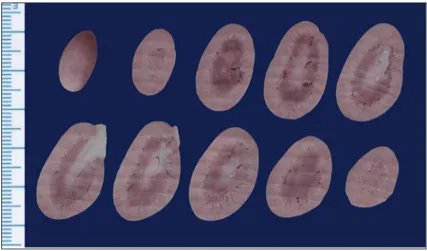
In order to be able to apply the Cavalieri’s method and to avoid disintegration of the kidneys during sectioning, the kidneys were plated with agar (Blood Agar Base LABM-LAB028). After boiling for 10 min, the solution was cooled to 60ºC and poured into special containers containing kidneys and the blocks were prepared [16]. The blocks were stood at room temperature for 24 h. Kidneys were cut with an electric salami slicing machine (SINBO SMS-5601) and depending on the size of the kidneys, 10 to 12 sections were obtained for volume estimation (Fig. 1). The mean slice thickness was 4.03 mm in the left kidney and 4.01 mm in the right kidney. The slicing process was carried out perpendicular to the craniocaudal length. The same faces of the sections were scanned at 600 dpi in JPG format using a horizontal scanner (hp Scanjet G4010).
In the volume calculations (kidney, renal cortex, renal medulla and renal pelvis), ImageJ program was used. The point counting frame with diff erent point frequencies was discarded on the section images with the grid command
of the software. In this counting frame, the area per point was set 1 mm2 for kidney, the cortex and medulla and 0.1 mm2 for the renal pelvis to reach a reliable coefficient of error (CE) (Fig. 2). For each area of interest, a diff erent marker was chosen and the points falling into the areas were counted separately. CE was calculated according to the relevant literature [17].
The volumes of the structures of interest in the sections were calculated separately using the formula V= a(p) xx t. In this formula, V refers to the volume of interest region a(p) is the area of the one point on the grid, Σp is the sum
of the points on the structure of interest and t is the section thickness [17,18]. Renal cortex, renal medulla and renal pelvis volume ratios were obtained by dividing related kidney section to the volume of total kidney.
Histological Analysis
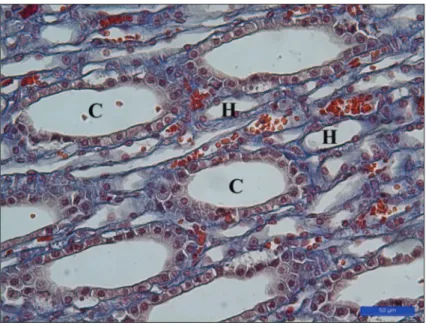
After the volume calculations, the kidneys were sampled at a rate of ½. The tissue samples were fixed in 10% buffered formaldehyde-saline solution, dehydrated, and embedded in paraffin blocks. The tissue sections taken from paraffin blocks in 6 μm thick were stained with Crossman’s trichrome staining. The cross-sections of the corpusculum renis, tubulus proximalis, tubulus distalis, Henle’s loop and number of glomerulus on the sections taken from the blocks were determined using light microscopy in the unit area (Fig. 3 and Fig. 4).
Statistical Analysis
Statistical analysis was performed using SPSS software version 21.0. The results of this study were compared by two sample t test. The values were expressed as mean and standard error (mean±SE). P<0.05 was considered statistically significant.
RESULTS
The weights of female and male New Zealand rabbits were 3254.4±169.9 g and 2714.2±77.6 g, respectively. The weight of the left kidney measured in female rabbits was 12.66±0.69 g and the right kidney weight was 12.19±0.54 g. In the male rabbit, these weights were 11.19±0.41 g and 10.86±0.37 g, respectively. It was found that the density of the left kidney was 1.04±0.06 g/mL and of the right kidney was 1.05±0.02 g/mL in the female rabbit. In the male rabbit, density measurements of left and right were 1.05±0.02 g/mL and 1.04±0.02 g/mL, respectively. There was no statistical diff erence between the renal densities.
Measurements of length and diameter of kidneys in female and male rabbits were given in Table 1. The dorsoventral and mediolateral diameters of right kidneys in females and males were found to be greater than those of the left ones. It was determined that the left and right mediolateral diameters of female rabbits were larger than those of the males, and the difference was statistically significant (P<0.05).
In the volume measurements of female rabbits performed with Archimedes’ principle, the left kidney was 12±0.76 mL and the right kidney was 11.11±0.68 mL. In males,
Fig 2. Renal cortex, renal medulla and renal pelvis counting on kidney with ImageJ
program (area per point = 1 mm2)
Fig 3. Histological appearance of the kidney in New Zealand Rabbits, D: Distal
these measurements were 10.5±0.41 mL in the left and 10.11±0.34 mL in the right. In the measurements made with the Cavalieri’s method, in female rabbits the left kidney was 12.26±0.66 mL and the right kidney was 11.66±0.54 mL, in male rabbits the left kidney was 10.69±0.42 mL and the right kidney was 10.49±0.49 mL. No statistical diff erence was found in volume measurements with Archimedes’ principle and Cavalieri’s method (P>0.05) (Fig. 5).
The calculated volumes with Cavalieri’s method for the
kidneys and subcomponents of kidneys in male and female rabbits were given in Table 2. It was determined that in female rabbits, 69.67% of the left kidney was composed of renal cortex, 29.35% of the renal medulla, 0.98% of the renal pelvis. On the right side, 77.98% of the kidney was renal cortex and 20.87% of the renal medulla 1.15% renal pelvis. In male rabbits, these rates were 77.58%, 21.57% and 0.85% in the left kidney, 77.13%, 22.03% and 0.84% in the right kidney, respectively. The left renal medulla was found to be larger than the right in male and female rabbits (P<0.05). No statistical diff erence in the volume of kidney and in the subcomponents of kidney was found between the female and the male New Zealand rabbits (P>0.05). The error coefficients were below 5% (Fig. 6).
The average counts of the glomerulus, proximal tubule, distal tubule, Henle’s loop, collecting tubule counts in the unit area of male and female New Zealand rabbit kidney were given in Table 3. It was determined that the number of left collecting tubules in female New Zealand rabbits was higher than that of males and it was statistically significant (P<0.05). There was no diff erence between counted histological kidney structures in the left and right kidneys (P>0.05).
DISCUSSION
In diagnosis of renal diseases, parameters such as volume and volume ratios, histomorphometric structure, and relative organ weight of kidneys are of great importance [4,19]. Changes in cortex and medulla of the kidney indicate pathological
changes. Kidney morphometry and the amount of nephron structures are influential on the potential functional capacity of the organ [10,20]. The knowledge of the volumes of structures of the healthy kidney is required for diagnosis of pathologies that alter renal volume and its structure. In this study, morphometric properties of kidney were determined in detail by making sexual dimorphism. In addition, a study showing the numbers of the glomerulus, proximal tubule, distal tubule, Henle’s loop and collecting tubule in New Zealand rabbits could not be determined in
Table 1. Kidney length and diameter measurements
Parameter Female P Value Male P Value Left
(Mean±SE) (Mean±SE)Right (Mean±SE)Left (Mean±SE)Right
Craniocaucaudal length (mm) 38.32±0.62 38.66±0.79 0.549 36.83±0.51 37.14±0.54 0.462
Dorsoventral diameter (mm) 21.37±0.56 19.57±0.47 0.005* 20.89±0.47 19.56±0.50 0.044*
Mediolateral diameter (mm) 24.99±0.54 25.92±0.46 0.009* 23.52±0.34 24.35±0.26 0.049*
* P<0.05
Fig 4. Histological appearance of the kidney in New Zealand Rabbits, C: Collecting
tubules, H: Henle’s loops, Crossman’s trichrome staining
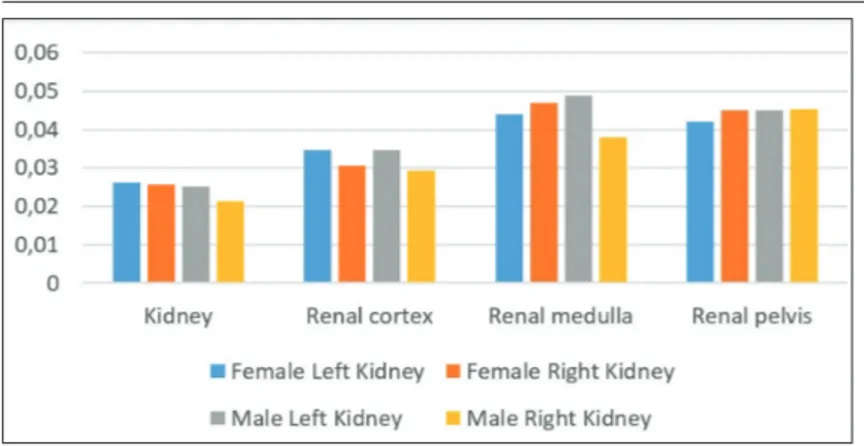
Fig 5. The volume of kidneys obtained using Archimedes’ principle and the
literature search. Therefore, this data will contribute to the existing anatomical knowledge.
In a study with 50 adult male and female rabbits without macroscopic renal pathology, Santos-Sousa et al.[2] was found no significant diff erence in any of the renal dimensions between the right and left kidneys in either sexes. In another study performed on 12 mature healthy rabbits, Dimitrov et al.[21] reported that the left kidney’s cranio-caudal length and mediolateral diameter were larger than that of the right kidney and the right kidney’s dorsoventral diameter was larger than that of the left. In a study with eight adult healthy rabbits of both sexes which used three dimensional reconstructions of multidetector computed tomography images, Eken et al.[22] reported that the dorso-ventral diameter, mediolateral diameter and craniocaudal length of the left kidney were larger than those of the right. Kidneys begin their development near the sacral region and move forward. The posture of the two kidneys
is asymmetrical. The right kidney is located more cranial than the left kidney [12]. Therefore, diff erences in right and left kidney’s diameters and lengths are expected. In the present study, dorsoventral and mediolateral diameters of left kidneys were found to be larger than those of the right kidney in females and males New Zealand rabbit, and females were found to be larger than males. However, no diff erence was detected in craniocaudal length. In the study conducted by Dimitrov et al.[21], the diff erence was thought to be due to the age diff erence of the rabbits used. The diff erence with Eken et al.[22] was thought to be due to the methodological diff erence.
In a study with nine male rabbits comparing fresh and fixed kidneys in formalin solution, Bolat et al.[12] did not detect any diff erence between the right and left kidney volumes. Renal cortex, renal medulla and renal pelvis volume ratios were 59.8%, 36.4%, 3.8% in left kidney and 61.8%, 34.7%, 3.4% in right kidney, respectively. The left kidney’s dorsoventral diameters were also found to be larger than that of the right. In the present study, it was found that the left renal medulla was larger than that of the right in male New Zealand rabbits. Volume fractions of left and right renal cortex, renal medulla and renal pelvis were estimated to be 77.58%, 21.57%, 0.85% and 77.13%, 22.03%, 0.84%, respectively. In the study, dorsoventral diameters of the left kidney as well as the left mediolateral diameter were found to be larger than right. It is thought that the diff erence between the two studies is due to the age diff erences of the rabbits used.
Bolat et al.[12] reported that left renal density of male New
Table 3. The number of glomerulus, proximal tubule, distal tubule, Henle’s loop, collecting tubule in per unit area (Mean±SE)
Item Female P Value Male P Value
Left Right Left Right
Glomerulus 19.89±1.79 17.44±1.81 0.281 20.00±1.69 17.33±1.44 0.110 Proximal tubule 262.22±14.17 270.11±17.66 0.723 266.44±6.24 257.33±9.48 0.259 Distal tubule 117.33±7.12 132.89±10.48 0.300 107.78±6.05 105.33±8.77 0.863 Henle’s loop 433.44±41.54 456.56±35.79 0.733 468.44±26.18 473.00±49.44 0.942 Collecting tubule 184.78±15.26 157.56±16.85 0.303 119.22±11.66 140.11±13.21 0.208 * P<0.05
Table 2. Volume measurements of left and right kidneys
Item Female P Value Male P Value Left (mL) (Mean±SE) Right (mL) (Mean±SE) Left (mL) (Mean±SE) Right (mL) (Mean±SE) Kidney 12.26±0.66 11.66±0.54 0.291 10.69±0.42 10.49±0.49 0.520 Renal cortex 8.54±0.61 8.63±0.52 0.856 7.28±0.34 7.42±0.27 0.674 Renal medulla 3.59±0.12 2.93±0.18 0.005* 3.31±0.27 2.97±0.26 0.044* Renal pelvis 0.12±0.02 0.10±0.01 0.285 0.10±0.01 0.10±0.01 0.477 * P<0.05
Zealand rabbits was 0.97, and the right renal density was 1. There was no statistical difference between left and right renal density. In present study, renal density was found to be 1.04±0.06 for the left kidney and 1.05±0.02 for the right kidney in female rabbit. In male rabbit, left and right was 1.05±0.02 and 1.04±0.02, respectively. No statistical difference was found in the presented study (P>0.05). In the literature search, data on the renal density of female New Zealand rabbits could not be found.
One of the most important steps of stereological studies is the determination of the error coefficient. The quality of the numerical measurements made by stereological studies and the accuracy of the sampling plan can be observed by calculating the error coefficient (CE). Although the error coefficient in stereological studies does not correspond to a real biological value, it is a value indicating the quality of the sampling strategy [23]. In order for the results of stereological studies to be considered as reliable, the error coefficient should be 5% or less [17]. In the present study, the error coefficients were below 5% (Fig. 6).
The morphometric data of the New Zealand rabbits’ kidney and its subcomponents determined by using stereological methods and the data obtained by counting in unit area will provide insight for the investigation and comparison of renal hypertrophy, atrophy and tumor formation. Furthermore, in order to complete our study, the structures of functional subcomponents as a result of diseases should be examined by electron microscopy in New Zealand rabbits. It is thought that this study will guide the future studies methodologically.
REFERENCES
1. Pazvant G, Sahin B, Kahvecioglu KO, Gunes H, Gezer Ince N, Bacinoglu D: The volume fraction method for the evaluation of kidney: A
stereological study. Ankara Üniv Vet Fak Derg, 56, 233-239, 2009.
2. Santos-Sousa C, Stocco A, Mencalha R, Jorge S, Abidu-Figueiredo M: Morphometry and vascularization of the rabbit kidneys (Oryctolagus
cuniculus). Int J Morphol, 33 (4): 1293-1298, 2015.
3. Cheung CM, Shurrab AE, Buckley DL, Hegarty J, Middleton RJ, Mamtora H, Kalra PA: MR-derived renal morphology and renal function
in patients with atherosclerotic renovascular disease. Kidney Int, 69 (4): 715-722, 2006. DOI: 10.1038/sj.ki.5000118
4. Michael B, Yano B, Sellers RS, Perry R, Morton D, Roome N, Johnson JK, Schafer K: Evaluation of organ weights for rodent and
non-rodent toxicity studies: A review of regulatory guidelines and a survey of current practices. Toxicol Pathol, 35 (5): 742-750, 2007. DOI: 10.1080/01926230701595292
5. Lødrup AB, Karstoft K, Dissing TH, Nyengaard JR, Pedersen M:
The association between renal function and structural parameters: A pig study. BMC Nephrol, 9:18, 2008.
6. Isotani S, Shimoyama H, Yokota I, Noma Y, Kitamura K, China T,
Saito K, Hisasue SI, Ide H, Muto S, Yamaguchi R, Ukimura O, Gill IS, Horie S: Novel prediction model of renal function after nephrectomy from
automated renal volumetry with preoperative multidetector computed tomography (MDCT). Clin Exp Nephrol, 19 (5): 974-981, 2015. DOI: 10.1007/ s10157-015-1082-6
7. Beland MD, Walle NL, Machan JT, Cronan JJ: Renal cortical thickness
measured at ultrasound: Is it better than renal length as an indicator of renal function in chronic kidney disease? Am J Roentgenol, 195 (2): W146-W149, 2010.
8. Akbari M, Goodarzi N, Tavafi M: Stereological assessment of normal
Persian squirrels (Sciurus anomalus) kidney. Anat Sci Int, 92 (2): 267-274, 2017. DOI: 10.1007/s12565-016-0332-3
9. Graur D, Duret L, Gouy M: Phylogenetic position of the order
Lagomorpha (rabbits, hares and allies). Nature, 379, 333-335, 1996. DOI: 10.1038/379333a0
10. Tsamouri MM, Rapti M, Kouka P, Nepka C, Tsarouhas K, Soumelidis A, Koukoulis G, Tsatsakis A, Kouretas D, Tsitsimpikou C: Histopathological
evaluation and redox assessment in blood and kidney tissues in a rabbit contrast-induced nephrotoxicity model. Food Chem Toxicol, 108, 186-193, 2017. DOI: 10.1016/j.fct.2017.07.058
11. Bertram JF, Soosaipillai MC, Ricardo SD, Ryan GB: Total numbers of
glomeruli and individual glomerular cell types in the normal rat kidney.
Cell Tissue Res, 270 (1): 37-45, 1992.
12. Bolat D, Bahar S, Selcuk M, Tıpırdamaz S: Morphometric investigations
fresh and fixed rabbit kidney. Eurasian J Vet Sci, 27 (3): 149-154, 2011.
13. Knepper M, Danielson RA, Saidel GM, Post RS: Quantitative analysis
of renal medullary anatomy in rats and rabbits. Kidney Int, 12, 313-323, 1977.
14. Al-Sharoot HA: Morphological & histological study of the kidney in
guinea pig. Int J Recent Sci Res, 5 (11): 1973-1976, 2014.
15. Flecknell P: Laboratory Animal Anaesthesia. Academic Press, Oxford,
2015.
16. Zarow C, Kim T-S, Singh M, Chui H: A standardized method for
brain-cutting suitable for both stereology and MRI-brain co-registration.
J Neurosci Methods, 139 (2): 209-215, 2004.
17. Gundersen HJG, Jensen EBV, Kieu K, Nielsen J: The efficiency of
systematic sampling in stereology reconsidered. J Microsc, 193 (3): 199-211, 1999. DOI: 10.1046/j.1365-2818.1999.00457.x
18. Bolat D, Selcuk ML: Stereological and biochemical evaluation of
diclofenac-induced acute nephrotoxicity in rats. Revue Med Vet, 164, 290-294, 2013.
19. Kiss N, Hamar P: Histopathological evaluation of contrast-induced
acute kidney injury rodent models. Biomed Res Int, 2016:3763250, 2016. DOI: 10.1155/2016/3763250
20. Jeon HG, Lee SR, Joo DJ, Oh YT, Kim MS, Kim YS, Yang SC, Han WK: Predictors of kidney volume change and delayed kidney function
recovery after donor nephrectomy. J Urol, 184 (3): 1057-1063, 2010. DOI: 10.1016/j.juro.2010.04.079
21. Dimitrov R, Kostov D, Stamatova K, Yordanova V:
Anatomo-topographical and morphological analysis of normal kidneys of rabbit
(Oryctolagus cuniculus). Trakia J Sci, 10 (2): 79-84, 2012.
22. Eken E, Çorumluoğlu Ö, Paksoy Y, Beşoluk K, Kalaycı İ: A study on
evaluation of 3D virtual rabbit kidney models by multidetector computed tomography images. Anatomy, 3 (1): 40-44, 2009.
23. Slomianka L, West MJ: Estimators of the precision of stereological
estimates: An example based on the CA1 pyramidal cell layer of rats.
Neuroscience, 136 (3): 757-767, 2005. DOI: 10.1016/j.neuroscience.


