77
Case Report / Olgu Sunumu
Introduction
Disc hemorrhage, a round or splinter hemorrhage present on the optic disc or in the peripapillary retina extending to the disc rim, has been described in people with glaucoma. The prevalence of disc hemorrhages is about 0.9% to 1.4% in the normal population and 1.9% to 13.8% in patients with glaucoma.1,2
Other conditions associated with disc hemorrhage include diabetes mellitus, anticoagulation use, advanced age, female gender, and recent posterior vitreous detachment.3,4
Here, we report a case of glaucoma with an atypical optic disc hemorrhage, which was observed in the optic disc cup.
Case Report
A 73-year-old male patient presented for routine follow-up with a history of diabetes mellitus, which had been well controlled with medication. He was also taking acetylsalicylic acid. He had bilateral primary open-angle glaucoma and had been using latanoprost and timolol maleate fix combination for
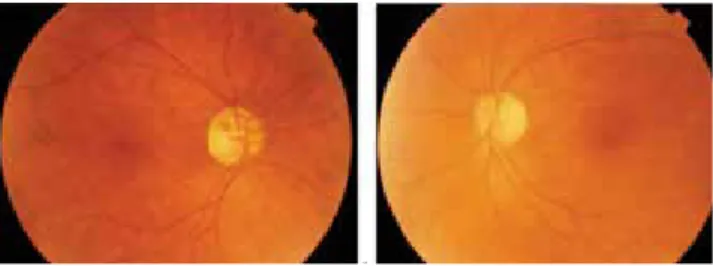
nine years. There were no ocular symptoms and no preceding trauma. Best-corrected visual acuity was 20/20 in both eyes. Intraocular pressures were 16 mmHg in the right eye and 14 mmHg in the left eye. The central corneal thickness was 540 μm in the right eye and 552 μm in the left eye. Slit-lamp examination was normal. Dilated fundus examination demonstrated a cup-to-disc ratio of 0.7 with a narrow temporal and superior neuroretinal rim and cup hemorrhage but absence of signs of papillitis or retinal vascular abnormalities in the right eye, and a cup-to-disc ratio of 0.6 with narrow temporal neuroretinal rim in the left eye (Figure 1a, 1b). These findings were not mentioned in his previous documentation, however, the optic coherence tomography (OCT) that was taken at the time showed the progression of the superotemporal retinal nerve fiber layer (RNFL) defect clearly when compared with the previous OCT from 7 months earlier (Figure 2a, 2b). Fluorescein angiography showed no abnormalities related to diabetic retinopathy. The patient has been under our follow-up for 9 more months, and the disc hemorrhage still exists.
Address for Correspondence/Yazışma Adresi: Sirel Gür Güngör MD, Başkent University Faculty of Medicine, Department of Ophthalmology, Ankara, Turkey
Phone: +90 312 212 68 68 E-mail: [email protected] Received/Geliş Tarihi: 01.02.2014 Accepted/Kabul Tarihi: 30.04.2014
A 73-year-old man presented for routine follow-up. There were primary open-angle glaucoma, diabetes mellitus, and usage of acetylsalicylic acid in patient’s history. Dilated fundus examination demonstrated cup hemorrhage in the right eye. Because of the progression of the superotemporal retinal nerve fiber layer defect in the last seven months, we think that the disk hemorrhage could be associated with glaucoma. (Turk J Ophthalmol 2015; 45: 77-8)
Key Words: Intrapapillary hemorrhage, glaucoma, retinal nerve fiber layer
Yetmiş üç yaşında erkek hasta rutin göz kontrolü için kliniğimize başvurdu. Hastanın hikayesinde primer açık açılı glokom, diyabetes mellitus ve asetilsalisilik asit kullanımı mevcuttu. Dilate fundus muayenesinde sağ gözde optik disk çukurluğunda kanama mevcuttu. Son 7 ayda üst temporal retinal sinir lifi tabakasındaki incelmede progresyon olduğundan disk kanamasının glokom ile ilişkili olabileceği düşünüldü. (Turk J Ophthalmol 2015; 45: 77-8)
Anah tar Ke li me ler: İntrapapiller kanama, glokom, retina sinir lifi tabakası
Summary
Özet
Başkent University Faculty of Medicine, Department of Ophthalmology, Ankara, Turkey
Sirel Gür Güngör, Gülce Gökgöz Özışık, Ahmet Akman, Leyla Asena
Atypical Intrapapillary Hemorrhage in a
Patient with Glaucoma
Glokomlu Bir Hastada Atipik İntrapapiller Kanama
TJO 45; 2: 2015
78
Discussion
The pathogenesis of disc hemorrhage remains unclear, although both mechanical and vascular theories have been proposed. Optic disc hemorrhage can be caused not only by ischemic microinfarction in the optic disc, but also by mechanical rupture of small blood vessels arising from structural changes at
the level of the lamina cribrosa.5 Vasculopathy associated with
systemic hypertension and diabetes can cause microinfarctions and ischemic changes in optic disc vessels, making these vessels vulnerable to mechanical rupture.6
In this study, we report a 73-year-old male patient who presented with optic disc hemorrhage. The interesting point of this case was that the hemorrhage occurred in the cup of the optic disc rather than a more common splinter type hemorrhage. There were primary open-angle glaucoma, diabetes mellitus, and usage of acetylsalicylic acid in patient’s history. We did not think that the disc hemorrhage was associated with diabetes mellitus because the fundus examination showed no findings related to diabetic retinopathy. The possible reason of the persistence of the hemorrhage in this patient might be related to his usage of acetyl salicylic acid. Because of the progression of the superotemporal RNLF defect in the last seven months, we think that the disk hemorrhage could be associated with glaucoma, rather than diabetes mellitus in this patient.
Conflict of interest: The authots reported no conflict of
interest related to this article.
References
1. Healey PR, Mitchell P, Smith W, Wang JJ. Optic disc hemorrhages in a population with and without signs of glaucoma. Ophthalmology. 1998;105:216-223.
2. Klein BE, Klein R, Sponsel WE, Franke T, Cantor LB, Martone J, Menage MJ. Prevalence of glaucoma. The Beaver Dam Eye Study. Ophthalmology. 1992;99:1499-1504.
3. Soares AS, Artes PH, Andreou P, Leblanc RP, Chauhan BC, Nicolela MT. Factors associated with optic disc hemorrhages in glaucoma. Ophthalmology. 2004;111:1653-1657.
4. Roberts TV, Gregory-Roberts JC. Optic disc hemorrhages in posterior vitreous detachment. Aust N Z J Ophthalmol. 1991;19:61-63.
5. Quigley HA, Addicks EM, Green WR. Optic nerve damage in human glaucoma. II. The site of injury and susceptibility to damage. Arch Ophthalmol. 1981;99:635-649.
6. Cooper ME, Bonnet F, Oldfield M, Jandeleit-Dahm K. Mechanisms of diabetic vasculopathy: an overview. Am J Hypertens. 2001;14:475-486.
Figure 1. Fundus photograph of the right eye showing a cup-to-disc ratio of 0.7
with a narrow temporal and superior neuroretinal rim and cup hemorrhage in the right eye (a), and a cup-to-disc ratio of 0.6 with narrow temporal neuroretinal rim in the left eye (b)
Figure 2. Optical coherence tomography demonstrating the progression of the
superotemporal retinal nerve fiber layer defect clearly when compared with the previous OCT from 7 months earlier (a: 7 months prior to detection of the cup hemorrhage; b: at the time of detection of cup hemorrhage)