Downloaded from https://journals.lww.com/menopausejournal by BhDMf5ePHKav1zEoum1tQfN4a+kJLhEZgbsIHo4XMi0hCywCX1AWnYQp/IlQrHD3tIQ5gQCIeyxoHNtHYz46siWQ+R7s7imnRJ+LIMpDF/MfOCX5owmBhQ== on 07/31/2018 Downloadedfrom https://journals.lww.com/menopausejournalby BhDMf5ePHKav1zEoum1tQfN4a+kJLhEZgbsIHo4XMi0hCywCX1AWnYQp/IlQrHD3tIQ5gQCIeyxoHNtHYz46siWQ+R7s7imnRJ+LIMpDF/MfOCX5owmBhQ==on 07/31/2018 Vol. 17, No. 6, pp. 1188/1193 DOI: 10.1097/gme.0b013e3181e04a18
* 2010 by The North American Menopause Society
Effects of raloxifene on serum macrophage colony-stimulating
factor and interleukin-18 levels in postmenopausal women younger
than 60 years
Efser Oztas, MD,
1and Gulay Kurtay, MD
2Abstract
Objective: Macrophage colony-stimulating factor (M-CSF) and interleukin-18 (IL-18) are cytokines expressed predominantly in atheromatous plaque, and overproduction of these has been found to be associated with coronary artery disease. The aim of this study was to investigate the effect of raloxifene, a selective estrogen receptor modulator, on serum M-CSF and IL-18 levels, cytokines that are presumably involved in the pathogenesis of atherosclerosis.
Methods: A total of 70 postmenopausal women (age, 56.45 T 1.52 y) without previously confirmed
cardio-vascular disease were enrolled in a 6-month prospective, randomized, controlled study. Women were randomly assigned to two groups: 35 women received oral administration of 60 mg/day raloxifene for 6 months and 35 were in the control group and received no medications. Serum lipid concentrations and high-sensitivity C-reactive protein (hs-CRP), M-CSF, and IL-18 levels were measured at baseline and at the sixth month in both groups.
Results: Compared with the control group, the raloxifene group had a significant decrease in serum IL-18 concentrations and a 25.29% reduction in serum hs-CRP concentrations. M-CSF levels were reduced by 5.94% in the raloxifene group, but the difference was not statistically significant. At the sixth month, 60 mg/day of raloxifene significantly decreased the median serum total cholesterol and low-density lipoprotein cholesterol levels when compared with the baseline levels.
Conclusions: Raloxifene reduces serum total cholesterol, low-density lipoprotein cholesterol, hs-CRP, and IL-18 levels. According to the results of our study, it is suggested that raloxifene may have a favorable effect on the prevention of cardiovascular disease in healthy postmenopausal women younger than 60 years.
Key Words: Raloxifene Y Macrophage colony-stimulating factor Y Interleukin-18.
A
therosclerosis, the main etiological factor forcardi-ovascular disease (CVD), is not only a degenerative condition as previously assumed but is also now considered to be a chronic inflammatory process. It is now well established that from the earliest lesion of atheroscle-rosis to the plaque formation, various cellular and molecular
inflammatory mediators participate in the disease process.1
Overproduction of interleukin-6 (IL-6), IL-1A, and tumor necrosis factor-> (TNF->) has been found to be associated
with CVD.2
Interleukins are considered to be key players in the chronic vascular inflammatory response that is typical for atheroscle-rosis. Recently, a review of studies about the relationship of cytokines with atherosclerosis in mice pointed out that there
are sufficient consistent data allowing the classification of only a few cytokines as typically proatherogenic: IL-1, IL-12, IL-18, macrophage migration inhibitory factor, interferon-F,
TNF->, and macrophage colony-stimulating factor (M-CSF).3
M-CSF is known to promote atherogenesis by inducing monocyte-macrophage activation, foam cell formation, and the release of other cytokines such as IL-1A, monocyte chemo-tactic protein 1, and IL-6 by vascular cells, which lead to
hepatic C-reactive protein (CRP) production.4 IL-18, also
known as IF-F inducing factor, is responsible for the up-regulation of adhesion molecules, as well as other
inflamma-tory cytokines like IL-1A, IL-8, and TNF->; inhibits collagen
synthesis by smooth muscle cells; and promotes T helper 1
responses that dominate during human atherogenesis.5
Fur-thermore, the beneficial effect of inhibiting IL-18 on plaque progression and composition has been shown in animal
models.6 Therefore, increased M-CSF and IL-18 levels are
thought to be representative of other proinflammatory cyto-kines. In addition, an increasing number of investigations on humans have demonstrated that M-CSF and IL-18, cytokines expressed predominantly in atheromatous plaque, play an important role in atherogenesis and contribute to plaque
in-stability, thrombosis, and acute coronary syndrome.7<9
Received March 1, 2010; revised and accepted March 29, 2010. From the1Department of Obstetrics and Gynecology, Ufuk University Faculty of Medicine, Ankara, Turkey; and 2Department of Obstetrics
and Gynecology, Ankara University Faculty of Medicine, Ankara, Turkey.
Financial disclosure/conflicts of interest: None reported.
Address correspondence to: Efser Oztas, MD, Mevlana BulvarN No. 86-88 Konya yolu, Balgat, Ankara, Turkey 06590. E-mail: efseroztas@ yahoo.com
Raloxifene, a nonsteroidal benzothiophene selective estro-gen receptor modulator, has beneficial effects on the arterial system by decreasing total cholesterol (TC) and low-density lipoprotein cholesterol (LDL-C) and the levels of some
in-flammatory markers such as CRP and homocysteine10<12and
induces endothelial-dependent vasodilatation due to a
reduc-tion in the risk factors causing endothelial damage.13In
addi-tion, recently, some studies have suggested that raloxifene has effects on circulating levels of IL-4, IL-6, IL-7, and TNF->,
but the results were discrepant.14<18Furthermore, the results
of two randomized, placebo-controlled studies seem to be con-flicting, because these suggested that raloxifene did not affect
the risk of CVD.19,20The Multiple Outcomes of Raloxifene
Evaluation (MORE) trial investigated the effect of raloxifene on cardiovascular events as a secondary endpoint and reported that raloxifene therapy for 4 years did not significantly affect the overall risk of cardiovascular events but did significantly reduce cardiovascular events in women with increased risk for
cardiovascular events.19In addition, according to the results of
the Raloxifene Use for the Heart (RUTH) study, raloxifene did not significantly affect the risk of coronary heart disease, but when the incidence of coronary events among subgroups of participants were evaluated, it was realized that in women younger than 60 years, the incidence of coronary events was significantly lower in those assigned to raloxifene compared
with those assigned to placebo.20,21 Therefore, it is unclear
whether raloxifene plays a role in protection against athero-sclerosis and CVDs. In addition, there are insufficient data about the effects of raloxifene on other cytokines that are proved to be proatherogenic.
The aim of the current clinical study was to evaluate the effects of raloxifene on serum M-CSF and IL-18 levels in relation to changes in serum lipids and high-sensitivity CRP (hs-CRP) levels in healthy postmenopausal women younger than 60 years.
METHODS Participants
This prospective randomized open-label study was con-ducted at the University of Ankara, School of Medicine. A total of 70 postmenopausal women with an intact uterus between 55 and 59 years of age recruited from the patients referred to the menopause clinic were eligible to participate in the study. The range of years past menopause was 5 to 10 years. Postmenopause status was confirmed by measure-ment of serum follicle-stimulating hormone levels greater than 40 IU/L and serum estradiol levels less than 20 pg/mL. All participants had amenorrhea for at least 12 months. Body
mass index was required to be between 18 and 31 kg/m2. All
women underwent gynecological examination and transvag-inal ultrasonography before recruitment. Exclusion criteria in the study were smoking and having any history of hyperten-sion, venous thromboembolism, cerebral or coronary events, any endocrinopathy, impaired renal or liver function, breast cancer, gynecological malignancy, or chronic inflammatory disease. Women treated with hypolipidemic drugs, systemic
corticosteroids, and nonsteroidal antiinflammatory drugs within 6 months of entry and those who received hormone therapy or raloxifene at any time in the past were also excluded.
The procedures used in this study were in accordance with the guidelines of the Declaration of Helsinki on human ex-perimentation. The study was approved by the ethical review board of the University of Ankara. The purpose of the study protocol was explained to the women before they entered the study, and their informed consent was obtained.
Study design
This prospective, randomized, controlled study was con-ducted at the University of Ankara, School of Medicine. A total of 70 eligible participants were randomly assigned to two groups: 35 women received oral administration of 60 mg raloxifene HCl (Evista, Eli Lilly) daily for 6 months, and 35 were in the control group and received no medications. Randomization was performed using a computer-generated random number table. During the follow-up period, it was formally assessed that none of the women started any kind of medications including hormones, lipid-altering therapy, and analgesics.
After the women fasted for 12 hours, venous blood samples were collected at the baseline and at the sixth month of raloxifene treatment. Samples were obtained, centrifuged within 30 minutes of collection at 3,000g for 5 minutes, and stored at j70-C until assayed.
Analytical techniques
At baseline and after 6 months, we measured serum TC, high-density lipoprotein cholesterol (HDL-C), and triglyceride (TG) levels with the enzymatic color test method. LDL-C was calculated by the Friedewald formula. Serum CRP levels were measured by a nephelometric method using the Beckman Coulter Immage Immunochemistry System. The sensitivity level of the CRP kits was 0.002 mg/dL. Concentrations of M-CSF and IL-18 in serum samples were measured using re-spective human enzyme-linked immunosorbent assay (ELISA) kits. The sensitivity level of the M-CSF ELISA kits (R&D Systems, Minneapolis, MN) was 9 pg/mL, and that of the IL-18 ELISA kits (MBL, Nagoya, Japan) was 12.5 pg/mL. Moreover, according to the manufacturer guidelines, assay characteristics for the measurement of hs-CRP, IL-18, and M-CSF were as follows: intra-assay coefficients of variation, 5%, 4.3%, and 3.4%, respectively; and interassay coefficients of variation, 7.5%, 5.21%, and 3.1%, respectively.
Statistical analysis
Evaluation of the data was performed using SPSS v 11.5 (SPSS Inc., Chicago, IL). The results were expressed as means and medians. To determine whether any changes are present in the parameters at baseline and at the sixth month, a paired t test was used when the parameters are homogenically distributed and the Wilcoxon signed-rank test was used when the parameters are nonhomogenically distributed. Compar-isons between the raloxifene and the control groups were
performed using an unpaired t test or the Mann-Whitney U test as appropriate. The differences regarding primary outcomes between groups were evaluated by both
intention-to-treat analysis and per protocol analysis. P G 0.05 was
considered to be statistically significant. RESULTS Background characteristics
A total of 70 women were originally enrolled in the study and were randomized to treatment. The intent-to-treat popu-lation consisted of 70 women (35 in the raloxifene group and 35 in the control group), 90% (n = 63) of whom completed the 6-month follow-up period. One of the 35 women who received raloxifene dropped out of the study at 2 months because of increased hot flushes as an adverse effect. Six women in the control group dropped out of the study because of the following reasons: four received medications including lipid-lowering drugs and bisphosphonates, and two withdrew for personal reasons. Of the 63 women who completed the study, 34 were in the raloxifene group (60 mg raloxifene daily) and 29 were in the control group (received no medications).
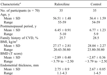
Baseline demographic characteristics according to assign-ment are presented in Table 1. There were no significant differences among the two groups for age, duration of the postmenopausal period, family history of CVD, body mass index, bone mineral density, and endometrial thickness. In addition, baseline lipid profiles and levels of serum M-CSF, IL-18, and hs-CRP were not significantly different in women in both groups who completed the study (Table 2).
After a 6-month follow-up period, serum TC, LDL-C, TG, HDL-C, M-CSF, IL-18, and hs-CRP concentrations were reevaluated.
Changes in serum concentrations of lipids
At the sixth month, 60 mg of daily raloxifene decreased
the median serum TC levels by 12 mg/dL (PG 0.0001) and
the median LDL-C levels by 9 mg/dL (P = 0.025) when com-pared with the baseline levels (Table 3). At the sixth month of the follow-up period, we found that, compared with the control group, the raloxifene group had significantly decreased
serum TC, TG, and LDL-C levels (PG 0.0001, P = 0.015, and
P = 0.009, respectively) and significantly increased serum HDL-C levels (P = 0.009; Table 4).
TABLE 1. Baseline characteristics of the intent-to-treat population
Characteristica Raloxifene Control
No. of participants (n = 70) 35 35 Age, y MeanT SD 56.51T 1.48 56.4T 1.59 Range 55-59 54-59 Postmenopausal period, y MeanT SD 6.45T 0.91 6.77T 1.23 Range 5-10 5-9 Family history of CVD, % 25.7 28.5 BMI, kg/m2 MeanT SD 27.17T 2.81 28.04T 2.27 Range 20.43-30.80 23.80-30.80 BMD (T score) MeanT SD j2.89 T 0.40 j2.87 T 0.36 Range j3.79 to j2.50 j3.79 to j2.50 Endometrial thickness, mm MeanT SD 2.75T 0.9 2.67T 0.85 Range 1.1-4.3 1-4.5
CVD, cardiovascular disease; BMI, body mass index; BMD, bone mineral density.
aNo statistically significant difference with respect to baseline characteristics among the groups (P9 0.05).
TABLE 2. Baseline lipid, M-CSF, IL-18, and hs-CRP levels of the women enrolled in the study
Raloxifene (n = 34) Control (n = 29) Pa TC, mg/dL MeanT SD 203.64T 28.95 199.20T 31.73 Median (range) 202 (136-268) 201 (148-279) 0.564 LDL-C, mg/dL MeanT SD 122.29T 26.02 114.06T 24.76 Median (range) 121 (82-196) 115 (56-164) 0.206 Triglyceride, mg/dL MeanT SD 158.11T 51.38 154.75T 45.23 Median (range) 153 (81-294) 160 (75-234) 0.786 HDL-C, mg/dL MeanT SD 47.23T 10.54 49.51T 10.15 Median (range) 46.00 (24-72) 49 (27-70) 0.387 M-CSF, pg/mL MeanT SD 377.67T 517.84 376.12T 279.41 Median (range) 247.52 (44.15-2,895.45) 249.65 (30.15-1,007.40) 0.486 IL-18, pg/mL MeanT SD 222.57T 195.51 198.73T 230.10 Median (range) 206.60 (0-877.10) 115.45 (0-911.80) 0.339 CRP, mg/L MeanT SD 3.48T 2.15 3.19T 2.88 Median (range) 3.18 (0.54-10.40) 2.7 (0.4-14.20) 0.270 M-CSF, macrophage colony-stimulating factor; IL-18, interleukin-18; hs-CRP, high-sensitivity C-reactive protein; TC, total cholesterol; LDL-C, low-density lipoprotein cholesterol; HDL-C, high-low-density lipoprotein cholesterol. a
No statistically significant difference with respect to baseline characteristics among the groups (P9 0.05).
TABLE 3. Comparison of baseline and sixth-month values in the raloxifene group (n = 34)
Baseline Sixth month Pa
TC, mg/dL MeanT SD 203.64T 28.95 188.88T 31.36 Median 202 (136-268) 190 (124-248) G0.001 LDL-C, mg/dL MeanT SD 122.29T 26.02 113.94T 24.84 Median 121 (82-196) 112 (71-181) 0.041 Triglyceride, mg/dL MeanT SD 158.11T 51.38 151.67T 44.27 Median 153 (81-294) 144 (90-261) 0.193 HDL-C, mg/dL MeanT SD 47.23T 10.54 48.52T 9.994 Median 46.00 (24-72) 48.00 (26-74) 0.188 M-CSF, pg/mL MeanT SD 377.67T 517.84 250.90T 126.54 Median 247.52 (44.15-2,895.45) 247.00 (0-497.75) 0.48 IL-18, pg/mL MeanT SD 222.57T 195.51 184.40T 195.15 Median 206.60 (0-877.10) 130.76 (0-846.25) 0.176 CRP, mg/L MeanT SD 3.48T 2.15 2.53T 1.49 Median 3.18 (0.54-10.40) 2.57 (0.48-7.08) 0.009
TC, total cholesterol; LDL-C, low-density lipoprotein cholesterol; HDL-C, high-density lipoprotein cholesterol; M-CSF, macrophage colony-stimulating factor; IL-18, interleukin-18; CRP, C-reactive protein.
a
Changes in serum concentrations of hs-CRP, M-CSF, and IL-18
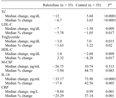
The serum hs-CRP levels were unchanged in the control group after 6 months (Table 4). In contrast, raloxifene sig-nificantly decreased hs-CRP levels when compared with the baseline levels (P = 0.009) (Table 3). In the raloxifene group, the median value of hs-CRP was decreased from 3.18 to 2.57 mg/L, and when compared with the control group, the difference was statistically significant (P = 0.001; Table 5).
As can be seen in Table 5, raloxifene induced a significant decrease in serum IL-18 levels when compared with the control group (P = 0.005). Median percentage changes in
serum M-CSF concentrations were Y5.94% in the raloxifene
group and 44.75% in the control group at the sixth month of the therapy when compared with the baseline levels. However, this difference was not statistically significant (P = 0.083; Table 5). Among the 63 women who participated in the study, serum levels of IL-18 in 13 women and M-CSF in 1 woman were less than the sensitivity levels of the assay.
DISCUSSION
In the present study, the group given 60 mg/day of raloxifene had significantly decreased serum levels of IL-18 and hs-CRP when compared with the control group over a 6-month treatment period. However, M-CSF levels tended to decrease in the raloxifene group, but the difference was not statistically significant. To the best of our knowledge, there has been no report evaluating the effects of raloxifene on serum M-CSF and IL-18 concentrations in healthy post-menopausal women younger than 60 years.
According to the MORE study, 60 mg/day of raloxifene did not differ from a dose of 120 mg/day when compared for changes in lipid concentrations, risk of cardiovascular events,
and cumulative risk of new vertebral fractures.19In addition,
women who participated in the RUTH study were assigned
to 60 mg of raloxifene daily or placebo.20 Therefore, we
preferred a dose of 60 mg/day of raloxifene to compare our results efficiently with those RUTH, a randomized study investigating the effect of raloxifene on coronary heart dis-ease as a primary outcome.
The results of the current study showed that 60 mg/day of raloxifene reduces serum concentrations of LDL-C, TG, and TC in healthy postmenopausal women. These results are in
accordance with the previous reports.11,22<24
hs-CRP is an independent marker for the risk of CVD in postmenopausal women without clinically evident coronary
artery disease (CAD).25Previous studies regarding the effect
of raloxifene on hs-CRP were discrepant. Some of them found no or little effect on CRP in postmenopausal women, whereas others showed that raloxifene exerted favorable
effects by decreasing CRP levels.26<30 On the basis of data
from the study of Ridker et al reported in 2002, in which 27,939 initially healthy women participated, women with a CRP level of 1 to 3 mg/L were established as at average risk, and those with a CRP level greater than 3 mg/L were
estab-lished as at high risk for a first cardiovascular event.31When
the results of Ridker et al are considered, it is evident that the raloxifene group in our study had been in the high-risk group for CVD at baseline and became included in the average-risk group at the sixth month (median hs-CRP levels at the baseline and at the sixth month were 3.18 and 2.57 mg/L,
TABLE 5. Comparison of the median baseline, median change, and median percent change values of the raloxifene and control
groups in the intent-to-treat population
Raloxifene (n = 35) Control (n = 35) Pa TC Median change, mg/dL j12 5.68 G0.0001 Median % change j6.7 3.65 G0.0001 LDL-C Median change, mg/dL j7 j1.58 0.009 Median % change j5.78 j1.05 0.017 Triglyceride Median change, mg/dL j3.0 7.0 0.015 Median % change j1.63 3.22 0.02 HDL-C Median change, mg/dL 1.0 j2.68 0.009 Median % change 2.32 j4.28 0.017 M-CSF Median change, pg/mL j26.75 10.74 0.315 Median % change j5.94 44.75 0.083 IL-18 Median change, pg/mL j33.17 73.98 G0.0001 Median % change j17.0 42.56 0.005 CRP Median change, mg/L j0.84 0.99 0.001 Median % change j25.29 57.14 0.001
TC, total cholesterol; LDL-C, low-density lipoprotein cholesterol; HDL-C, high-density lipoprotein cholesterol; M-CSF, macrophage colony-stimulating factor; IL-18, interleukin-18; CRP, C-reactive protein.
a
When PG 0.05, the result is statistically significant.
TABLE 4. Comparison of baseline and sixth-month values in the control group (n = 29)
Baseline Sixth month Pa
TC, mg/dL MeanT SD 199.20T 31.73 204.89T 35.62 Median 201 (148-279) 197 (143-291) 0.31 LDL-C, mg/dL MeanT SD 114.06T 24.76 112.48T 28.23 Median 115 (56-164) 119 (52-162) 0.65 Triglyceride, mg/dL MeanT SD 154.75T 45.23 162.86T 43.33 Median 160 (75-234) 168 (76-241) 0.074 HDL-C, mg/dL MeanT SD 49.51T 10.15 46.82T 9.69 Median 49 (27-70) 47 (25-76) 0.063 M-CSF, pg/mL MeanT SD 376.12T 279.41 386.87T 256.52 Median 249.65 (30.15-1,007.40) 301.00 (11.45-1,171.05) 0.38 IL-18, pg/mL MeanT SD 198.73T 230.10 272.71T 367.68 Median 115.45 (0-911.80) 171.35 (0-1,675.40) 0.013 CRP, mg/L MeanT SD 3.19T 2.88 4.19T 3.47 Median 2.7 (0.4-14.20) 3.9 (0.75-14.90) 0.114
TC, total cholesterol; LDL-C, low-density lipoprotein cholesterol; HDL-C, high-density lipoprotein cholesterol; M-CSF, macrophage colony-stimulating factor; IL-18, interleukin-18; CRP, C-reactive protein.
a
respectively). In the study mentioned above, it is suggested that CRP was superior to LDL-C in predicting the risk of first CVD. The results of the current study show that raloxifene causes a significant reduction in both CRP and LDL-C levels; thus, in agreement with the study of Ridker et al, we propose that raloxifene decreases the most important cardiovascular risk predictors.
CVD is the most common cause of death in postmeno-pausal women, and the risk of CAD is increased markedly in women older than 50 years, which coincides with the ap-pearance of menopause. During the menopausal transition, changes in cytokines and inflammatory markers, as well as changes in serum lipid levels are known to be involved in the pathogenesis, development, and progression of
postmeno-pausal atherosclerosis. Welsh et al32demonstrated that
post-menopausal women not taking hormone therapy had very
significantly higher levels of IL-18. In addition, Yasui et al33
showed a significant increase in serum concentrations of IL-2, IL-4, granulocyte-macrophage CSF, and granulocyte CSF in postmenopausal women. These alterations might be respon-sible for the increased cardiovascular events after menopause in women. Recently, a review of studies on the relationship of cytokines with atherosclerosis in mice pointed out that there are sufficient consistent data allowing the classifica-tion of only a few cytokines as typically proatherogenic: IL-1, IL-12, IL-18, macrophage migration inhibitory factor,
interferon-F, TNF->, and M-CSF.3
We first found that raloxifene reduced serum IL-18 levels when compared with the control group. IL-18, also known as IF-F inducing factor, is a member of the IL-1 family, like IL-1A. IL-18 has been found to up-regulate the expression of intercellular adhesion molecule 1 and other cytokines
in-cluding IL-1A, IL-6, and IL-8,34and it induces the production
of vascular cell adhesion molecule 1.35 Indeed, Mallat et al6
demonstrated IL-18 in atherosclerotic plaques in human carotids. In addition, serum levels of IL-18 have been iden-tified as a strong predictor of cardiovascular death in stable
and unstable angina.8 The only available report examining
the effect of raloxifene on serum IL-18 levels associated with
osteoporosis is by Maugeri et al36and suggested that
ralox-ifene has been found to have minimal effects on IL-18; how-ever, the patient population was small and the study included women older than 60 years.
M-CSF, also known as CSF-1, is the primary regulator of the survival, proliferation, and differentiation of mononuclear phagocytes. Plasma levels of M-CSF have been found to be elevated in patients with chronic CAD, unstable angina,
and myocardial infarction.37 Furthermore, circulating levels
of M-CSF were found to be an independent predictor of
car-diac events38,39 and are associated with the process of
ath-erosclerosis in hemodialysis patients.40 We could not find
any reports investigating the effects of raloxifene on serum
M-CSF levels. However, an in vitro study by Devaraj et al41
suggested that when human aortic endothelial cells are incubated with CRP and M-CSF release is examined, CRP increased M-CSF mRNA in a significant and dose-dependent
manner. In the current study, the reduction in M-CSF levels was not statistically significant, but both M-CSF and hs-CRP levels were reduced in the raloxifene group, whereas these were increased in the control group. This condition sug-gests that in a larger study population and also for a longer treatment period, the difference may tend to be statistically significant.
However, the results of two randomized, prospective,
con-trolled studies19,20seemed to be conflicting because they
sug-gested that raloxifene did not affect the risk of CVD. But when the secondary outcomes of the MORE trial were eval-uated, it is evident that raloxifene did significantly reduce the cardiovascular events in a subset of women with increased cardiovascular risk. But the results of the RUTH trial did not support this hypothesis. In addition, when the results are ex-amined in detail, it can be seen that the baseline characteristics of the groups in the RUTH trial were not similar because the raloxifene group had a higher cardiovascular risk score and the number of women with established CAD were also higher in the raloxifene group than those in the placebo group. In our study, the mean age of the participants was 57.66 years, whereas in the MORE and RUTH trials mean age was approx-imately 67 years; in other words, participants were already at increased risk for CVD. This may be another reason leading to the different results. On the other hand, when the incidence of coronary events among subgroups of participants was evaluated, it was realized that in women older than 60 years, the incidence of coronary events was significantly lower in those assigned to raloxifene compared with those assigned to
placebo.20,21This is in accordance with our results suggesting
that raloxifene may have a favorable effect on cardiovascular events only when used in the early postmenopausal stage.
CONCLUSIONS
In our study, circulating levels of IL-18 and hs-CRP were reduced in postmenopausal women younger than 60 years who received raloxifene. These results are consistent with those of the subgroup analysis of the RUTH study. In con-clusion, we suggest that raloxifene may decrease the risk of cardiovascular events in younger postmenopausal women. Physicians should evaluate postmenopausal osteoporotic women, considering the individual risks and benefits, and should be aware of the fact that raloxifene treatment may be suitable, especially for women younger than 60 years. Further randomized, placebo-controlled, multicenter studies are required to arrive at a final decision.
REFERENCES
1. Von der Thusen JH, Kuiper J, Van Berkel TJ, Biessen EA. Interleukins in atherosclerosis: molecular pathways and therapeutic potential. Pharmacol Rev 2003;55:133-166.
2. Tedgui A, Mallat Z. Cytokines in atherosclerosis: pathogenic and regu-latory pathways. Physiol Rev 2006;86:515-581.
3. Kleemann R, Zadelaar S, Kooistra T. Cytokines and atherosclerosis: a comprehensive review of studies in mice. Cardiovasc Res 2008;79: 360-376.
4. Ikonomidis I, Stamatelopoulos K, Lekakis J, Vamvakou GD, Kremastinos DT. Inflammatory and non-invasive vascular markers: the multimarker approach for risk stratification in coronary artery disease. Atherosclerosis 2008;199:3-11.
5. Gerdes N, Sukhova GK, Libby P, et al. Expression of interleukin-18 and functional IL-18 receptor on human vascular endothelial cells, smooth muscle cells, and macrophages: implications for atherogenesis. J Exp Med 2002;195:245-257.
6. Mallat Z, Corbaz A, Scoazec A, et al. Expression of interleukin-18 in human atherosclerotic plaques and relation to plaque instability. Circula-tion 2001;104:1598-1603.
7. Seshiah PN, Kereiakes DJ, Vasudevan SS, et al. Activated monocytes induce smooth muscle cell death. Role of macrophage colony-stimulating factor and cell contact. Circulation 2002;105:174-180.
8. Blankeberg S, Tiret L, Bickel C, et al. Interleukin-18 is a strong pre-dictor of cardiovascular death in stable and unstable angina. Circulation 2002;106:24-30.
9. Haraguchi K, Kubo M, Saito T, et al. Serum level of macrophage colony-stimulating factor and atherosclerosis in hemodialysis patients. Nephron Clin Pract 2006;102:14-20.
10. Blumenthal RS, Baranowski B, Dowsett SA. Cardiovascular effects of raloxifene: the arterial and venous systems. Am Heart J 2004;147: 783-789.
11. Walsh BW, Kuller LH, Wild RA, et al. Effects of raloxifene on serum lipids and coagulation factors in healthy postmenopausal women. JAMA 1998;279:1445-1451.
12. Saitta A, Morabito N, Frisina N. Cardiovascular effects of raloxifene hydrochloride. Cardiovasc Drug Rev 2001;19:57-74.
13. Colacurci N, Manzella D, Fornaro F, Carbonella M, Paolisso G. Endothelial function and menopause: effects of raloxifene administra-tion. J Clin Endocrinol Metab 2003;88:2135-2140.
14. Gianni W, Ricci A, Gazzaniga P, et al. Raloxifene modulates interleukin-6 and tumor necrosis-> synthesis in vivo: results from a pilot clinical study. J Clin Endocrinol Metab 2004;89:6097-6099. 15. Kumru S, Yildiz FM, Godekmerdan A, Kutlu S, Yilmaz B, Gurates B.
Effects of raloxifene and hormone replacement therapy on serum TH2 and TH3 type cytokine concentrations in healthy postmenopausal women: a randomised controlled trial. Arch Gynecol Obstet 2008;277:489-493. 16. Ozmen B, Kirmaz C, Aydin K, Kafesciler SO, Guclu F, Hekimsoy Z.
Influence of the selective estrogen receptor modulator (raloxifene hydrochloride) on IL-6, TNF->, TGF-A1 and bone turnover markers in the treatment of postmenopausal osteoporosis. Eur Cytokine Netw 2007; 18:148-153.
17. Yasui T, Uemura H, Hyodo S, et al. Raloxifene reduces circulating levels of interleukin-7 and monocyte chemoattractant protein-1 in post-menopausal women. Atherosclerosis 2009;204:471-475.
18. Walsh BW, Cox DA, Sashegyi A, Dean RA, Tracy RP, Anderson PW. Role of tumor necrosis factor-> and interleukin-6 in the effects of hormone replacement therapy and raloxifene on C-reactive protein in postmenopausal women. Am J Cardiol 2001;88:825-828.
19. Barrett-Connor E, Grady D, Sashegyi A, et al. Raloxifene and cardio-vascular events in osteoporotic postmenopausal women: four-year results from the MORE (Multiple Outcomes of Raloxifene Evaluation) ran-domized trial. JAMA 2002;287:847-857.
20. Barrett-Connor E, Mosca L, Collins P, et al. Effects of raloxifene on cardiovascular events and breast cancer in postmenopausal women. N Engl J Med 2006;355:125-137.
21. Collins P, Mosca L, Geiger MJ, et al. Effects of selective estrogen re-ceptor modulator raloxifene on coronary outcomes in the raloxifene use for the heart trial: results of subgroup analyses by age and other factors. Circulation 2009;119:922-930.
22. Griffiths KA, Sader MA, Skilton MR, Harmer JA, Celermajer DS. Effects of raloxifene on endothelium-dependent dilation, lipoproteins, and markers of vascular function in postmenopausal women with coronary artery disease. J Am Coll Cardiol 2003;42:698-704.
23. De Leo W, la Marca A, Morgante G, Lanzetta D, Setacci C, Petraglia F. Randomized control study of the effects of raloxifene on serum lipids and homocysteine in older women. Am J Obstet Gynecol 2001;184:350-353. 24. Delmas PD, Bjarnason NH, Mitlak BH, et al. Effects of raloxifene on bone mineral density, serum cholesterol concentrations, and uterine endo-metrium in postmenopausal women. N Engl J Med 1997;337:1641-1647. 25. Ridker PM, Buring JE, Shih J, Matias M, Hennekens CH. Prospective study of C-reactive protein and the risk of future cardiovascular events among apparently healthy women. Circulation 1998;98:731-733. 26. Herrington DM, Brosnihan KB, Pusser BE, et al. Differential effects of
E and droloxifene on C-reactive protein and other markers of inflam-mation in healthy postmenopausal women. J Clin Endocrinol Metab 2001;86:4216-4222.
27. Walsh BW, Paul S, Wild RA, et al. The effects of hormone replacement therapy and raloxifene on C-reactive protein and homocysteine in healthy postmenopausal women: a randomized, controlled trial. J Clin Endocrinol Metab 2000;85:214-218.
28. Blum A, Cannon RO. Selective estrogen receptor modulator effects on serum lipoproteins and vascular function in postmenopausal women and in hypercholesterolemic men. Ann N Y Acad Sci 2001;949:168-174. 29. de Valk-de Roo GW, Stehouwer CD, Meijer P, et al. Both raloxifene
and estrogen reduce major cardiovascular risk factors in healthy post-menopausal women: a 2-year, placebo-controlled study. Arterioscler Thromb Vasc Biol 1999;19:2993-3000.
30. Eilertsen AL, Sandvik L, Steinsvik B, Sandset PM. Differential impact of conventional-dose and low-dose postmenopausal hormone therapy, tibolone and raloxifene on C-reactive protein and other inflammatory markers. J Thromb Haemost 2008;6:928-934.
31. Ridker PM, Rifai N, Rose L, Buring JE, Cook NR. Comparison of C-reactive protein and low-density lipoprotein cholesterol levels in the prediction of first cardiovascular events. N Engl J Med 2000;347: 1557-1565.
32. Welsh P, Woodward M, Rumley A, Lowe G. Associations of plasma pro-inflammatory cytokines, fibrinogen, viscosity and C-reactive protein with cardiovascular risk factors and social deprivation: the fourth Glasgow MONICA study. Br J Haematol 2008;141:852-861.
33. Yasui T, Maegawa M, Tomita J, et al. Changes in serum cytokine concen-trations during the menopausal transition. Maturitas 2007;56:396-403. 34. Dinarello CA. IL-18: a TH1-inducing, proinflammatory cytokine and new
member of the IL-1 family. J Allergy Clin Immunol 1999;103:11-24. 35. Vidal-Vanaclocha F, Fantuzzi G, Mendoza L, et al. 18 regulates
IL-1A-dependent hepatic melanoma metastasis via vascular cell adhesion molecule-1. Proc Natl Acad Sci U S A 2000;97:734-739.
36. Maugeri D, Mamazza C, Lo Giudice F, et al. Interleukin-18 (IL-18) and matrix metalloproteinase-9 (MMP-9) in post-menopausal osteoporosis. Arch Gerontol Geriatr 2005;40:299-305.
37. Ikonomidis I, Andreotti F, Economou E. Increased proinflammatory cytokines in patients with chronic stable angina and their reduction by aspirin. Circulation 1999;100:793-798.
38. Rallidis LS, Zolidaki MG, Manioudaki HS, Laoutaris NP, Velissaridou AH, Papasteriadis EG. Prognostic value of C-reactive protein, fibrinogen, interleukin 6 and macrophage colony stimulating factor in severe unstable angina. Clin Cardiol 2002;25:461-466.
39. Ikonomidis I, Lekakis J, Revela I, Andreotti F, Nihoyannopoulos P. Increased circulating C-reactive protein and macrophage-colony stimu-lating factor are complementary predictors of long-term outcome in patients with chronic coronary artery disease. Eur Heart J 2005;26: 1618-1624.
40. Kihara T, Miyata Y, Furukawa M, et al. Predictive value of serum macrophage colony-stimulating factor for development of aortic calci-fication in haemodialysis patients: a 6 year longitudinal study. Nephrol Dial Transplant 2005;20:1647-1652.
41. Devaraj S, Yun JM, Duncan-Staley C, Jialal I. C-reactive protein induces M-CSF release and macrophage proliferation. J Leukoc Biol 2009;85: 262-267.