Zurich Open Repository and Archive University of Zurich Main Library Strickhofstrasse 39 CH-8057 Zurich www.zora.uzh.ch Year: 2018
Postoperative Pain Intensity after Single- versus Two-visit Nonsurgical Endodontic Retreatment: A Randomized Clinical Trial
Erdem Hepsenoglu, Yelda ; Eyuboglu, Tan F ; Özcan, Mutlu
Abstract: INTRODUCTION The aim of this study was to evaluate postoperative pain after single-visit and 2-visit non-surgical endodontic retreatments with 2 different intracanal medicaments. METHODS A total of 150 patients with asymptomatic root canal-treated teeth in need of nonsurgical endodontic retreatment were randomly divided into 3 groups (n = 50). Patients were selected randomly from among those without preoperative pain. Patients in group 1 (single visit) were treated in a single visit. Patients in group 2 and group 3 were treated in different visits with calcium hydroxide and chlorhexidine (CHX) as intracanal medicaments. The presence of postoperative pain was assessed 1, 2, 3, and 7 days and 1 month after treatment. All 2-visit treatments were completed 1 week after the initial visit. Data were analyzed using the Mann-Whitney U, Kruskal-Wallis, and Pearson chi-square tests ( = 0.01, 0.05). RESULTS Postoperative pain was significantly higher in the CHX group in comparison with the single-visit group (P .05) on the first day of assessment. On the second day, postoperative pain was significantly less in the single-visit group (P < .05) than in the other 2 groups. There were no significant differences among the groups on the third and seventh days of assessment. At the 1-month assessment, postoperative pain was significantly higher in both the calcium hydroxide group (P < .05) and the CHX group (P < .05) in comparison with the single-visit group. CONCLUSIONS Single-visit nonsurgical endodontic retreat-ment presented fewer incidences of postoperative pain in comparison with 2-visit nonsurgical endodontic retreatment based on assessments ranging from 1 day to 1 month.
DOI: https://doi.org/10.1016/j.joen.2018.05.017
Posted at the Zurich Open Repository and Archive, University of Zurich ZORA URL: https://doi.org/10.5167/uzh-162748
Journal Article Accepted Version
The following work is licensed under a Creative Commons: Attribution-NonCommercial-NoDerivatives 4.0 International (CC BY-NC-ND 4.0) License.
Originally published at:
Erdem Hepsenoglu, Yelda; Eyuboglu, Tan F; Özcan, Mutlu (2018). Postoperative Pain Intensity after Single- versus Two-visit Nonsurgical Endodontic Retreatment: A Randomized Clinical Trial. Journal of Endodontics, 44(9):1339-1346.
1 Article type: A Randomized Clinical Trial
Postoperative Pain Intensity after Single versus Two-visit Non-surgical Endodontic Retreatment: A Randomized Clinical Trial
Hepsenoglu Erdem Y1● Eyuboglu TF1● Ozcan M2
1Istanbul Medipol University, School of Dentistry, Department of Endodontics, Istanbul, Turkey,
2University of Zürich, Dental Materials Unit, Center for Dental and Oral Medicine, Clinic for Fixed and
Removable Prosthodontics and Dental Materials Science, Plattenstrasse 11, CH-8032, Zürich, Switzerland
Keywords: Intracanal medicament ● Multiple visit root canal ● Postoperative pain ● Retreatment● Single-visit root canal treatment
Running title: Single-visit root canal retreatments
Acknowledgement: The authors deny any conflicts of interest related to this study.
Author’s address:
Istanbul Medipol University, Unkapanı, Atatürk Bulvarı, No: 27 Fatih 34083 İstanbul Turkey Phone: +90 212 453 48 48;
Fax: +90 212 521 04 26;
2 ABSTRACT
Aim: This study aims to evaluate postoperative pain after single-visit and two-visit non-surgical endodontic retreatments with two different intracanal medicaments.
Methodology: A total of 150 patients with asymptomatic root canal treated teeth in need of non-surgical endodontic retreatment were randomly divided into three groups (n=50). Patients were selected randomly from among those without preoperative pain. Patients in Group 1 (single visit) were treated in a single visit. Patients in Group 2 (Ca (OH)2) and Group 3 (CHX) were treated in
different -visits with Ca(OH)2 and chlorhexidine (CHX) as intra-canal medicaments. The presence of
postoperative pain was assessed 1, 2, 3, 7 days and 1 month after the treatment. All two-visit treatments were completed 1 week after the initial visit. Data were analysed using the Mann-Whitney U, Kruskall Wallis and Pearson’s chi- squared tests (alpha=0.01, 0.05).
Results Postoperative pain was significantly higher in Group CHX in comparasion to Group single visit p< 0.05 on the first day of assessment. On the second day, postoperative pain was significantly less in Group single visit (p< 0.05) than in the other two groups. There were no significant differences among the groups on the third and seventh days` of assessment. At the 1-month assessment, postoperative pain was significantly higher in both Group Ca(OH)2 (p< 0.05) and Group CHX (p< 0.05) in comparasion to
Group single visit.
Conclusions: Single-visit non-surgical endodontic retreatment presented fewer incidences of postoperative pain in comparasion to two-visit non-surgical endodontic retreatment based on assessments oranging from 1 day to 1 month.
INTRODUCTION
Root canal treatment (RCT) is a dental procedure that consists the of removal of infectious tissue followed by, cleaning and shaping of the remaining tooth structure based on the original root canal. With novel techniques and materials, RCT can be completed safely in single visit instead of multiple visits. Retreatment is a type of procedure that is applied when previous RCT have failed. Postoperative pain after endodontic retreatment is an undesirable occurrence for patients and clinicians (1).
3 Postoperative pain is the result of acute inflammation in the periradicular tissue caused by penetration of microorganisms from the root canal during endodontic retreatment (2). Postoperative pain is associated with the number of visits as well as preoperative factors, preoperative complications, Periapical Index (PAI ) score, the size of the radiolucency, the quality of the coronary restoration and intraoperative factors, the intracanal medications, tooth localization, inadequate instrumentation, extrusion of intracanal medicament, age, gender, periapical pathosis and, apical debris extrusion and irrigant extrusion (3, 4).
Calcium hydroxide (Ca(OH)2) has been recommended as a very effective intracanal medicament
to control infection. It reduced the incidence of interappointment symptoms more effectively than traditional medications, such as camphorated paramonochlorophenol iodine, potassium iodide and formocresol. The exact mechanism of action of (Ca(OH)2) is not clearly understood. Most of its
favourable properties have been correlated with its high alkalinity (5, 6).
However, (Ca(OH)2) is not effective against all microorganisms found in the root canal system
(7).It has been reported that E. Feacalis shows a resistance to elevated pH, it has the, ability to penetrate dentinal tubules and to adapt to different environmental conditions (8). Therefore, different intracanal medicaments have been used inside the root canal to overcome the disadvantages of (Ca(OH)2).
Chlorhexidine (CHX) is another commonly used intracanal material in endodontic therapy that has significant antibacterial effects on intracanal microorganisms (9). The gel form of CHX was introduced as a root canal medicament due to its wide ranging antimicrobial activity and low toxicity, which makes it an ideal medicament for endodontic purposes (2).
Over the past several years, there has been a growing concern about the urgency of multiple appointments in endodontic treatments because no significant differences in antimicrobial efficacies have been reported between single-visit and multiple-visit treatments (9). The recent novelty of rotary nickel-titanium systems and developments in the understanding of irrigation dynamics have simplified the mechanical instrumentation and disinfection of the root canal, which makes a single-appointment treatment a more practical and acceptable treatment regime than multiple appointments.
Single-visit RCT has been recommended for use in cases with purulent inflammation, traumatic pulpal exposure or necrotic pulp with present sinus tract (10).
4 A Single-visit RCT are more advantageous than multiple-visit RCT in terms of time and cost. Thus, it is a treatment plan that is more amenable to the needs of busy patients (11, 12).
In addition, RCT carried out over the course of multiple visits has negative clinical consequences, such as the inability of the intracanal medicament to come into contact with the residual microorganisms within the dentinal tubules, isthmus or lateral canals due to the complicated anatomical structure of the root canal, or the ineffectiveness of the medicament to fight these microorganisms even if the medicament comes into contact with them (6). Moreover, dentin resistance is reduced in multiple-visit RCT due to the fragile state of the crown with a temporary filling and the caustic effect of some intracanal medicaments, such as Ca(OH)2. This can result in a high risk of fractures during or after the
treatment procedure (13).
Therefore, the present study aimed to compare the incidence of postoperative pain for single-visit and multiple-single-visit primary non-surgical endodontic retreatments with two different intracanal medicaments, Ca(OH)2 and CHX, in asymptomatic teeth. The hypothesis is that the intensity of
postoperative pain is lower in single-visit retreatments than in multiple-visit retreatments.
A number of confounding factors were evaluated, including gender, age, number of visits, dental arch (upper or lower), tooth position (anterior or posterior), PAI score, preoperative periapical radiolucency, preoperative coroner restoration quality, preoperative root canal filling density and length- and sealer and gutta-percha extrusion with different intracanal medicaments in asymptomatic teeth.
MATERIALS AND METHODS
This clinical study was approved (10840098-604.01.01-E.14947) by the Research Ethics Committee at the Medipol University of Science and Technology in Istanbul, Turkey. The study population was selected from those patients requiring conventional endodontic retreatment who presented at the Medipol University Endodontics Clinic from January 21, 2015 through November 11, 2015. All the patients read and signed forms giving their consent to participate before they were included in the study.
5 A patient was excluded from the study if one or more of the following conditions were observed: complicating systemic disease, severe pain and/or acute apical abscess, under 18 years of age, antibiotic or corticosteroid use, and multiple teeth that required pretreatment to eliminate the possibility of pain referral. In total, this study included 150 teeth from 150 patients between the ages of 18 and 75 years old. The patients were consecutively distributed into three different groups as follows:
Group 1: Single-visit retreatment (n=50).
Group 2: Multiple-visit retreatment with the interappointment application of calcium hydroxide
[Ca(OH)2] (n=50).
Group 3: Multiple-visit retreatment with the interappointment application of chlorhexidine
(CHX) gel (n=50).
Radiographic Evaluation
The diagnoses of the relevant teeth were made using panoramic radiographs (Kodak 9000; Carestream Health, Inc., Rochester, NY, USA) and periapical radiographs (Kodak RVG 5100; Carestream Health, Inc.) with a paralleling technique, exposure time of 0.16 s, and exposure dose of 1.22 mGy. A periapical radiograph of the relevant tooth was taken immediately after the retreatment using a paralleling technique with the same digital radiograph. The postoperative and control film data were recorded in the database.
Periapical Index (PAI)
The PAI is a basic radiographic method of interpretation consisting of a scale from 1 to 5. It was first described by Ørstavik et al. in 1986 (14). For each subject, the periapical tissue was assessed radiographically using the PAI as follows:
PAI 1: Normal periapical structure.
PAI 2: Small changes in the bone structure not pathognomonic of apical periodontitis. PAI 3: Changes in the bone structure with mineral loss characteristic of apical periodontitis. PAI 4: Well-defined apical radiolucency characteristic of apical periodontitis.
6 The quality of the existing root canal fillings and the status of the periapical tissues were determined according to the PAI by 1 author using the periapical radiographs. The measurements were taken using the paralleling technique. The PAI scores were dichotomized to reflect the absence (PAI ≤ 2) or presence (PAI > 2) of apical periodontitis (44). Those teeth with multiple root canals were scored based on the root canal with the highest PAI score.
Retreatment
The endodontic retreatment was conducted according to the contemporary standards of endodontic therapy. Each patient was anesthetized with 40 mg of articaine hydrochloride + 0.006 mg/ml of epinephrine hydrochloride (Ultracaine DS Forte; Aventis Pharma, Istanbul, Turkey). All the patients were anesthetized to provide maximum comfort. The standard procedure for each group at the first appointment included rubber dam isolation and the removal of the previous coronal restorations and root canal filling materials. We achieved patency in all the canals. After gaining access to the previously obturated root canals, #1, #2, and #3 Gates Glidden burs (Mani Inc., Tochigi, Japan) were used on the coronal two-thirds of the canal, while a #15 Kerr file (Dentsply Maillefer, Ballaigues, Switzerland) was used to gain access to the apical third of the root canal. During the removal of the root canal filling material, a copious amount of a 2.5% sodium hypochlorite solution was used as irrigation. No chemical solvents were used to remove the gutta-percha or the sealer. Apical patency was achieved in all the root canals prior to the cleaning and shaping, which were performed by employing a crown-down technique using hand files and nickel-titanium rotary instruments (Revo-S; Micro-Mega, Besançon, France). After measuring the root lengths with an apex locater (Apex Pointer; Micro-Mega), each tooth was prepared up to an AS 40 file, 0.5 mm short of the apex. Irrigation was performed with 2.5% NaOCl (Wizard; Rehber Chemistry, Istanbul, Turkey) after the use of each instrument in all the cases. At the end of the instrumentation, the final irrigation was performed using 2.5 ml of 5% ethylenediaminetetraacetic acid (EDTA) (Wizard; Rehber Chemistry, Istanbul, Turkey), 2.5 ml of 2.5% NaOCl, and 5 ml of distilled water, respectively, and the root canals were dried with paper points.
In the Ca(OH)2 group, after removing the excess irrigant with paper points, a calcium hydroxide
7 Lentulo spiral as the 7-day interappointment medication. In the third group, the root canals were medicated with a 2% CHX gel (GLUCO-CHeX 2% gel; PPH Cerkamed, Stalowa Wola, Poland) for 7 days. The teeth in this group were closed with a sterile dry cotton pellet and a minimum of 3 mm of temporary restorative material (Cavit; ESPE Dental, Seefeld, Germany). When the patient came in for the second visit after 7 days, the medicaments in the root canal walls were removed mechanochemically. At the end of the instrumentation, the final irrigation was carried out using 2.5 ml of 5% EDTA, 2.5 ml of 2.5% NaOCl, and 5 ml of distilled water, respectively. All the root canals were dried with paper points (SU 40, Revo-S; Micro-Mega) prior to the root canal filling procedure. The root canal filling paste (AH Plus; Dentsply, DeTrey, Konstanz, Germany) was introduced into the root canal with master cones using a brushing motion. Accessory gutta-percha cones (SU 40, Revo-S; Micro-Mega) were used, when needed, via a noncompaction technique.
A total-etch technique (Single Bond 2 3M ESPE, St. Paul, MN, USA) was used according to the manufacturer’s instructions for the coronal restorations. A flowable resin composite (Filtek Ultimate; 3M ESPE) was introduced into the pulp chamber as a base material in order to seal the root canal orifices prior to incrementally building up the permanent restoration with composite filling material (Filtek; 3M ESPE). If needed, the tooth was treated using a fiber post (Cytec Blanco HT-Glasfiber; E. Hahnenkratt GmbH, Königsbach-Stein, Germany), luting agent, and composite core (RelyX U200 self-adhesive resin cement; 3M Deutschland GmbH, Neuss, Germany) prior to the prosthetic restoration.
Periapical x-rays were taken before and immediately after the retreatment.
Postoperative Pain Analysis
At the beginning of the second appointment, each patient was asked about the presence or absence of pain between visits, as well as its intensity. The postoperative pain was recorded using a verbal rating scale (VRS) with well-defined categories at the five time intervals after obturation: 1, 2, 3, 7, and 30 days. The postoperative pain assessment was defined as no pain, mild pain, moderate pain, and severe pain or flare-up, suggesting the acute exacerbation of an asymptomatic pulpal and/or periradicular pathological condition occurring after the root canal treatment (42). With regard to the level of discomfort, each patient was asked to categorize their pain according to the following criteria:
8
No pain: The treated tooth felt normal.
Mild pain: The tooth involved was slightly painful for a time, regardless of the duration, but
there was no need to take analgesics.
Moderate pain: The tooth involved caused discomfort and/or pain, which was either tolerable
or was rendered tolerable by analgesics.
Severe pain: The pain caused by the treated tooth disturbed normal activity or sleep, and
analgesics had little or no effect.
For the purposes of this study, a specific questionnaire was designed, including the patient’s name, gender, age, preoperative complications (file separations, perforations), tooth type, preoperative PAI score, size of the periapical radiolucency, and quality of coronary restoration. It also included the intraoperative factors, such as the apical extrusion of the sealing material and gutta-percha. The patients were informed about the possible occurrence of pain after the procedure, and analgesics were suggested for mild to moderate pain. In the cases of severe pain that did not respond to analgesics or swelling, the patients were advised to immediately report back to the clinic. The postoperative pain scores were recording using a VRS. Each patient was recalled and asked about the occurrence of postoperative pain 1, 2, 3, 7, and 30 days after the initial appointment.
Statistical Analysis
The 2007 Number Cruncher Statistical System (NCSS Statistical Software, Kaysville, Utah, USA) was used for the statistical analysis. During the evaluation of the study data, regarding the quantitative data comparisons and descriptive statistical methods (mean, standard deviation, median, frequency, and ratio), the Kruskal-Wallis test was used for the intergroup comparisons of the parameters without normal distributions. The Mann-Whitney U test was used in determining the group causing the difference, and in the evaluation of two groups. The Yates correction for continuity test, chi-squared test, Fisher’s exact test, Fisher-Freeman-Halton test, and Pearson chi-squared test were used to compare the qualitative data. The results were evaluated using 95% confidence intervals, and the level of significance was p < 0.05.
9 RESULTS
The results obtained from the study are summarized in Tables 1, 2, and 3. A total of 150 teeth of 150 patients who were diagnosed and scheduled for nonsurgical retreatment were divided into three different treatment groups (n=50). Several different factors were taken into consideration while evaluating the postoperative pain throughout the groups (Table 1).
With regard to age, the pain incidence was higher in the women 45 years old than in those > 45 years old at 30 days (p<0.05) (Table 2). On the first day of observation, the postoperative pain was significantly higher in the females than the males (p<0.05) (Table 2). Moreover, the postoperative pain results were significantly higher on the first day of the measurement in the teeth with preoperative pain than in the teeth with no preoperative pain (p<0.05). With regard to the tooth type and pain incidence, there were no significant differences between the five groups (p>0.05). Additionally, there was no correlation between the PAI score (PAI ≤ 2 indicated no signs or symptoms or presence of apical periodontitis and PAI > 2 indicated signs or symptoms) and the postoperative pain in the study (p>0.05). Those periapical lesions with diameters larger than 2 mm demonstrated significantly higher postoperative pain than the lesions smaller than 2 mm (p<0.05). On the first day, with regard to the root filling length, the incidence of pain was higher in the short root filling teeth than in the adequate root filling and over teeth (p<0.05) (Table 2). The root filling material density and gutta-percha extrusion exhibited no significant effects on the postoperative pain (p>0.05). On the third day, with regard to the quality of the coronal restoration, the postoperative pain incidence was higher in the teeth with marginal defects in the coronal restorations (p=0.007) (Table 2). When considering sealer extrusion, the postoperative pain incidence was high on the second day (p<0.05) (Table 2).
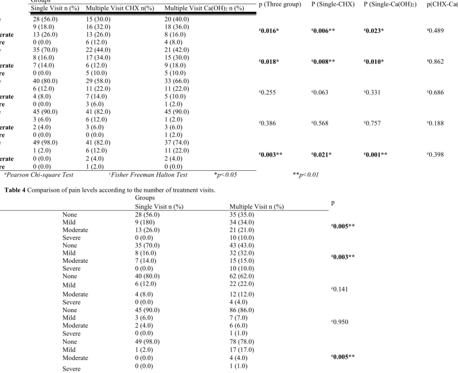
In the single visit group, 28 (56%) patients reported no pain after 24 hours, 9 (18%) experienced mild pain, and 13 (26%) reported moderate pain, but none of the patients reported severe pain. After 48 hours, 35 (70%) patients reported no pain, 8 (16%) reported mild pain, and 7 (14%) reported moderate pain, but none of them reported severe pain. After 72 hours, 40 (80%) patients reported no pain, 6 (12%) reported mild pain, and 4 (8%) reported moderate pain, but none reported severe pain. Seven days after the retreatment, 45 (90%) individuals reported no pain, 3 (6%) reported mild pain, and 2 (4%) reported moderate pain. Thirty days after the retreatment, 49 (98%) patients reported no pain, and only 1 (2%)
10 reported mild pain (Table 3).
In the two-visit CHX group after 24 hours, 15 (30%) patients reported no pain, 16 (32%) reported mild pain, 13 (26%) reported moderate pain, and 6 (12%) reported severe pain. After 48 hours, 22 (44%) patients reported no pain, 17 (34%) reported mild pain, 6 (12%) reported moderate pain, and 5 (10%) reported severe pain. After 72 hours, 29 (58%) patients reported no pain, 11 (22%) reported mild pain, 7 (14%) reported moderate pain, and 3 (6%) reported severe pain. Seven days after the retreatment, 41 (82%) individuals reported no pain, 6 (12%) reported mild pain, and 3 (6%) reported moderate pain, but none reported severe pain. Thirty days after the retreatment, 41 (82%) patients reported no pain, 6 (12%) reported mild pain, and 2 (4%) reported severe pain (Table 3).
In the two-visit Ca(OH)2 group after 24 hours, 20 (40%) patients reported no pain, 18 (36%)
reported mild pain, 8 (16%) reported moderate pain, and 5 (10%) reported severe pain. After 48 hours, 21 (42%) patients reported no pain, 15 (30%) reported mild pain, 6 (12%) reported moderate pain, and 5 (10%) reported severe pain. After 72 hours, 33 (66%) patients reported no pain, 11 (22%) reported mild pain, 5 (10%) reported moderate pain, and 1 (2%) reported severe pain. Seven days after the retreatment, 45 (90%) individuals reported no pain, 1 (2%) reported mild pain, 3 (6%) reported moderate pain, and 1 (2%) reported severe pain. Thirty days after the retreatment, 37 (74%) patients reported no pain, 11 (22%) reported mild pain, and 2 (4%) reported moderate pain, but none reported severe pain (Table 3).
On the third and seventh days, no specific differences between the pain categories (none, mild, moderate, or severe) were identified (p>0.05). When the incidence of pain was compared between the single and multiple-visit groups (Table 4), it was found that the single-visit group experienced significantly less pain than the multiple-visit group on the 1st, 2nd, and 30th days (p<0.05).
Overall, there were no statistically significant differences between the two medications with regard to the incidence of postoperative pain in any of the comparisons (Table 3).
When considered together, on the first day, the results of the 150 cases revealed that 63 (42%) teeth exhibited no postoperative pain. On the second day, 78 (52%) teeth exhibited no postoperative pain. On the third day, 102 (68%) teeth exhibited no postoperative pain. On the seventh day, 131 (87%) teeth exhibited no postoperative pain, and on the thirtieth day, 127 (84%) teeth exhibited no
11 postoperative pain (Table 3).
In this study, no flare-ups were observed in any of the groups.
DISCUSSION
It has been reported previously that the sensitivity of panoramic radiographs is lower than that of periapical radiographs, especially in the anterior region of the jaws; therefore, periapical radiographs should be used to evaluate the periapical tissues (15, 16). In this study, periapical film was used when the postoperative and control films were taken.
A person’s pain perception is influenced by many factors, so it varies widely according to the amount of preoperative pain, number of appointments, use of intracanal medication, tooth localization, pulpal vitality, microbial factors, change in the periapical tissue pressure, chemical mediators, change in the cyclic mediators, and various physiological factors. Many different scales and methods have been used to detect the pain that occurs after root canal treatment (1, 17-21).
The postoperative pain severity was evaluated numerically, grading the pain into none, slight, moderate, severe, and agonizing categories using a VRS (22, 23). A VRS can be used for both the identification and measurement of pain. In addition, a visual analogue scale (VAS) is considered to be a valid and reliable scale for measuring pain. A VAS can accurately predict the pain intensity and effect along a ratio, not an interval. Some studies have used VASs and some studies have used VRSs (19, 24). However, pain is affected by many different factors; therefore, in this study, the level of discomfort was measured using a VRS that was classified into only 4 categories in order to simplify the pain rating (1). With regard to the postoperative pain collection methods, the VRS was used because it is considered to be the most adequate method for reporting the pain experienced by a patient (25).
Di Renzo et al. evaluated the postoperative pain at 6, 12, 24, and 48 hours after single and multiple-visit root canal treatments (19). In addition, El Mubarak et al. observed postoperative pain during the first 12 and 24 hours after the patients had completed their treatments (26). In this study, the level of discomfort was rated in only 4 categories 1, 2, 3, 7, and 30 days after the root canal treatment.
In a recent study, Ertan et al. reported that the postoperative pain in the molar teeth was greater than that in the premolar and anterior teeth (27). Salma et al. found that the postoperative pain in the
12 premolar teeth was greater than the pain in the anterior teeth (28). In our study, no differences were seen between the localizations and the postoperative pain levels. Moreover, the incidence of pain in relation to gender was significantly higher in the women than in the men. In agreement with our results, Kim et
al. also reported that the women exhibited more postoperative pain than the men (29, 30). Furthermore,
there was no significant association between the postoperative pain and any of the tooth types included in our study. These findings are incompatible with some studies (19), but in agreement with others (20). The age factor showed no significant relationships with the postoperative pain as reported by the patients on the 1st, 2nd, 3rd, and 7th days. These findings are consistent with the results of another
study (31). On the 30th day, while the number of patients 45 years old who reported postoperative pain
was higher among the groups, statistically significant differences could not be demonstrated. Overall, there was less postoperative pain due to the greater sensitivity in the younger patients and reduced blood flow in the elderly patients.
Repeated endodontic treatment is a very interesting endodontic problem that requires a complex analysis of the indications and excellent procedural practice. Ørstavik et al. (14) introduced the PAI system for the radiographic assessment of the periapical status, and this was used in our study. This system allows for the easier tracking of periodic changes and a significant comparison of the outcomes of retreatment in clinical studies.
An aseptic technique and intracanal medication with calcium hydroxide must be complemented with a 2% CHX solution in order to decrease the number of microorganisms (32). Yoldas et al. (1) conducted a clinical study to compare the efficacy of one-visit vs. two-visit retreatments using a medication that combined calcium hydroxide and a 2% CHX solution. They showed that the two-visit retreatment was more effective for reducing postoperative pain and any potential flare-ups. In this study, there were no flare-ups observed in any of the groups. Previous studies have suggested that CHX gel is an effective intracanal medication, which is in agreement with our results. However, CHX is not an effective intracanal barrier, and it is also radiolucent, making it hard to visualize while it is inside the canal (33). Neelakantan et al. (34) investigated the antimicrobial activity of several canal medicaments against Porphyromonas gingivalis and Prevotella intermedia, indicating that the effect of the calcium hydroxide was significantly reduced after 48 hours, while the CHX gel lasted for 72 hours.
13 Previous studies have shown that the use of an intracanal medicament in symptomatic teeth can significantly reduce the incidence of flare-ups and postoperative pain (1). Moreover, Sjögren et al. (35) showed that there may be high error rates in root canal disinfection in single-visit root canal treatments. Siqueira et al. (36) and Maatscheck et al. (37) found that there were no significant differences in the postoperative pain between the retreatment and the primary root canal treatment in their studies. In these studies, different medicaments were used for the root canal treatments, and the teeth were treated in two or more visits.
Some researchers have reported that the application of intracanal medicament reduces postoperative pain. However, they found no significant differences in the postoperative pain after one week of medicament administration between calcium hydroxide and 0.2% CHX (38). Because of the postoperative pain, several intracanal medicaments are used to temporarily fill the root canal, such as CHX or Ca(OH)2, and they can play important roles in suppressing the recontamination of the root canal
between visits (36). However, the apical extrusion of contaminated debris and medicaments may also cause postoperative pain (1). Walton et al. reported that there was no statistical difference in the postoperative pain with regard to the frequency and quantity of Ca(OH)2 used as an intracanal
medicament (18). Fox et al. (39) and Roane et al. (20) argued that the postoperative pain percentages in single-visit root canal treatments were lower than those in multiple-visit root canal treatments.
Peckruhn reported that 1,140 teeth of 918 patients were treated in single visits. When the patients were recalled 1 year later, there was less failure reported in the single-visit root canal treatments (40). In a 2008 study of dissatisfaction scores, it was reported that a single-visit root canal treatment was preferred by the patients to a multiple-visit root canal treatment, but the Australian endodontists were reluctant to accept single-visit root canal treatments (41). In this study, on the second day, the postoperative pain rate in the single-visit root canal retreatment group (30%) was significantly lower than that in the multiple-visit retreatment group (54%).
An increase in the pain incidence at the 1 month follow-up was observed in the patients with multiple-visit retreatments, which was not the case in the single-visit retreatments. Although the patients are still being followed up for further evaluation, it was strongly suspected that the introduction of the root canal medicaments into the root canal space may have resulted in the extrusion of some of the
14 material into the periapical area. This may have coupled with the healing process, and therefore, resulted in the increased incidence of pain at the 1 month follow-up (18). Although the caregiver paid extreme attention and tried not to extrude any intracanal medicament into the periapical area, this may not have been the case in every patient. The disrupted periapical anatomy due to a previous root canal treatment and the status of the periapical tissue prior to retreatment may result in the extrusion of intracanal medicament into the periapical area (26).
The presence of a periapical lesion is a risk factor for the development of postoperative pain. In the study by Alves et al., there was more postoperative pain in the teeth with periapical radiolucency (42). When the full-scale PAI scores were evaluated individually, no significant correlation was recorded between the preoperative PAI scores and the incidence of postoperative pain. Even after the PAI scores were dichotomized to reflect the absence (PAI ≤ 2) or presence (PAI > 2) of apical periodontitis according to previous studies (14, 44), there was still no correlation between the preoperative PAI scores and the incidence of postoperative pain. Although the baseline PAI score was reported to impair the outcome results due to the strong predictive value, this study was not an outcomes study, and the preoperative PAI scores were recorded for the purpose of determining a correlation between the preoperative PAI scores and postoperative pain. Moreover although the mentioned study criticized the PAI scores, no better method has been suggested. With cone-beam computed tomography being out of question due to ethical issues in Turkey (higher exposure values), we were left with PAI scoring for further evaluation (43).
On the second day, there was a correlation between the periapical radiolucency and the postoperative pain; the teeth with periapical lesions exhibited greater postoperative pain. Our findings are compatible with the study conducted by Eyuboglu et al. (44).
Sari and Duruturk reported that the complete resorption of the amount of extruded AH Plus sealer in 56.09% of the successfully treated canals at the end of a 4-year follow-up showed that any excess AH Plus filling material at the periapex disappears over time (45). In this study, we used AH plus as the root canal filling material. On the second day, there was a relationship between the sealing extrusion and the postoperative pain, but there was no significant difference between the extruded gutta-percha and the postoperative pain.
15 CONCLUSIONS
Based on the results of this study, it was found that the postoperative pain incidence in the single-visit endodontic retreatments without intracanal medicaments was less than that in the multiple-single-visit endodontic retreatments. When the medicaments were compared among themselves, the pain intensity was higher in the CHX group.
Conflict of Interest
16 REFERENCES
1. Yoldas O, Topuz A, Isci AS, Oztunc H. Postoperative pain after endodontic retreatment: single-versus two-visit treatment. Oral Surg Oral Med Oral Pathol Oral Radiol Endod. 2004;98:483-7. 2. Petrović V, Opačić-Galić V, Živković S. Postoperative pain after primary endodontic treatment and
retreatment of asymptomatic teeth. Stomatološki glasnik Srbije. 2011;58:75-81.
3. Shahi S, Asghari V, Rahimi S, Lotfi M, Samiei M, Yavari H, Shakouie S, Nezafati S. Postoperative pain after endodontic treatment of asymptomatic teeth using rotary instruments: a randomized clinical trial. Iran Endod. 2016;11:38.
4. Nekoofar MH, Sheykhrezae MS, Meraji N, Jamee A, Shirvani A, Jamee J, Dummer PM. Comparison of the effect of root canal preparation by using Wave One and ProTaper on postoperative pain: a randomized clinical trial. J Endod. 2015;41:575-8.
5. Siqueira JF, de Uzeda M. Intracanal medicaments: evaluation of the antibacterial effects of chlorhexidine, metronidazole, and calcium hydroxide associated with three vehicles. J Endod. 1997;23:167-9.
6. Barbosa CA, Goncalves RB, Siqueira JF, De Uzeda M. Evaluation of the antibacterial activities of calcium hydroxide, chlorhexidine, and camphorated paramonochlorophenol as intracanal medicament. A clinical and laboratory study. J Endod. 1997;23:297-300.
7. Rôças IN, Siqueira JF, Santos KR. Association of Enterococcus faecalis with different forms of periradicular diseases. J Endod. 2004;30:315-320.
8. Love RM. Enterococcus faecalis–a mechanism for its role in endodontic failure. Int Endod J. 2001;34:399-405.
9. Kvist T, Molander A, Dahlén G, Reit C. Microbiological evaluation of one- and two-visit endodontic treatment of teeth with apical periodontitis: a randomized, clinical trial. J Endod. 2004;30:572-6.
10. Calhoun RL, Landers RR. One-appointment endodontic therapy: a nationwide survey of endodontists. J Endod. 1982;8:35-40.
11. Soltanoff W. A comparative study of the single-visit and the multiple-visit endodontic procedure. J Endod. 1978;4:278-281.
17 12. Jurcak JJ, Bellizzi R, Loushine RJ. Successful single-visit endodontics during Operation Desert
Shield. J Endod. 1993;19:412-3.
13. de Almeida Gomes BPF, Vianna ME, Sena NT, Zaia AA, Ferraz CCR, de Souza Filho FJ. In vitro evaluation of the antimicrobial activity of calcium hydroxide combined with chlorhexidine gel used as intracanal medicament. Oral Surg Oral Med Oral Pathol Oral Radiol and Endodont. 2006;102:544-550.
14. Ørstavik D, Kerekes K, Eriksen HM. The periapical index: a scoring system for radiographic assessment of apical periodontitis. Dent Traumatol. 1986;2:20-34.
15. Boucher Y, Matossian L, Rilliard F, Machtou P. Radiographic evaluation of the prevalence and technical quality of root canal treatment in a French subpopulation. Int Endod J. 2002;53:229-238. 16. Allard U, Palmqvist S. A radiographic survey of periapical conditions in elderly people in a Swedish
county population. Dent Traumatol. 1986;2:103-8.
17. Siqueira JF Jr, Rjcas IN, Favieri A, et al. Incidence of postoperative pain after intracanal procedures based on an antimicrobial strategy. J Endod. 2002;28:457-60.
18. Walton RE, Holton IF, Michelich R. Calcium hydroxide as an intracanal medication: effect on posttreatment pain. J Endod. 2003;29:627-9.
19. DiRenzo A, Gresla T, Johnson BR, Rogers M, Tucker D, BeGole E. Postoperative pain after 1- and 2-visit root canal therapy. Oral Surg Oral Med Oral Pathol Oral Radiol and Endodont. 2002;93:605-10.
20. Roane JB, Dryden JA, Grimes EW. Incidence of postoperative pain after single- and multiple-visit endodontic procedures. Oral Surg Oral Med Oral Pathol Oral Radiol Endod. 1983;55:68-72. 21. Anjaneyulu K, Nivedhitha MS. Influence of calcium hydroxide on the post-treatment pain in
endodontics: Aa systematic review. J Conservat Cent. 2014;17:200-7.
22. Houde RW, Wallenstein SL. A method for evaluating analgesics in patients with chronic pain. Drug Addiction & Narcotics Bull. 1953:660-82.
23. Revill SI, Robinson JO, Rosen M, Hogg MIJ. The reliability of a linear analogue for evaluating pain. Anaesthesia. 1976;31:1191-8.
18 24. Price DD, McGrath PA, Rafii A, Buckingham BÇ. The validation of visual analogue scales as ratio
scale measures for chronic and experimental pain. Pain. 1983;17:45-56.
25. Stinson JN, Kavanagh T, Yamada J, Gill N, Stevens B. Systematic review of the psychometric properties, interpretability and feasibility of self-report pain intensity measures for use in clinical trials in children and adolescents. Pain. 2006;125:143-57.
26. El Mubarak AHH, Abu-bakr NH, Ibrahim YE. Postoperative pain in multiple-visit and single-visit root canal treatment. J Endod. 2010;36:36-39.
27. Ertan T, Şahinkesen G, Tunca YM. Evaluation of postoperative pain in root canal treatment. The J Turkish Soc Algol. 2010;22:159-64.
28. Salma J. A study of post obturation pain following single visit root canal treatment. Chattagram Maa-O-Shishu Hosp Med Coll J. 2013;12:16-19.
29. Saunders WP. A prospective clinical study of periradicular surgery using mineral trioxide aggregate as a root-end filling. J Endod. 2008;34:660-5.
30. Kim E, Song JS, Jung IY, Lee SJ, Kim S. Prospective clinical study evaluating endodontic microsurgery outcomes for cases with lesions of endodontic origin compared with cases with lesions of combined periodontal–endodontic origin. J Endod. 2008;34:546-51.
31. Abbott PV. Factors associated with continuing pain in endodontics. Aust Dent J. 1994;39:157-61. 32. Stuart CH, Schwartz SA, Beeson TJ, Owatz CB. Enterococcus faecalis: its role in root canal
treatment failure and current concepts in retreatment. J Endod. 2006;32:93-98.
33. Souza-Filho FJD, Soares ADJ, Vianna ME, Zaia AA, Ferraz CCR, Gomes BPFDA. Antimicrobial effect and pH of chlorhexidine gel and calcium hydroxide alone and associated with other materials. Braz Dent J. 2008;19:28-33.
34. Neelakantan P, Sanjeev K, Subbarao CV. Duration-dependent susceptibility of endodontic pathogens to calcium hydroxide and chlorhexidine gel used as intracanal medicament: an in vitro evaluation. Oral Surg Oral Med Oral Pathol Oral Radiol Endod. 2007;104:138-41.
35. Sjögren U, Figdor D, Persson S, Sundqvist G. Influence of infection at the time of root filling on the outcome of endodontic treatment of teeth with apical periodontitis. Int Endod J. 1997;30:297-306.
19 36. Siqueira JF, Lopes HP, de Uzeda M. Recontamination of coronally unsealed root canals medicated with camphorated paramonochlorophenol or calcium hydroxide pastes after saliva challenge. J Endod. 1998;24:11.
37. Mattscheck DJ, Law AS, Noblett WC. Retreatment versus initial root canal treatment: factors affecting posttreatment pain. Oral Surg Oral Med Oral Pathol Oral Radiol and Endod. 2001;92:321-4.
38. Menakaya IN, Oderinu OH, Adegbulugbe IC, Shaba OP. Incidence of postoperative pain after use of calcium hydroxide mixed with normal saline or 0.2% chlorhexidine digluconate as intracanal medicament in the treatment of apical periodontitis. The Saudi Dent J. 2015;27:187-193.
39. Fox J, Atkinson JS, Dinin AP, Greenfield E, Hechtman E, Reeman CA, Todaro CJ. Incidence of pain following one-visit endodontic treatment. Oral Surg Oral Med Oral Pathol Oral Radiol Endod. 1970;30:123-30.
40. Pekruhn RB. The incidence of failure following single-visit endodontic therapy. J Endod. 1986;12:68-72.
41. Sathorn C. Effectiveness and efficiency: systematic reflections on single and multiple visit root canal treatment. University of Melbourne, School of Dental Science, Thesis. 2008.
42. de Oliveira Alves V. Endodontic flare-ups: a prospective study. Oral Surg Oral Med Oral Pathol Oral Radiol Endod. 2010;110:68-72.
43. Kirkevang LL, Ørstavik D, Bahrami G, Wenzel A, Væth M. Prediction of periapical status and tooth extraction. Int Endod J. 2017;50:5-14.
44. Eyuboglu TF, Olcay K, Özcan M. A clinical study on single-visit root canal retreatments on consecutive 173 patients: frequency of periapical complications and clinical success rate. Clin Oral Investig. 2017;21(5):1761-8.
45. Sari Ş, Durutűrk L. Radiographic evaluation of periapical healing of permanent teeth with periapical lesions after extrusion of AH Plus sealer. Oral Surg Oral Med Oral Pathol Oral Radiol Endod. 2007;104:54-59.
20 CAPTIONS FOR TABLES:
Tables
Table 1. Distribution of prognostic factors, inception cohort, study sample and p values (univariate analysis).
Table 2. Effect of preoperative intraoperative and postoperative factors on postoperative pain. Table 3. Frequency and percentage of postoperative pain in retreatment groups.
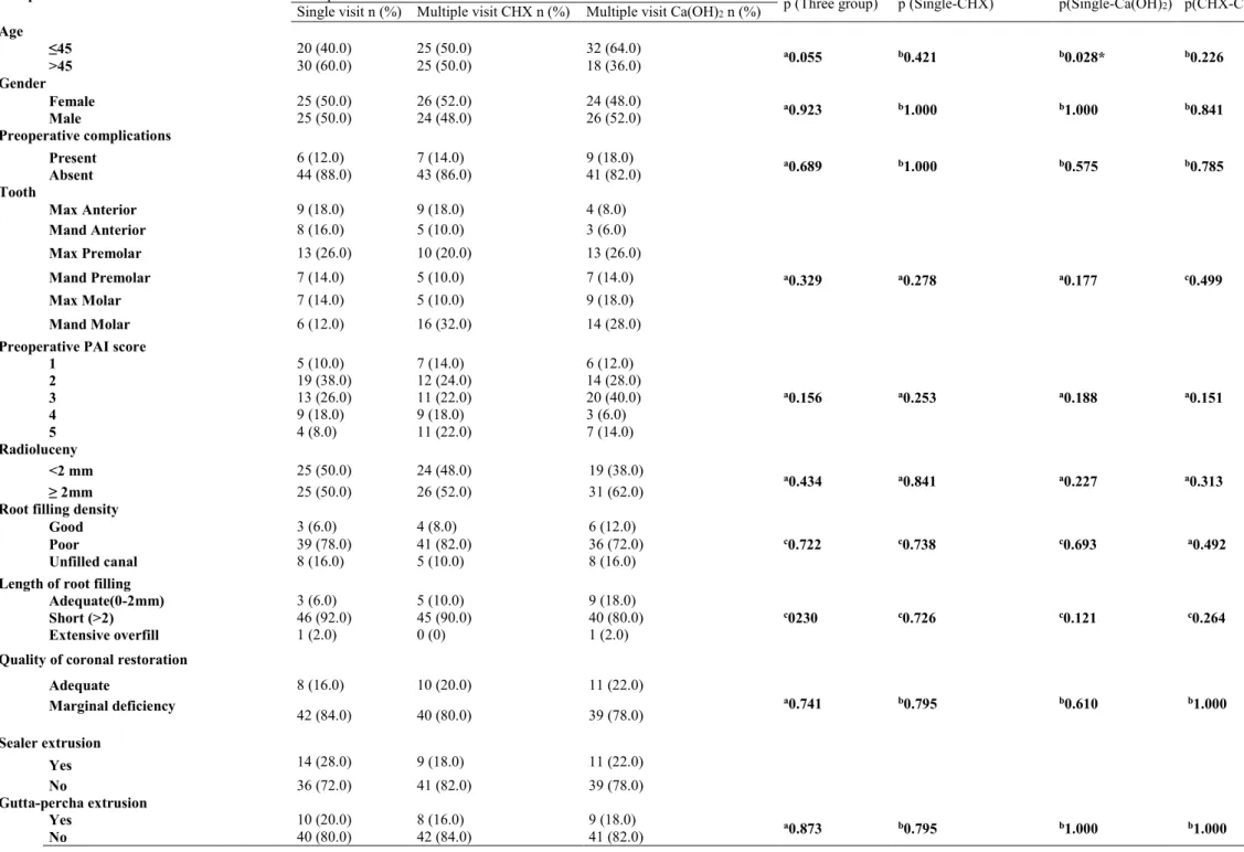
Table 1 Distribution of prognostic factors, inception cohort, study sample and p values (univariate analysis).
Preoperative Factors Groups
p (Three group) p (Single-CHX) p(Single-Ca(OH)2) p(CHX-Ca(OH)2) Single visit n (%) Multiple visit CHX n (%) Multiple visit Ca(OH)2 n (%)
Age ≤45 20 (40.0) 25 (50.0) 32 (64.0) a 0.055 b0.421 b0.028* b0.226 >45 30 (60.0) 25 (50.0) 18 (36.0) Gender Female 25 (50.0) 26 (52.0) 24 (48.0) a 0.923 b1.000 b1.000 b0.841 Male 25 (50.0) 24 (48.0) 26 (52.0) Preoperative complications Present 6 (12.0) 7 (14.0) 9 (18.0) a0.689 b1.000 b0.575 b0.785 Absent 44 (88.0) 43 (86.0) 41 (82.0) Tooth Max Anterior 9 (18.0) 9 (18.0) 4 (8.0) Mand Anterior 8 (16.0) 5 (10.0) 3 (6.0) Max Premolar 13 (26.0) 10 (20.0) 13 (26.0) Mand Premolar 7 (14.0) 5 (10.0) 7 (14.0) a0.329 a0.278 a0.177 c0.499 Max Molar 7 (14.0) 5 (10.0) 9 (18.0) Mand Molar 6 (12.0) 16 (32.0) 14 (28.0)
Preoperative PAI score
1 5 (10.0) 7 (14.0) 6 (12.0) a0.156 a0.253 a0.188 a0.151 2 19 (38.0) 12 (24.0) 14 (28.0) 3 13 (26.0) 11 (22.0) 20 (40.0) 4 9 (18.0) 9 (18.0) 3 (6.0) 5 4 (8.0) 11 (22.0) 7 (14.0) Radioluceny <2 mm 25 (50.0) 24 (48.0) 19 (38.0) a0.434 a0.841 a0.227 a0.313 ≥ 2mm 25 (50.0) 26 (52.0) 31 (62.0)
Root filling density
Good 3 (6.0) 4 (8.0) 6 (12.0)
Poor 39 (78.0) 41 (82.0) 36 (72.0) c0.722 c0.738 c0.693 a0.492
Unfilled canal 8 (16.0) 5 (10.0) 8 (16.0)
Length of root filling
Adequate(0-2mm) 3 (6.0) 5 (10.0) 9 (18.0)
Short (>2) 46 (92.0) 45 (90.0) 40 (80.0) c0230 c0.726 c0.121 c0.264
Extensive overfill 1 (2.0) 0 (0) 1 (2.0)
Quality of coronal restoration
Adequate 8 (16.0) 10 (20.0) 11 (22.0) a0.741 b0.795 b0.610 b1.000 Marginal deficiency 42 (84.0) 40 (80.0) 39 (78.0) Sealer extrusion Yes 14 (28.0) 9 (18.0) 11 (22.0) No 36 (72.0) 41 (82.0) 39 (78.0) Gutta-percha extrusion Yes 10 (20.0) 8 (16.0) 9 (18.0) a 0.873 b0.795 b1.000 b1.000 No 40 (80.0) 42 (84.0) 41 (82.0)
Table 2 Effect of preoperative and intraoperative factors on postoperative pain.
1.day pain p 2.day pain p 3. day pain p 7.day pain p 30.day pain p
Age ≤45 0.439 0.241 0.188 0.888 0.038* >45 Gender Female 0.013* 0.251 0.863 0.198 0.818 Male Preoperative complications Present 0.039* 1.000 0.220 0.279 1.000fe Absent Tooth Max Anterior 0.906 0.343 0.947 0.399 0.382 Mand Anterior Max Premolar Mand Premolar Max Molar Mand Molar
Preoperative PAI score ≤2
0.395 0.098 0.620 0.654 0.911 >2
Radiolucency <2 mm
0.507 0.039* 0.280 0.503 0.634 ≥ 2mm
Root filling density Good
0.316 0.514 0.286 0.657 0.846 Poor
Unfilled canal
Length of root filling Adequate (0-2)
0.026* 0.133 0.057 1.000 0.614
Short (>2) Extensive overfill
Intraoperative
Quality of coronal restoration
Adequate
1.000 0.129 0.007** 0.325 0.141
Marginal deficiency
Intraoperative
Quality of root canal filling
Dense and tapered
0.692 0.917 0.903 0.583 0.544 Voids present Poorly condensed Intraoperative Sealer extrusion Present 0.091 0.036* 0.157 0.764 0.784 Absent Intraoperative
Gutta-percha extrusion Present 0.884 0.271 0.936 0.539 0.263
Absent
Table 3 Frequency and percentage of postoperative pain in retreatment groups.
Pain Levels Groups p (Three group) P (Single-CHX) P (Single-Ca(OH)2) p(CHX-Ca(OH)2) Single Visit n (%) Multiple Visit CHX n(%) Multiple Visit Ca(OH)2 n (%)
1. Day None 28 (56.0) 15 (30.0) 20 (40.0) c0.016* c0.006** c0.023* a0.489 Mild 9 (18.0) 16 (32.0) 18 (36.0) Moderate 13 (26.0) 13 (26.0) 8 (16.0) Severe 0 (0.0) 6 (12.0) 4 (8.0) 2. Day None 35 (70.0) 22 (44.0) 21 (42.0) c0.018* c0.008** c0.010* a0.862 Mild 8 (16.0) 17 (34.0) 15 (30.0) Moderate 7 (14.0) 6 (12.0) 9 (18.0) Severe 0 (0.0) 5 (10.0) 5 (10.0) 3. Day None 40 (80.0) 29 (58.0) 33 (66.0) c0.255 c0.063 c0.331 c0.686 Mild 6 (12.0) 11 (22.0) 11 (22.0) Moderate 4 (8.0) 7 (14.0) 5 (10.0) Severe 0 (0.0) 3 (6.0) 1 (2.0) 7. Day None 45 (90.0) 41 (82.0) 45 (90.0) c0.386 c0.568 c0.757 c0.188 Mild 3 (6.0) 6 (12.0) 1 (2.0) Moderate 2 (4.0) 3 (6.0) 3 (6.0) Severe 0 (0.0) 0 (0.0) 1 (2.0) 30. Day None 49 (98.0) 41 (82.0) 37 (74.0) c0.003** c0.021* c0.001** c0.398 Mild 1 (2.0) 6 (12.0) 11 (22.0) Moderate 0 (0.0) 2 (4.0) 2 (4.0) Severe 0 (0.0) 1 (2.0) 0 (0.0)
aPearson Chi-square Test cFisher Freeman Halton Test *p<0.05 **p<0.01
Table 4 Comparison of pain levels according to the number of treatment visits.
Pain levels Groups p
Single Visit n (%) Multiple Visit n (%)
1. Day None 28 (56.0) 35 (35.0) c0.005** Mild 9 (180) 34 (34.0) Moderate 13 (26.0) 21 (21.0) Severe 0 (0.0) 10 (10.0) 2. Day None 35 (70.0) 43 (43.0) c0.003** Mild 8 (16.0) 32 (32.0) Moderate 7 (14.0) 15 (15.0) Severe 0 (0.0) 10 (10.0) 3. Day None 40 (80.0) 62 (62.0) c0.141 Mild 6 (12.0) 22 (22.0) Moderate 4 (8.0) 12 (12.0) Severe 0 (0.0) 4 (4.0) 7. Day None 45 (90.0) 86 (86.0) c0.950 Mild 3 (6.0) 7 (7.0) Moderate 2 (4.0) 6 (6.0) Severe 0 (0.0) 1 (1.0) 30. Day None 49 (98.0) 78 (78.0) c0.005** Mild 1 (2.0) 17 (17.0) Moderate 0 (0.0) 4 (4.0) Severe 0 (0.0) 1 (1.0)