Research Article
Investigation of NF-
𝜅B1 and NF-𝜅BIA Gene Polymorphism in
Non-Small Cell Lung Cancer
Y. M. Oltulu,
1E. Coskunpinar,
1G. Ozkan,
1E. Aynaci,
2P. Yildiz,
3T. Isbir,
4and I. Yaylim
11Department of Molecular Medicine, Institute of Experimental Medicine Research, Istanbul University DETAE,
P.O. Box 7, 34390 C¸ apa/˙Istanbul, Turkey
2Department of Chest Diseases, Faculty of Medicine, Medipol University, Istanbul, Turkey
3Third Clinics, Yedikule Chest Diseases and Thoracic Surgery Training Research Hospital, Istanbul, Turkey 4Department of Medical Biology, School of Medicine, Yeditepe University, Istanbul, Turkey
Correspondence should be addressed to I. Yaylim; [email protected]
Received 20 October 2013; Revised 13 December 2013; Accepted 9 January 2014; Published 23 February 2014 Academic Editor: Sherven Sharma
Copyright © 2014 Y. M. Oltulu et al. This is an open access article distributed under the Creative Commons Attribution License, which permits unrestricted use, distribution, and reproduction in any medium, provided the original work is properly cited. Lung cancer is a complex, multifactorial disease which is the leading cause of cancer death in both men and women. NF-𝜅B is a transcription factor which is known to affect the expression of more than 150 genes related to inflammation, lymphocyte activation, cell proliferation, differentiation, and apoptosis, as well as contributing to cell apoptosis and survival. However, NF-𝜅BIA (I𝜅B𝛼) is the inhibitor of the transcription factor. The -94ins/delATTG polymorphism of the NF-𝜅B1 gene promoter region which causes a functional effect and NF-𝜅BIA 3UTR A→ G polymorphism has been shown to be related to various inflammatory diseases and
cancer. Ninety-five NSCLC patients and 99 healthy controls were included in study. The NF-𝜅B1 -94ins/delATTG and NF-𝜅BIA 3UTR A→ G polymorphism have been studied by using PCR-RFLP method. It was found that the NF-𝜅B1 -94ins/delATTG DD genotype and D allele frequencies were higher in patients than healthy controls and the presence of the DD genotype has a 3.5-fold increased risk of the disease (P: 0.014). This study is the first to investigate the NF-𝜅B1 -94ins/delATTG and NF-𝜅BIA 3UTR A→ G
polymorphism together in the Turkish population. According to the results, the NF-𝜅B1 -94ins/del ATTG promoter polymorphism may have a role in lung carcinogenesis and prognosis.
1. Introduction
Non-small cell lung cancer (NSCLC), which includes squa-mous cell carcinoma, adenocarcinoma, and large-cell carci-noma, is the most common lung cancer possessing approxi-mately 80–85% rates in the prevalence of lung cancer case [1]. Despite all advances in the current treatments including sur-gical resection, chemotherapy, and radiation therapy alone or in combination, the disease is rarely curable prognosis and remains poor [2]. As all of these facts are considered, recent researches are tending to understand molecular, biological, and genetic factors and to find other prognostic factors providing long-term survival and intending new targeted therapies [3,4]. Lung cancer cells manage to escape from the signal transduction pathways to facilitate their own survival and proliferation by using multiple mechanisms [5]. Carcino-gens and inflammatory cytokines contributing substantially
to cancer development are involved in activation of common cell survival signaling pathways. The one of this cell survival signal is nuclear factor-kappaB (NF-𝜅B) which is involved in multiple steps in carcinogenesis and in cancer cell’s resistance to chemo- and radiotherapy [6]. Recently, many studies with animal models and cell culture systems indicate the interplay between NF-𝜅B and lung carcinogenesis, which emphasizes the importance of targeting the NF-𝜅B signaling pathway for lung cancer treatment and chemoprevention [7]. NF-𝜅B, a nuclear transcription factor [8], was first identified in 1986 by Sen and Baltimore [9]. It was initially observed to be a transcription factor binding to the intronic enhancer of the kappa light chain gene (the 𝜅B site) in B cells, but it was later shown to be present in every cell type [10]. Afterwards, NF-𝜅B emerged as a major regulator of more than 200 genes involved in diverse process such as cell survival and
Volume 2014, Article ID 530381, 6 pages http://dx.doi.org/10.1155/2014/530381
cell adhesion, inflammation, differentiation, and growth [11]. NF-𝜅B is activated by phosphorylation of I𝜅B𝛼 which is catalyzed by an I𝜅B𝛼 Kinase (IKK) complex consisting of IkK-𝛼, IkK-𝛽, IkK-𝛾 (also called NEMO), and other proteins yet to be identified [12,13]. After activation of NF-𝜅B, I𝜅B𝛼 is degraded and p50–p65 heterodimer is translocated to the nucleus, binds to the DNA (at the promoter region), and activates gene [14,15]. Currently, studies show that activation of the transcription factor nuclear factor (NF)𝜅B is a novel mechanism of chemoresistance in NSCLC and other tumors [16,17]. NF-𝜅B1 is inhibited by IkB proteins (e.g., NF-𝜅BIA) [18]. Phosphorylation of serine residues on the I-kappa-B proteins, by kinases and marks them for degradation, thereby allowing activation of the NF-𝜅B complex [19]. As the first potential functional NF-𝜅B1 genetic variation was identified 94 insertion/deletion ATTG located between two received key promoter regulatory elements in the NF-𝜅B1 gene, ATTG deletion causes the loss of binding to nuclear proteins, which leads to reduced promoter activity [20]. The NF𝜅B -94ins/delATTG polymorphism of the NF-𝜅B1 gene promoter region which causes a functional effect and NF-𝜅BIA 3UTR A→ G polymorphism have been shown to be associated with various inflammatory diseases and cancers [21]. The aim of this study is firstly to investigate the NF-𝜅B1 -94ins/delATTG and NF-𝜅BIA 3UTR A→ G polymorphism together in the
Turkish population.
2. Materials and Methods
2.1. Study Groups. Ninety-five primary non-small cell lung
cancer (NSCLC) patients and 99 healthy individuals were included in the study. NSCLC patients were recruited from the Yedikule Chest Diseases and Thoracic Surgery Training Research Hospital, Istanbul. The diagnosis of NSCLC was made by the pathologist based on histopathological exam-ination. In NSCLC group, all subjects were diagnosed and confirmed with histopathological examination. They were all newly diagnosed without a history of prior radiotherapy and/or chemotherapy. Exclusion criteria included primary extra pulmonary malignancy, small cell lung cancer, a his-tory of malignant disease, and withdrawal of consent and patient aged less than 18. Pathological staging information on all NSCLC cases was confirmed by manual review of the pathology reports and clinical charts. Nodal status was categorized as no regional lymph nodes affected (N0) or at least one nodal metastasis. The mean ages of the patients and controls were61.2 ± 9.83 years and 57.49 ± 10.83 years, respectively. The percentage of females was 6.3% for patients and 32.3% for controls, and percentage of males was 93.7% for patients and 67.7% for controls. 99 healthy subjects without any malignancy were selected for the control group that is comprised only of individuals with a negative family history of cancer. The patient and control groups were matched for age. All participants signed an informed consent before enrollment and Institutional Ethical committee approval was obtained for the study.
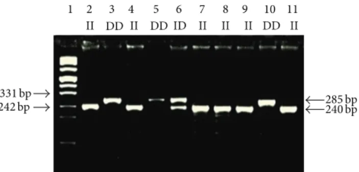
331 bp
242 bp 285 bp240 bp
II DD II DD ID II II II DD II
1 2 3 4 5 6 7 8 9 10 11
Figure 1: Representative genotypes of NF-𝜅B1 -94ins/delATTG polymorphism. Lane 1: marker DNA ladder; Lanes 2, 4, 7, 8, 9, and 11: ins/ins (ATTG2/ATTG2) genotypes; Lanes 3, 5, and 10: del/del (ATTG1/ATTG1) genotypes; Lane 6: heterozygous del/ins (ATTG1/ATTG2) genotypes. 1 2 GG AA3 GG4 AG5 AG6 400 bp 300 bp 100 bp 424 bp 316 bp 108 bp
Figure 2: Representative genotypes of NF-𝜅BIA 3UTR A→G
polymorphism. Lane 1: marker DNA ladder; Lanes 2 and 4: GG mutant genotypes; Lane 3: AA wild-type homozygous genotypes; Lanes 5 and 6: AG heterozygous genotypes.
2.2. Polymorphism Analysis. Blood samples from all study
participants were collected in EDTA-containing tubes. Genomic DNA was extracted from peripheral whole blood according to kit protocol (High Pure PCR Template Preparation Kit, REF 11796828001 (Roche, Diagnostics GmbH, Mannheim, Germany). Genotyping was performed by polymerase chain reaction (PCR) and restriction fragment length polymorphism (RFLP); the procedures of PCR-RFLP are given in Table 1. Two separate PCR reactions were used to detect the two types of polymorphism in NF-𝜅B gene, namely, NF-𝜅B1 -94ins/delATTG polymorphism and NF-𝜅BIA 3UTR A→ G polymorphism. The appropriate primers were used to amplify the corresponding gene of the subjects by PCR and the reaction products were digested by using the appropriate enzyme and incubated at 37∘C overnight. The digested products were analyzed on 3% agarose gel, stained with ethidium bromide, and examined under transillumination (Figures1and2). Each gel was read by two observers, unaware of the subject’s status. In order to verify our PCR-RFLP results, we repeated PCR-RFLP stage 2 times for each of selected subject. The expected results after restriction for each gene were also given inTable 1.
2.3. Statistical Analysis. Statistical analyses were performed
Table 1: PCR and RFLP procedures and products of NF-𝜅B1 -94ins/delATTG and NF-𝜅BIA 3UTR A→ G.
Primers (forward and reverse) PCR product Restriction enzyme Restriction products NF-𝜅B1
5-TGGGCACAAGTCGTTTATGA-3
285 bp Van91l (PflMI)
ATTG2/ATTG2(ins/ins): 240 bp, 45 bp
5-CTGGAGCCGGTAGGGAAG-3 ATTG1/ATTG1(del/del): 281
ATTG2/ATTG1(ins/del): 281, 240, 45
NF-𝜅BIA 5-GGCTGAAAGAACATGGACTTG-3 424 bp HaeIII AA wild type: 424 5-GTACACCATTTACAGGAGGG-3 AG heterozygous: 316, 108 GG mutant: 424, 316, 108
Table 2: Distribution of NF-𝜅B1 and NF-𝜅BIA genotypes and allele in NSCLC patients and controls.
Genotypes/alleles Controls𝑛 (%) Patients𝑛 (%) O.R (95% CI) 𝑃 value
NF-𝜅B1 II 46 (46.47) 35 (36.84) Reference ID 47 (47.47) 44 (46.32) 1.23 (0.67–2.25) 0.500 DD 6 (6.06) 16 (16.84) 3.50 (1.24–9.87) 0.014 ID + DD 53 (53.53) 60 (63.16) 1.49 (0.84–2.64) 0.174 I allele 139 (70.2) 114 (60) Reference D allele 59 (29.8) 76 (40) 1.57 (1.03–2.39) 0.035 NF-𝜅BIA AA 21 (21.21) 17 (17.9) Reference AG 45 (45.46) 45 (47.36) 1.24 (0.58–2.65) 0.587 GG 33 (33.33) 33 (34.74) 1.24 (0.55–2.75) 0.605 AG + GG 78 (78.79) 78 (82.1) 1.24 (0.61–2.52) 0.561 A allele 87 (43.94) 79 (41.58) Reference G allele 111 (56.06) 111 (58.42) 1.10 (0.74–1.65) 0.639
O.R: Odds ratio; CI: confidence interval.
Chicago, IL, USA). Data were expressed as means ± SD. Differences in the distribution of NF-𝜅B1 -94ins/delATTG and NF-𝜅BIA 3UTR A→ G genotypes or alleles between cases and controls were tested using the Chi-square statistic, respectively (Table 2). Differences in characteristics between NSCLC patients and controls were assessed with Fisher’s exact test, as well as disparities in genotype and allele frequencies. Relative risk at 95% confidence intervals (CI) was calculated as the odds ratio (OR). Values𝑃 < 0.05 were considered statistically significant. A multivariate logistic regression model was performed to investigate possible effects of genotypes and alleles after adjustment for age.
3. Results
In this study, we examined 194 volunteers, 95 NSCLC (89 males; 6 females) patients, and 99 healthy people (67 males; 32 females) detecting any chronic disease or any evidence of malignancy. Distribution of NF-𝜅B1 and NF-𝜅BIA genotypes according to clinic features in NSCLC patients is shown inTable 3. The distribution of the NF-𝜅B1 -94ins/delATTG genotypes in control and NSCLC patients was found to be significantly different (𝑃: 0.048). It was evaluated that individuals carrying DD genotype had 3.5-fold increased risk for NSCLC (𝑃: 0.014 𝜒2: 5.605, O.R: 3.50, %95 CI: 1.24–9.87). No statistically significant differences between groups were
observed when the NF-𝜅BIA 3UTR A→ G genotypes distri-butions were compared (𝑃: 0.844). A significant correlation between genotype combinations of NF-𝜅B1 and NF-𝜅BIA (DDAG genotype) and NSCLC risk was found compared to all other combinations (𝑃: 0.025; O.R: 5.035; 95% CI: 1.067– 24.14) and DDAG genotype had increased risk for NSCLC. The prevalence of IIAA genotype combinations versus to all other combinations was 5.3% in patients and 12.1% in the control group, but there are no statistically significant differences between groups (𝑃: 0.091; O.R: 0.403; 95% CI: 0.136–1.191). The results of multivariate logistic regression analysis are presented in Table 4. Gender, age (<57/≥57), and NF-𝜅B1 DD genotype were associated with NSCLC in univariate analysis, and additionally these were associated with this disease in multivariate logistic regression analysis.
4. Discussion
A functional polymorphism in the NF-𝜅B1 gene promoter region (-94ins/delATTG) has been identified and associated with both chronic inflammatory diseases and malignant diseases [22]. NF-𝜅B is inactivated in the cytoplasm by I𝜅B𝛼, 𝛽, or 𝛾 and the most common protein of this family is the NF-𝜅B inhibitor 𝛼 (NF-𝜅BIA) [23]. -94ins/delATTG polymorphism has evidence from two independent func-tional assays, in vitro promoter activity and differential an unidentified nuclear protein binding, that the specific allele
Table 3: Distribution of NF-𝜅B1 and NF-𝜅BIA genotypes with clinic features in NSCLC patients. NF-𝜅B1 NF-𝜅BIA II ID DD 𝑃 value AA AG GG 𝑃 value 𝑛 (%) 𝑛 (%) 𝑛 (%) 𝑛 (%) 𝑛 (%) 𝑛 (%) Sex Men 33 (37.10) 41 (46.10) 15 (16.80) 0.980 15 (16.90) 43 (48.30) 31 (34.80) 0.570 Women 2 (33.30) 3 (50.00) 1 (16.70) 2 (33.30) 2 (33.30) 2 (33.30) Age <57 10 (29.4) 17 (50) 7 (20.6) 0.496 5 (14.7) 19 (55.9) 10 (29.4) 0.567 ≥57 27 (41.5) 27 (41.5) 11 (16.9) 12 (18.5) 29 (44.6) 24 (36.9) Smoking (box/year) <50 25 (48.10) 24 (46.20) 3 (5.80) 0.02 10 (19.20) 25 (48.10) 17 (32.70) 0.876 ≥50 10 (23.30) 20 (46.50) 13 (30.20) 7 (16.30) 20 (46.50) 16 (37.20) Alcohol consumption No 17 (31.50) 28 (51.90) 9 (16.70) 0.405 6 (11.10) 31 (57.40) 17 (31.50) 0.044 Yes 18 (43.90) 16 (39.00) 7 (17.10) 11 (26.80) 14 (34.10) 16 (39.00) Histopathology Squamous 16 (45.70) 16 (45.70) 3 (8.60) 0.179 7 (20.00) 20 (57.10) 8 (22.90) 0.173 Nonsquamous 19 (31.70) 28 (46.70) 13 (21.70) 10 (16.70) 25 (41.70) 25 (41.70) Total protein <6 g/L 2 (50.00) 0 (0) 2 (50.00) 0.087 0 (0) 3 (75.00) 1 (25.00) 0.452 ≥6 g/L 31 (38.30) 38 (46.90) 12 (14.80) 16 (19.80) 37 (45.70) 28 (34.60) Albumin <3 g/L 6 (54.50) 1 (9.10) 4 (36.40) 0.019 1 (9.10) 6 (54.50) 4 (36.40) 0.635 ≥3 g/L 26 (34.70) 39 (52.00) 10 (13.30) 16 (21.30) 35 (46.70) 24 (32.00) Calcium <10 mg/Dl 30 (40.00) 30 (40.00) 15 (20.00) 0.001 16 (21.30) 35 (46.70) 24 (32.00) 0.506 ≥10 mg/dL 0 (0) 11 (100) 0 (0) 1 (9.10) 7 (63.60) 3 (27.30) LDH <250 U/L 22 (36.10) 28 (45.90) 11 (18.00) 0.644 13 (21.30) 31 (50.80) 17 (27.90) 0.249 ≥250 U/L 10 (47.60) 8 (38.10) 3 (14.30) 3 (14.30) 8 (38.10) 10 (47.60)
Table 4: The results of multivariate logistic regression.
Covariates 𝑃 value Exp (B) 95% C.I for Exp (B)
Gender <0.001 7.866 2.915–21.231
Age (<57/≥57) <0.001 5.074 2.633–9.776
NF-𝜅B1 DD genotype 0.035 3.167 1.086–9.234
inherited likely has functional consequences [24]. NF-𝜅BIA 3UTR A→ G polymorphism may affect mRNA stability and translational efficacy or conduces to differential nuclear RNA processing, or export also cannot be completely excluded. Many studies have been conducted to investigate a possible association between NF-𝜅B1 -94ins/delATTG and NF-𝜅BIA 3UTR A→ G polymorphism and both inflammatory dis-eases and various cancer types [25]. However no data are available in the English literature to report the association with NSCLC to date. Our study is the initial report on these two forms of polymorphism (both NF-𝜅B1 -94ins/delATTG and NF-𝜅BIA 3UTR A→ G) studied together in NSCLC
patients to our knowledge. The genotypic combinations of NF-𝜅B1 and NF-𝜅B2 polymorphism have been shown to be
associated with the development of common inflammatory diseases including ulcerative colitis (UC), Crohn’s disease, and Type I diabetes, as well as susceptibility of several cancers, such as oral squamous cell carcinoma and colorectal cancer [26]. It can be concluded that previous studies have conflicting results [27]. Oliver et al. suggest that the NF-𝜅B1 -94ins/delATTG gene variation, previously associated with UC susceptibility in North Americans, does not influence either susceptibility or phenotype of UC in the Spanish population [28]. In this study, we performed a risk association between the NF-𝜅B1 -94ins/delATTG promoter polymor-phism and NSCLC. The -94ins/delATTG polymorpolymor-phism has been shown as a first potential functional NF-𝜅B1 polymor-phism by Karban et al. Nuclear proteins from normal human colon tissue showed significant binding to -94insATTG but not to -94delATTG containing oligonucleotides. NF-𝜅B1 promoter/exon 1 luciferase reporter plasmid constructs containing the -94delATTG allele and transfected into either HeLa or HT-29 cell lines showed low promoter activity more than comparable constructs containing the -94insATTG allele. Therefore, it is known that D allele promoter activ-ity is low and I allele promoter activactiv-ity is high. Previous
studies have suggested that D allele may result in decreased NF-𝜅B1 message and hence decreased p50/p105 NF-𝜅B pro-tein production leads to increased inflammatory response. Otherwise, a potential explanation of decreased NF-𝜅B1 D allele gene expression may be the resulting decreases in NF-𝜅B p50/p65 heterodimers that are major mediators of inflammation [29].
Defects in components that regulate NF-𝜅B release from I𝜅B𝛼 result in constitutive or decreased NF-𝜅B activation. These components may be any of the kinases, phosphatases, or other signal transducers, normally involved in NF-𝜅B-activation pathways [30]. Sonenshein suggests that alterations of NF-𝜅B1 expression play an important role in the protec-tion of cells from apoptosis [31]. NF-𝜅B1 activity has been observed in various types of cancer, as well as colorectal cancer and breast cancer, to contribute to tumor angiogenesis, invasion, and progression [32]. Therefore, the variants of the NF-𝜅B1 gene could be expected to have an effect on cell death and thus carcinogenesis. NF-𝜅B1 3UTR A→ G
polymorphism has functional effects on expression of the NF-𝜅BIA gene and altered NF-𝜅B transcription [33]. There are many studies with different results on NF-𝜅BIA 3UTR
polymorphism in the literature [34]. Our results suggested that NF-𝜅BIA polymorphism has no effect on risk of NSCLC. In conclusion, we here clearly demonstrated that NF𝜅B1 -94ins/delATTG promoter polymorphism and the presence of the DD genotype might have a risk factor for NSCLC pathogenesis in our ethnic population. Larger trials that included different ethnic groups are necessary to define objectively the correlation between NF-𝜅B1 -94ins/delATTG promoter and development of NSCLC as well as prognosis of disease.
Conflict of Interests
The authors declare that there is no conflict of interests regarding the publication of this paper.
Acknowledgments
The present work was supported by a Grant from the Scientific Research Projects Coordination Unit of Istanbul University (Project no. 10537). The authors would like to thank M.Sc. Allison P. Eronat, B.Sc. Nesibe Selma Guler, and Ph.D. Ayla Karimova for their understanding and suggestions in English grammar of our article and the Editor and anony-mous reviewers for their valuable comments and suggestions, which were helpful in improving the paper.
References
[1] A. Jemal, R. Siegel, E. Ward et al., “Cancer statistics, 2006,” CA
Cancer Journal for Clinicians, vol. 56, no. 2, pp. 106–130, 2006.
[2] S. Sun, J. H. Schiller, M. Spinola, and J. D. Minna, “New molecularly targeted therapies for lung cancer,” The Journal of
Clinical Investigation, vol. 117, no. 10, pp. 2740–2750, 2007.
[3] M. Fleischhacker, T. Beinert, and K. Possinger, “Molecular genetic characteristics of lung cancer—useful as “real” tumor markers?” Lung Cancer, vol. 25, no. 1, pp. 7–24, 1999.
[4] X. Tang, D. Liu, S. Shishodia et al., “Nuclear factor-𝜅B (NF-𝜅B) is frequently expressed in lung cancer and preneoplastic lesions,” Cancer, vol. 107, no. 11, pp. 2637–2646, 2006.
[5] R. Doll and A. B. Hill, “The mortality of doctors in relation to their smoking habits: a preliminary report,” British Medical
Journal, vol. 1, no. 4877, pp. 1451–1455, 1954.
[6] M. Koti, R. J. Gooding, P. Nuin et al., “Identification of the IGF1/PI3K/NF𝜅B/ERK gene signalling networks associated with chemotherapy resistance and treatment response in high-grade serous epithelial ovarian cancer,” BMC Cancer, vol. 13, article 549, 2013.
[7] W. Chen, Z. Li, L. Bai, and Y. Lin, “NF-𝜅B in lung cancer, a carcinogenesis mediator and a prevention and therapy target,”
Frontiers in Bioscience, vol. 16, no. 3, pp. 1172–1185, 2011.
[8] P. H. Salim, M. Jobim, M. Bredemeier et al., “Interleukin-10 gene promoter and NFKB1 promoter insertion/deletion polymorphisms in systemic sclerosis,” Scandinavian Journal of
Immunology, vol. 77, no. 2, pp. 162–168, 2013.
[9] R. Sen and D. Baltimore, “Multiple nuclear factors interact with the immunoglobulin enhancer sequences,” Cell, vol. 46, no. 5, pp. 705–716, 1986.
[10] G. Sethi, B. Sung, and B. B. Aggarwal, “Nuclear factor-𝜅B activation: from bench to bedside,” Experimental Biology and
Medicine, vol. 233, no. 1, pp. 21–31, 2008.
[11] M. S. Hayden and S. Ghosh, “Signaling to NF-𝜅B,” Genes &
Development, vol. 18, no. 18, pp. 2195–2224, 2004.
[12] M. Karin, “The beginning of the end: I𝜅B kinase (IKK) and NF-𝜅B activation,” The Journal of Biological Chemistry, vol. 274, no. 39, pp. 27339–27342, 1999.
[13] S. Khan, Z. Lopez-Dee, R. Kumar, and J. Ling, “Activation of NFkB is a novel mechanism of pro-survival activity of glucocorticoids in breast cancer cells,” Cancer Letters, vol. 337, no. 1, pp. 90–95, 2013.
[14] A. C. Bharti and B. B. Aggarwal, “Nuclear factor-𝜅 B and cancer: its role in prevention and therapy,” Biochemical Pharmacology, vol. 64, no. 5-6, pp. 883–888, 2002.
[15] D. D. Datta, A. Datta, S. Bhattacharjya, and S. Roychoudhury, “NF-𝜅B mediated transcriptional repression of acid modifying hormone gastrin,” PLoS ONE, vol. 8, no. 8, Article ID e73409, 2013.
[16] D. R. Jones, R. M. Broad, L. V. Madrid, A. S. Baldwin Jr., and M. W. Mayo, “Inhibition of NF-𝜅B sensitizes non-small cell lung cancer cells to chemotherapy-induced apoptosis,” The Annals of
Thoracic Surgery, vol. 70, no. 3, pp. 930–936, 2000.
[17] C.-Y. Wang, J. C. Cusack Jr., R. Liu, and A. S. Baldwin Jr., “Control of inducible chemoresistance: enhanced anti-tumor therapy through increased apoptosis by inhibition of NF-𝜅B,”
Nature Medicine, vol. 5, no. 4, pp. 412–417, 1999.
[18] M. Adamzik, S. Sch¨afer, U. H. Frey et al., “The NFKB1 promoter polymorphism (−94ins/delATTG) alters nuclear translocation of NF-𝜅B1 in monocytes after lipopolysaccharide stimulation and is associated with increased mortality in sepsis,”
Anesthe-siology, vol. 118, no. 1, pp. 123–133, 2013.
[19] H. Nakshatri, P. Bhat-Nakshatri, D. A. Martin, R. J. Goulet Jr., and G. W. Sledge Jr., “Constitutive activation of NF-𝜅B during progression of breast cancer to hormone-independent growth,”
Molecular and Cellular Biology, vol. 17, no. 7, pp. 3629–3639, 1997.
[20] D. M. Hegazy, D. A. O’Reilly, B. M. Yang, A. D. Hodgkinson, B. A. Millward, and A. G. Demaine, “NF𝜅B polymorphisms and susceptibility to type 1 diabetes,” Genes & Immunity, vol. 2, no. 6, pp. 304–308, 2001.
[21] A. F. Gazdar, “DNA repair and survival in lung cancer—the two faces of Janus,” The New England Journal of Medicine, vol. 356, no. 8, pp. 771–773, 2007.
[22] J. Vangsted, T. W. Klausen, N. Abildgaard et al., “Single nucleotide polymorphisms in the promoter region of the IL1B gene influence outcome in multiple myeloma patients treated with high-dose chemotherapy independently of relapse treat-ment with thalidomide and bortezomib,” Annals of Hematology, vol. 90, no. 10, pp. 1173–1181, 2011.
[23] S. Song, D. Chen, J. Lu et al., “NF𝜅B1 and NF𝜅BIA polymor-phisms are associated with increased risk for sporadic colorectal cancer in a southern Chinese population,” PLoS ONE, vol. 6, no. 6, Article ID e21726, 2011.
[24] M. Liang, X. Xu, Y. Gong, Y. Tang, and L. Lin, “Risk association between the NF-𝜅B1 −94ins/delATTG promoter polymorphism and inflammatory bowel diseases: a meta-analysis,” Digestive
Diseases and Sciences, vol. 57, no. 9, pp. 2304–2309, 2012.
[25] C. W. Cheng, J. L. Su, C. W. Lin et al., “Effects of NFKB1 and NFKBIA gene polymorphisms on hepatocellular carcinoma susceptibility and clinicopathological features,” PLoS ONE, vol. 8, no. 2, Article ID e56130, 2013.
[26] D. Vu, E. Tellez-Corrales, P. Sakharkar et al., “Impact of NF-𝜅B gene polymorphism on allograft outcome in Hispanic renal transplant recipients,” Transplant Immunology, vol. 28, no. 1, pp. 18–23, 2013.
[27] Y.-F. Zou, F.-L. Yuan, X.-L. Feng et al., “Association between NFKB1−94ins/delATTG promoter polymorphism and cancer risk: a meta-analysis,” Cancer Investigation, vol. 29, no. 1, pp. 78– 85, 2011.
[28] J. Oliver, M. G´omez-Garc´ıa, L. Paco et al., “A functional polymorphism of the NFKB1 promoter is not associated with ulcerative colitis in a Spanish population,” Inflammatory Bowel
Diseases, vol. 11, no. 6, pp. 576–579, 2005.
[29] A. S. Karban, T. Okazaki, C. I. M. Panhuysen et al., “Functional annotation of a novel NFKB1 promoter polymorphism that increases risk for ulcerative colitis,” Human Molecular Genetics, vol. 13, no. 1, pp. 35–45, 2004.
[30] R. C. Bargou, F. Emmerich, D. Krappmann et al., “Constitutive nuclear factor-𝜅B-RelA activation is required for proliferation and survival of Hodgkin’s disease tumor cells,” The Journal of
Clinical Investigation, vol. 100, no. 12, pp. 2961–2969, 1997.
[31] G. E. Sonenshein, “Rel/NF-𝜅B transcription factors and the control of apoptosis,” Seminars in Cancer Biology, vol. 8, no. 2, pp. 113–119, 1997.
[32] X. Dolcet, D. Llobet, J. Pallares, and X. Matias-Guiu, “NF-𝜅B in development and progression of human cancer,” Virchows
Archiv, vol. 446, no. 5, pp. 475–482, 2005.
[33] D. Glavac, M. Ravnik-Glavac, S. J. O’Brien, and M. Dean, “Polymorphisms in the 3untranslated region of the I𝜅B/MAD-3 (NFKBI) gene located on chromosome 14,” Human Genetics, vol. 93, no. 6, pp. 694–696, 1994.
[34] J. Gao, D. Pfeifer, L.-J. He et al., “Association of NFKBIA polymorphism with colorectal cancer risk and prognosis in Swedish and Chinese populations,” Scandinavian Journal of
Submit your manuscripts at
http://www.hindawi.com
Hindawi Publishing Corporation
http://www.hindawi.com Volume 2014
Anatomy
Research International
Peptides
Hindawi Publishing Corporation
http://www.hindawi.com Volume 2014
Hindawi Publishing Corporation http://www.hindawi.com
International Journal of
Volume 2014
Zoology
Hindawi Publishing Corporation
http://www.hindawi.com Volume 2014
Molecular Biology International
Genomics
International Journal of Hindawi Publishing Corporation
http://www.hindawi.com Volume 2014
The Scientific
World Journal
Hindawi Publishing Corporation
http://www.hindawi.com Volume 2014
Hindawi Publishing Corporation
http://www.hindawi.com Volume 2014
Bioinformatics
Advances inMarine Biology
Journal ofHindawi Publishing Corporation
http://www.hindawi.com Volume 2014
Hindawi Publishing Corporation
http://www.hindawi.com Volume 2014
Signal Transduction
Journal ofHindawi Publishing Corporation
http://www.hindawi.com Volume 2014
BioMed
Research International
Evolutionary Biology International Journal of
Hindawi Publishing Corporation
http://www.hindawi.com Volume 2014
Hindawi Publishing Corporation
http://www.hindawi.com Volume 2014 Biochemistry Research International
Archaea
Hindawi Publishing Corporation
http://www.hindawi.com Volume 2014
Hindawi Publishing Corporation
http://www.hindawi.com Volume 2014 Genetics
Research International
Hindawi Publishing Corporation
http://www.hindawi.com Volume 2014 Advances in
Virology
Hindawi Publishing Corporation http://www.hindawi.com
Nucleic Acids
Journal ofVolume 2014
Stem Cells
International
Hindawi Publishing Corporation
http://www.hindawi.com Volume 2014
Hindawi Publishing Corporation
http://www.hindawi.com Volume 2014
Enzyme
Research
Hindawi Publishing Corporation
http://www.hindawi.com Volume 2014
International Journal of