Abstract
The aim of this study was to evaluate the alterations in acute phase proteins, cytokines and hemostatic parameters in dogs with sepsis and to determine the importance of these parameters in diagnosis of the sepsis. Thirty dogs with sepsis and 9 healthy dogs were used in this study. Anorexia, depression, lethargy, hyperthermia, tachycardia, tachypnea, congestion in the mucosal membranes, prolonged capillary refill time, and leukocytosis or leucopenia were identified in the dogs with sepsis. The serum interleukin-1ß (IL-1ß), tumor necrosis factor α (TNF-α), interferon γ (INF-γ), C-reactive protein (CRP), serum amyloid A (SAA), prothrombin time (PT), activated partial thromboplastin time (aPTT), antithrombin III (AT III), fibrinogen, protein C (PC), and D-dimer levels were measured in all dogs. We found that the serum IL-1ß, TNF-α, INF-γ, CRP and SAA concentrations were significantly elevated in dogs with sepsis as compared with healthy controls. In addition, the plasma PT and APTT levels were notably prolonged, the plasma fibrinogen, D-dimers and protein C concentrations were significantly increased. However, the antithrombin III activity was significantly decreased in the dogs with sepsis. In conclusion, the results of this study indicate that the SAA, IL-1ß and TNF-α parameters play important roles in the inflammatory process in dogs with sepsis. The hemostatic abnormalities observed in dogs with sepsis may be due to the development of disseminated intravascular coagulation (DIC).
Keywords: Dogs, Sepsis, Acute phase protein, Cytokines, Coagulation profile
Sepsisli Köpeklerde Akut Faz Proteinler, Bazı Sitokinler ve
Hemostatik Parametrelerin Değerlendirilmesi
Özet
Bu çalışmanın amacı; sepsisli köpeklerde akut faz proteinler, bazı sitokinler ve hemostatik sistem parametrelerin değişimlerini değerlendirerek, hastalığın tanısında bu parametrelerin önemini ortaya koymaktır. Bu çalışmanın materyalini 30 sepsisli ve 9 sağlıklı köpek oluşturdu. Sepsisli köpeklerde iştahsızlık, durgunluk, depresyon, vücut ısısında artış, mukoz membranlarda konjesyon, kapiller dolum zamanında uzama, taşikardi, takipnea, lökositozis veya lökopeni belirlendi. Bütün köpeklerin interlökin-1ß (IL-1ß), tümör nekroz faktör α (TNF α), interferon γ (INF- γ), C-reaktif protein (CRP), serum amiloid A (SAA) ve protrombin zamanı (PT), aktive edilmiş parsiyel tromboplastin zamanı (aPTT), antitrombin III (AT III), fibrinojen, protein C ve D-dimer seviyeleri ölçüldü. Sepsisli köpeklerde serum IL-1ß, TNF-α, INF-γ, CRP ve SAA düzeylerinde önemli artış belirlendi. Sepsisli köpeklerde plazma PT ve APTT sürelerinde önemli uzama, fibrinojen, D-dimer ve protein C düzeylerinde önemli artış, AT-III düzeyinde ise önemli azalma tespit edildi. Sonuç olarak sepsisli köpeklerde SAA, IL-1ß ve TNFα paramterelerinin yangısel olaylarda önemli rol aldığı belirlendi. Sepsisli köpeklerde tespit edilen hemostatik anormallikler dissemine intravaskuler koagulasyon (DİK) gelişimi ile ilgili olabilir.
Anahtar sözcükler: Köpek, Sepsis, Akut faz proteinleri, Sitokinler, Koagulasyon profil
Evaluation of Acute Phase Proteins, Some Cytokines and
Hemostatic Parameters in Dogs with Sepsis
[1]Mahmut OK
1
Cenk ER
1Ramazan YILDIZ
2Ramazan ÇÖL
3Uğur AYDOĞDU
4İsmail ŞEN
1Hasan GÜZELBEKTEŞ
1[1] 1 2 3 4
This study was supported by the University of Selcuk, Scientific Research Project Office (SUBAP no: 13401038)
Selçuk University, Faculty of Veterinary Medicine, Department of Internal Medicine, TR- 42075 Konya - TURKEY Mehmet Akif Ersoy University, Faculty of Veterinary Medicine, Department of Internal Medicine, TR-15043 Burdur - TURKEY Selçuk University, Faculty of Veterinary Medicine, Department of Physiology, TR-42075 Konya - TURKEY
Cumhuriyet University, Faculty of Veterinary Medicine, Department of Internal Medicine, TR-58037 Sivas - TURKEY
INTRODUCTION
Sepsis is defined as a systemic inflammatory response against infection and characterized by fever, tachycardia, tachypnea and leukocytosis or leukopenia [1-3]. This disease
has a complex pathophsiological state and is stil associated with a high degree of mortality. Sepsis is considered to be a common cause of morbidity and mortality in both veterinary medicine and human medicine. The incidance of sepsis in dogs was increased from 1 per 1.000 hospital
İletişim (Correspondence)
+90 332 2233584
[email protected]develop multiple organ dysfunction syndrome, which carries a high mortality rate despite recent advances in critical care [4].
Acute phase proteins (APPs), cytokines and coagulation profiles might change in dogs with sepsis. Because sepsis is the clinical menifestation of body’s response to an inciting stimulus which is severe enough to cause systemic release of circulating inflammatory mediators. The acute phase reaction, which occurs in sepsis, is stimulated by the release of cytokines such as IL-1ß, interleukin-6 (IL-6) and TNF-α from macrophage and monocytes at the site of inflammatory lesions or infections [8,9]. IL-1ß, IL-6, TNF-α
and IFN, produced by inflammatory cells could induce local and systemic reactions [10]. It has been reported that
serum IL-1ß, IL-6 and TNF-α levels are increased in sepsis. In addition, IL-6 might serve as a valuable marker for the determination of the severity of a systemic bacterial infection [6] and the measurement of serum IL-6 and TNF-α
could be useful for evaluating septic patients [7].
Using acute-phase proteins for the assessment of healthy and sick animals has greatly increased in the last decade [11]. Acute-phase proteins are synthesized in the
liver in response to release of proinflammatory cytokines in diseases such as bacterial and viral infections, immuno-mediated disease, neoplasia, tissue injury (trauma), necrosis and burns [12-14]. Clinical applications for APPs have been
widely demostrated for prognostication as well as for detection of clinical disease and chronic iflammation [15].
Importantly, the APP assay has repeatedly demostrated its ability to enhance the diagnostic sensitivity for inflamatory processes [15]. Furthermore, CRP and SAA have been
used for diagnosing the presence of infection in dogs. It is known that CRP is a major APP in dogs. Increased CRP and SAA concentrations have been detected in dogs with systemic inflammation [10,16,17]. It was reported that CRP and
SAA levels were increased in dogs with pyometra [18].
The relationship between infection and coagulation is an area of intense investigation, with studies suggesting that hypercoagulability and subsequent microvascular trombosis contribute to multiple organ dysfonction during sepsis [2]. Dissemine intravascular coagulation (DIC), an
acquired syndrome representing a hypercoagulable state, haemorrhagic symptoms and multiple organ failure, might occuring sepsis [2,19]. Protrombin time, aPPT, D-dimer
and fibrinogen levels, AT III activity and trombosit count should be considered regarding to DIC [20]. It was reported
that hemostatic disorder occurs in dogs with sepsis [2,21].
The aim of this study was to evaluate the importance of the acute phase proteins, cytokines and haemostatic parameters for diagnosis of sepsis.
Authorization to conduct this study has been taken from S.U. Faculty of Veterinary Medicine Animal Ethics Comittee (2011/061).
The materials of this study consist of thirty dogs with sepsis (experiment group) and 9 healthy dogs (control group) aged from 1 and 4 years, were brought into the Faculty of Veterinary Medicine, Internal Medicine Department. First, routin clinical examinations were performed for all dogs. In clinical examination, body temprature, heart rate, respiratory rate, capillary refill time, mucosa, and mental and consciousness states were evaluated. Total white blood cells and thrombocyte of dogs were counted. Dogs having sepsis criteria were included in the study.
Sepsis criteria;
Hyperthermia (>39°C) or Hypothermia (<35°C) Tachycardia (heart rate >140 per minute)
Tachypnea (respiratory rate >20 breaths per minute) Leukocytosis (>16.000/µL, or >3% bands) or leukopenia (<6000/µL) [2]. Bacteriologic culture for confirmation of
infection was not performed.
Collection of Blood Samples
Blood samples were thereafter obtained through venipuncture of the cephalic or jugular vein then placed into a citrate tube (1 part 3.8% citrate: 9 part blood) and a serum tube (without coagulant). Blood (with anti-coagulant and without anti-anti-coagulant) was centrifuged for twenty minutes collection, after seperation of blood, plasma and serum samples, which would be used in the evaluation of coagulation profiles and acute phase proteins and cytokines, were kept in -80°C deep freeze until the measurement was completed.
Measurement of Leukocyte And Trombocyte
Leukocyte and trombocyte counts were measured by hemocell counter (Haematology analyser, MS4e, CFE 279, Melet Schlosing Laboratories, France).
Measurement of Serum Acute Phase Proteins
Canine C-reactive protein (Eastbiopharm, Cat. No.CK- E90977), canine serum amyloid A (Eastbiopharm, Cat. No.CK-E90978), canine protein C (TSZ ELISA Cat. No.CA 1033) and canine fibrinogen (Eastbiopharm, Cat. No.CK- E90979 concentrations were measured by ELISA method in Synergy HT multi-mode microplate reader (BioTek Inc. USA) device. Measurable sensitivity and test interval of CRP was 0.051 mg/L, and 0.1 mg/L - 30 mg/L,respectively. In addition, measurable sensitivity of SAA was 0.047 µg/ mL and test interval of SAA level was 0.1 µg/mL and 40
µg/mL. For PC, measurable sensitivity was less than 0.15 ng/mL and test interval was 0.15 ng mL and 40 ng/mL. Lastly, measurable sensitivity of fibrinogen was 0.023 mg/mL and test interval of fibrinogen level was 0.05 mg/ mL and 15 mg/mL.
Measurement of Serum Cytokines
Canine interleukin 1ß (Eastbiopharm, Cat. No. CK-E90800) canine tumor necrosis factor α (Eastbiopharm, Cat. No.CK-E90806) and canine interferon γ (Eastbiopharm, Cat. No.CK-E90877) levels were measured by ELISA method in Synergy HT multi-mode microplate reader (BioTek Inc. USA) device. Measurable sensitivity of IL-1ß was 0.1 pg/ mL, and the test interval of IL-1ß level was 0.2 pg/mL and 60 pg/mL, measurable sensitivity of TNF-α is 0.01 ng/L and test interval of TNF-α level was 0,03 ng/L and 9 ng/L and measurable sensitivity of INF-γ is 2.35 ng/mL and the test interval of INF-γ level was 5 ng/L and 1.000 ng/L.
Measurement of Plasma Coagulatıon Profile
Protrombin time, APPT, AT III activity and D-dimer levels were measured by coagulometric method (Coagulometric method, Sysmex CA 1500 device, Siemens, A-7799, Germany).
Statistical Analysis
Two sample student test was used to determine the differences between groups. SPSS 19.0 for Windows was
used to perform the test. P values <0.05 were considered statistically significant.
RESULTS
Clinical and Hematological Findings
Anorexia, depression, lethargy, hyperthermia, tachy- cardia, tachpnea, congession in mucosal membrans, prolonged capillary refill time were determined in dogs with sepsis. Leucocytosis in 28 of 30 dogs with sepsis was determined, but leucopenia in 2 of them was determined.
Acute Phase Proteins and Cytokines Findings
The concentrations of serum CRP (P<0.001), SAA (P<0.01), IL-1ß (P<0.001), TNF-α (P<0.001) and INF-γ (P<0.001) were significantly increased in dogs with sepsis as compared with healty dogs (Table 1).
Coagulation Profile Findings
Global coagulation times (PT and APTT, P<0.01) and plasma fibrinogen (P<0.001), D-dimer (P<0.01) and PC (P<0.001) levels were dramatically increased in dogs with sepsis as compared with healthy dogs, but AT-III (P<0.05) activity was remarkably reduced. Haemotologically, the blood leukocyte count was found to be significantly elevated in dogs with sepsis as compared with healhty dogs,
Table 1. Serum concentrations of IL-1ß, TNF-α, INF-γ, CRP and SAA of healthy dogs and dogs with sepsis (Mean±SE) Tablo 1. Sepsisli ve sağlıklı köpeklerde serum IL-1ß, TNF-α, INF-γ, CRP ve SAA düzeyleri (Mean±SE)
Parameters Healthy Dogs (n=9) Dogs with Sepsis (n= 30) P Value
IL-1ß pg/mL 9.30±0.53 22.3±3.34 P<0.001
TNF-α ng/L 0.24±0.78 2.94±0.60 P<0.001
INF-γ ng/L 104±0.86 404±68.9 P<0.001
CRP mg/L 2.27±0.19 9.89±2.10 P<0.001
SAA µg/mL 2.30±0.18 10.8±2.43 P<0.01
IL-1ß: interleukin-1ß; TNF-α: tumor necrosis factor α; INF-γ: interferon γ; CRP: C-reactive protein; SAA: serum amyloid A
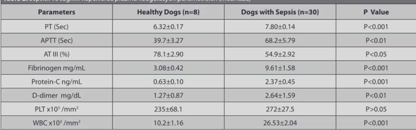
Table 2. Plasma coagulation parameters of of healthy dogs and dogs with sepsis (Mean±SE) Tablo 2. Sepsisli ve sağlıklı köpeklerde plazma koagulasyon parametreleri (Mean±SE)
Parameters Healthy Dogs (n=8) Dogs with Sepsis (n=30) P Value
PT (Sec) 6.32±0.17 7.80±0.14 P<0.001 APTT (Sec) 39.7±3.27 68.2±5.79 P<0.01 AT III (%) 78.1±2.90 54.9±2.92 P<0.05 Fibrinogen mg/mL 3.08±0.42 9.61±1.58 P<0.001 Protein-C ng/mL 0.63±0.10 2.37±0.45 P<0.001 D-dimer mg/dL 1.27±0.87 2.64±1.59 P<0.01 PLT x103 /mm3 235±68.1 272±27.5 P>0.05 WBC x103 /mm3 10.2±1.16 26.53±2.04 P<0.001
Sepsis has a high mortality. Therefore, a rapid diagnosis would be important to get good prognosis. This study showed significant changes of acute phase proteins, cytokines and haemostatic para-metersin dogs with sepsis. Therefore, measurement of these parameters in dogs with sepsis could be a valuable approach to evaluate septic stages.
Cytokines play an important role in the development and the regulation of immune response, thus, cytokine profiles contribute to the effect of immunity level in diseases [22]. The release of proinflammatory cytokines
such as IL-1ß, IL-6 and TNF-α by monocytes/macrophages and activeted T-lymphocytes is considered to be a key event in the development of sepsis [1]. Moreover, bacterial
toxins lead to release of inflammation mediators from mononuclear phagocytes in sepsis. The measurement of acute phase proteins, cytokines and coagulation profiles could be useful for determining the stage of sepsis in patients. IL-1ß and TNF-α, important inflammatory cyto-kines, are implicated in a variety of disease in dogs [23-26].
It has been reported that IL-6 and TNF-α levels are significantly increased in sepsis, and these parameters for evaluating of sepsis might be useful marker [6,7,27]. Serum
IL-1ß and TNF-α concentrations are usually increased in inflammatory disease of dogs [28,29]. Fransson et al.[30]
reported that IL-6, TNF-α and CRP levels were increased in dogs with pyometra and dogs with systemic inflammatory response syndrome. In this study, serum IL-1ß, TNF-α and INF-γ concentrations were dramatically elevated in dogs with sepsis as compared with healthy dogs (Table
1). The reason of this increase in IL-1ß, TNF-α and INF-γ
levels could be explained by the initiation of releasing of inflammatoric mediators against bacterial toxins that cause sepsis. Therefore, selected cytokines including IL-1ß, TNF-α and INF-γ maight be useful in assessing the clinical severity of sepsis in dogs. These results are consistent with published studies [3,6,28,29].
Acute phase proteins are synthesized in the liver in response to release of proinflammatory cytokines in diseases such as bacterial and viral infections, immuno-mediated disease, neoplasia, tissue injury (trauma), necrosis and burns [4,13]. These noninvasive markers might be
useful to determine the disease severity and more likely to response the treatment or prognosis, on a disease-spesific basis [11,13,31]. CRP and SAA are useful parameters
to indicate the inflammation in human and animals [32].In
addition, increased CRP and SAA concentrations have been detected in dogs with systemic inflammation [10,16,17,30].
Furthermore, CRP and SAA were shown to be significantly increased in dogs with parvovirus enteritis [33], ehrlichiosis [23]
in dogs with suspected sepsis as compared with healthy dogs. But, CRP level was normal range. The reason for higher concentrations of SAA in septic dogs may be related to inflammatory reactions and tissue damage. Similary, several studies have reported that CRP and SAA could be useful markers for diagnosis and prognosis in various disease [10,16,17,34,35]. However, CRP and SAA do not seem
to be very spesific markers for the detection of bacterial infections because it might increase in a variety of diseases, not particularly in bacterial infections [10,16,17,35]. Besides, in
this study, leukocytosis was found to be a common finding in dogs with sepsis. This observation agrees with the most studies [10,16]. In our study, SAA were detected to be specific
markers of systemic inflammation in dogs with sepsis. Disseminated intravascular coagulation a hemalotical syndrome, typically defined by the activation of intra-vascular coagulation resulting in excessive fibrin formation and consumption of caogulation factors [2,18,36] is a serious
problem that threats lives of both people and animals. DIC might be developed due to septic caogulation, viremia, parazitic infection, severe tissue damage, toxication, intra-vascular hemolizis, autoantibody, hepatitis, pancreatitis and neoplasma [19,21,37-40]. Prolongations in PT and APTT,
increase in FDP level, and decrease in AT-III activity and thrombosit count develop in dogs with DIC [41].
Prolongation in PT and APTT, increase in D-dimer, and decrease in AT-III level are detected in dogs with sepsis [2,42],
systemic inflammatory syndrome [43] or septic peritonitis [44].
In the present study, prolonged PT and APTT values as well as increased levels of fibrinogen, D-dimer and PC concentrations revealed hemostatic alteration in response to sepsis in dogs. In the present study, the indicators of the activated coagulation of DIC including prolongation of both PT and APTT as well as a decrease in AT III activity and also an increase in the level of D-dimer which shows fibrinolitic activition were defined. The occurrence of significant changes in the hemostatic system was determined. D-dimer concentration was significantly increased in dogs with sepsis, indicating the precence of fibrinolysis. Increased activity of fibrinolytic system has been associated with DIC [2,42]. Our results were in line with
previous studies [2,21,42-45].
Some authors [46-48] reported that the level of fibrinogen
might increase in the first period of DIC due to an inflamatory response; and then this level might decrease because of fibrinolisis. In contrast, other researchers [19,45]
reported a significant decrease in fibrinogen level. In this study, a significant increase in fibrinogen level in the dogs with sepsis was observed. This dramatic increase in fibrinogen level might be related to an elevation in acute phase proteins in spite of the presence of DIC in
dogs with sepsis. Therefore, our findings were in line with findings of earlier reporters [46-48].
It was reported that PC concentration was remarkably reduced in dogs with sepsis [4,49], and dogs with systemic
inflammatory syndrome [43]. Fourrier et al.[50] noticed
persistent decreases in PC activity in nonsurviving patients with sepsis and septic shock. However, de Laforcade et al.[2]
determined that the PC and AT activities were decreased in first 24 h and then this activities were gradually increased in dogs with sepsis. In this study, the activity of PC was dramatically increased in dogs with sepsis as compare with healthy dogs, most probably due to time dependent effects of diseases. This result was not coincidet with the results of reported by Laforcade et al.[4] and Yan and
Dhainaut [49], but this result was coincidet the results
of reported by de Laforcade et al.[2].
In conclusion, results of tis study indicated that SAA, IL-1ß, TNF-α , and INF- γ concentrations were significantly changed in dogs with sepsis. Particularly, SAA, IL-1ß and TNF-α parameters seem to be reliable markers for systemic inflammation in sepsis. Besides hemostatic abnormalities in dogs with sepsis may be due to developing disseminated intravascular coagulation.
REFERENCES
1. de Boer JP, Wolbink GJ, Thijs LG, Baars JW, Wagstaff J, Hack E:
Interplay of complement and cytokines in the pothohenesis of septic shock. Immunopharmacol, 24, 135-148, 1992. DOI: 10.1016/0162-3109(92)90019-9
2. de Laforcade AM, Freeman LMS, Shaw SP, Brooks MB, Rozanski EA, Rush JE: Hemostatic changes in dogs with naturally occuring sepsis. Vet
Intern Med, 17, 674-679, 2003. DOI: 10.1111/j.1939-1676.2003.tb02499.x
3. Nemzek JA, Agrodnia MD, Hauptman G: Breed-specific pro-
inflammatory cytokine production as a predisposing factor for susceptibility in the dogs. Vet Emerg Crit Care, 17, 368-372, 2007. DOI: 10.1111/j.1476-4431.2006.00215.x
4. de Laforcade AM, Rozanski EA, Freeman LM, Li W: Serial evalutaion
of protein C and antitrombin in dogs with sepsis. J Vet Intern Med, 22, 26- 30, 2008. DOI: 10.1111/j.1939-1676.2007.0021.x
5. Otto CM, Rieser TM, Brooks MB, Russell MW: Evidence of
hyper-coagulability in dogs with parvoviral enteritis. JAVMA, 217, 1500-1509, 2000. DOI: 10.2460/javma.2000.217.1500
6. Rau S, Kohn B, Richter C, Fenske N, Kuchenhoff H, Hartmann K, Hartle S, Kaspers B, Hirschberger J: Plasma interleukin-6 response is
predictive for severity and mortality in canine systemic inflammatory response syndrome and sepsis. Vet Clin Pathol, 36, 253-260, 2007. DOI: 10.1111/j.1939-165X.2007.tb00220.x
7. Song R, Kim J, Yu D, Park C, Park J: Kinetics of IL-6 and TNF-α
changes in canine models of sepsis induced by endotoxin. Vet Immunol
Immunopathol, 146, 143-149, 2012. DOI: 10.1016/j.vetimm.2012.02.008
8. Bochsler PN, Slauson DO: Inflammation and repair tissue. In,
Mechanism of Disease. A Textbook of Comparetive General Pathology. Third ed., 140-245, Mosby, St. Louis, 2002.
9. Cary C: Acute phase proteins in animals. Prog Mol Biol Transl Sci, 105,
113-150, 2011. DOI: 10.1016/B978-0-12-394596-9.00005-6
10. Jain S, Gautam V, Naseem S: Acute-phase proteins: As daignostic
tool. J Pram Bioall Sci, 1, 118-127, 2011. DOI: 10.4103/0975-7406.76489
11. Kjelgaard-Hansen M, Jacobsen S: Assay validation and diagnostic
applications of major acute-phase proteins testing in companion animals.
Clin Lab Med, 31, 51-70, 2011. DOI: 10.1016/j.cll.2010.10.002
12. Kocaturk M, Martinez S, Eralp O, Tvarijonaviciute A, Ceron J, Yilmaz Z: Prognostic value of serum acute-phase proteins in dogs with
parvoviral enteritis. J Small Anim Pract, 51, 478-483, 2010. DOI: 10.1111/ j.1748-5827.2010.00965.x
13. Murata H, Shimada N, Yoshioka M: Current research on acute phase
proteins in veterinary diagnosis: An overview. Vet J, 168, 28-40, 2004. DOI: 10.1016/S1090-0233(03)00119-9
14. Turgut K: Veteriner Klinik Laboratuvar Teşhis. İkinci Baskı, Bahçıvanlar
Basım Sanayi A.Ş. Konya, s.235-245, 2000.
15. Cary C: Biomarkers of inflammation in exotic pets. J Exotic Pet Med,
22, 245-250, 2013. DOI: 10.1053/j.jepm.2013.08.003
16. Gebhardt C, Hirschberger J, Rau S, Arndt G, Krainer K, Schweigert FJ, Brunnberg L, Kaspers B, Kohn B: Use of C-reaktive protein to predict
outcome in dogs with systemic inflammatory response syndrome or sepsis. Vet Emerg Crit Care, 19, 450-458, 2009. DOI: 10.1111/j.1476-4431.2009.00462.x
17. Langhorn CMB, Goddard R, Andreasen A, Moldal E, Tvarijonaviciute A, Kirpensteijn J, Jakobsen S, Persson F, Kjelgaard-Hansen M:
Comparison of serum amyloid A and C-reactive protein as diagnostic markers of systemic inflammation in dogs. Can Vet J, 55, 161-168, 2014.
18. Jitpean S, Holst BS, Höglund OV, Pettersson A, Olsson U, Strage E, Södersten F, Hagman R: Serum insulin-like factor-I, iron, C-reactive
protein, and serum amyloid A for prediction of outcome in dogs with pyometra. Theriogenelogy, 30, 1-6, 2014. DOI: 10.1016/j.theriogenology. 2014.02.014
19. Bruchim Y, Aroch I, Saragusty J, Waner T: Disseminated
intravascular coagulation. Compendium, 3, 1-16, 2008. DOI: 10.1056/ NEJM199908193410807
20. Caldin M, Furlanello T, Lubas G: Validation of an immunoturbidimetric
D-dimer assay in canine citrated plasma. Vet Clin Pathol, 29, 51-54, 2000. DOI: 10.1111/j.1939-165X.2000.tb00398.x
21. Esmon CT: Inflammation and thrombosis. J Thromb Haemost, 1,
1343-1348, 2003. DOI: 10.1046/j.1538-7836.2003.00261.x
22. Ceremi A: Inflammatory cytokines. Clin Immunol Immunolpathol,
62, 3-10, 1999.
23. Alsemgeest SP, Van- Klooster GA, Van Miert AS, Hulskamp-Koch CK, Gruys E: Primary bovine hepatocytes in the study of cytokine induced
acute-phase protein secretion in vitro. Vet Immunol Immunopathol, 53, 179-184, 1996. DOI: 10.1016/0165-2427(96)05602-4
24. Baggio V, Ott F, Fischer RW: Production of antibodies to canine
IL-1ß and canine TNF to assess the role of proinflammatory cytokines. Vet
Immunol Immunopathol, 107, 27-39, 2005.
25. Nakagawa-Tosa N, Morimatsu M, Kawasaki MN, Nakatsuji H, Syuto B, Saito M: Stimulation of haptoglobin synthesis by interleukin-6 and
tumor necrosis factor, but not by interleukin-1, in bovine primary cultured hepatocytes. J Vet Med Sci, 57, 219-223, 1995. DOI: 10.1292/jvms.57.219
26. Ohtsuka H, Ohki K, Tanaka T, Tajima M, Yoshino T, Takahashi K:
Circuling tumor necrosis factor and interleukin-1 after administration of lps in adult cows. J Vet Med Sci, 19, 227-229, 1997. DOI: 10.1292/ jvms.59.927
27. Nemzek JA, Siddiqui J, Remick DG: Development and optimization
of cytokine ELISAs using commercial antibody pairs. J Immunol Methods, 255, 149-157, 2001. DOI: 10.1016/S0022-1759(01)00419-7
28. Grone A, Aldinger S, Baumgartner W: Interleukin-1beta-6-12 and
tumor necrosis factor-alpha experission in barin of dogs with canine distemper virus infection. J Neuroimmunol, 110, 20-30, 2000. DOI: 10.1016/S0165-5728(00)00332-5
29. Miyamota T, Fujinaga T, Yamashita K, Hagio M: Changes of serum
cytokine activities and other parameters in dogs with experimentally induced endotoxic shock. Jpn J Vet Res, 44, 107-118, 1996. DOI: 10.14943/ jjvr.44.2.107
30. Fransson BA, Lagerstedt AS, Bergstrom A, Hagman R, Park JS, Evans MA, Ragle CA: C-reaktive protein, tumor necrosis factor α and
373-and inflammation in veterinary medicine. Vet J, 185, 23-27, 2010. DOI: 10.1016/j.tvjl.2010.04.009
33. Yule TD, Roth MB, Dreier K, Johnson AF, Palmer-Densmore M, Simmons K, Fanton R: Canine parvovirus vaccine elicits protection from
the inflammatory clinical consequences of the disease. Vaccine, 15, 720- 729, 1997. DOI: 10.1016/S0264-410X(96)00232-0
34. Mastrorilli C, Dondi F, Agnoli C: Clinopathologic features and
outcome predictors of leptospira interogans Australis serogroup infection in dogs: A retrospective study of 20 cases (2001-2004). J Vet Intern Med, 21, 3-10, 2007. DOI: 10.1892/0891-6640(2007)21[3:CFAOPO]2.0.CO;2
35. Mylonakis ME, Ceron JJ, Leontides L, Siarkou VI, Martinez S, Tvarijonaviciute A, Koutinas AF, Harrus S: Serum acute phase proteins
as clinical phase indicators and outcome predicators in naturally occuring canine monocytic ehrlichiosis. J Vet Intern Med, 25, 811-817, 2011. DOI: 10.1111/j.1939-1676.2011.0728.x
36. Saba HI, Morelli GA: The pathogenesis and management of
disseminated intravascular coagulation. Clin Adv Hematol Oncol, 4, 919-926, 2006.
37. Col R, Durgun Z: Sepsis, lökositler, sitokinler ve disseminant
intra-vasküler koagulasyon. Vet Bil Derg, 23, 97-106, 2007.
38. Goddard A, Leisewitz AL: Canine parvovirus. Vet Clin North Am: Small
Anim Pract, 40, 1041-1053, 2010. DOI: 10.1016/j.cvsm.2010.07.007
39. Levi M: Pathogenesis and treatment of DIC. Thromb Res, 1, 54-55,
2005.
42. Yilmaz Z, Yalçın E, Ilçol Y: Septik şoklu köpeklerde koagulasyon
profilinin değerlendirilmesi. Turk J Vet Anim Sci, 26, 171-176, 2002.
43. Bauer N, Moritz A: Coagulation response in dogs with and without
systemic inflammatory response syndrome-Preliminary results. Res Vet
Sci, 94, 123-121, 2013. DOI: 10.1016/j.rvsc.2012.07.029
44. Bentley AM, Mayhew PD, Culp WTN, Otto CM: Alterations in the
hemostatic profiles of dogs with naturally occuring septic peritonitis. Vet
Emerg Crit Care, 23, 14-22, 2013. DOI: 10.1111/vec.12013
45. Prins M, Schellens CJMM, van Leeuwen MW, Rothuizen J, Teske E:
Coagulation disorders in dogs with hepatic disease. Vet J, 185, 163-168, 2010. DOI: 10.1016/j.tvjl.2009.05.009
46. Er C, Ok M: Levels of cardiac biomarkers and coagulation profiles in
dogs with parvoviral enteritis. Kafkas Univ Vet Fak Derg, 21, 383-388, 2015. DOI: 10.9775/kvfd.2014.12575
47. Otto CM: Sepsis. In, Wingfield WE (Ed): The Veterinary ICU Book.
695-709, Jackson Hole, WY: Teton NewMedia, 2002.
48. Wada H: Disseminated intravascular coagulation. Clin Chim Acta, 344,
13-21, 2004. DOI: 10.1016/j.cccn.2004.02.015
49. Yan SB, Dhainaut JF: Activated protein C versus protein C in severe
sepsis. Crit Care Med, 29 (Suppl. 7): S69-S74, 2001.
50. Fourrier F, Goudemand J, Hendrycx S, Caron C, Rime A, Marey A, Lesteval P: Septic shock, multiple organ failure, and disseminated
intravascular coagulation. Chest, 101, 816-823, 1992. DOI: 10.1378/ chest.101.3.816