Cukurova Medical Journal
Cukurova Med J 2016;41(1):161-163ÇUKUROVA ÜNİVERSİTESİ TIP FAKÜLTESİ DERGİSİ DOI: 10.17826/cutf.147101
Yazışma Adresi/Address for Correspondence: Dr. Soner Solmaz, Adana Hospital of Başkent University, Department of Hematology, Adana, Turkey E-mail: [email protected]
Geliş tarihi/Received: 21.07.2015 Kabul tarihi/Accepted: 03.09.2015
OLGU SUNUMU/CASE REPORT
A rare hematological complication of visceral leishmaniasis:
hemophagocytic syndrome
Visseral leishmaniazisin nadir bir hematolojik komplikasyonu: hemofagositik
sendrom
Soner Solmaz
1, Can Boğa
1, İlknur Kozanoğlu
1, Süheyl Asma
1, Tuba Turunç
2, Yusuf Ziya
Demiroğlu
21Başkent University Adana Hospital, Department of Hematology, 2Department of Infectious Diseases, Adana, Turkey Cukurova Medical Journal 2016;41(1):161-163.
Abstract Öz
The term “hemophagocytosis” describes the pathologic finding of activated macrophages, engulfing erythrocytes, leukocytes, platelets and their precursor cells. This phenomenon is an important finding in patients with hemophagocytic syndrome. It is a distinct clinical entity characterized by fever, pancytopenia, splenomegaly and hemophagocytosis in bone marrow, liver and lymph nodes. Hemophagocytic syndrome can be classified according to the underlying etiology into either primary (genetic) or secondary (acquired). Severe infections, malignancies, rheumatologic disorders and some metabolic diseases can lead to secondary hemophagocytic syndrome. Infection-associated hemophagocytic syndrome implicating Leishmania is very rare and often difficult to diagnose. Therefore, we aimed to report a young boy with Visceral Leishmaniasis associated hemophagocytic syndrome.
Hemofagositoz terimi, aktive makrofajlari, fagosite edilmiş eritrosit, lökosit, trombosit ve onların öncül hücrelerini tarif etmektedir. Bu fenomen, hemofagositik sendromun önemli bir bulgusudur. Hemofagositik sendrom, ateş, pansitopeni, splenomegali ve kemik iliği, karaciğer ile lenf nodunda hemofagositoz ile karakterizedir. Hemofagositik sendrom, altta yatan etyolojiye göre primer (genetik) ve sekonder (kazanılmış) olmak üzere sınıflandırılabilir. Ağır enfeksiyonlar, kanserler, romatolojik hastalıklar ve bazı metabolik hastalıklar sekonder hemofagositik sendroma neden olabilmektedir. Leishmania’ nın neden olduğu enfeksiyon ilişkili hemofagositik sendrom çok nadirdir ve sıklıkla tanıda zorluklara yol açmaktadır. Bundan dolayı Visseral Leishmaniazis ilişkili hemofagotik sendromlu genç bir hasta sunulması amaçlanmıştır.
Key words: Leishmania, hemophagocytic syndrome, hemophagocytosis, zoonoses
Anahtar kelimeler: Leishmania, hemofagositik sendrom, hemofagositoz, zoonoz
INTRODUCTION
The term “hemophagocytosis” describes the pathologic finding of activated macrophages, engulfing erythrocytes, leukocytes, platelets, and their precursor cells1. This phenomenon is an
important finding in patients with hemophagocytic syndrome, more properly referred to as hemophagocytic lymphohistiocytosis (HLH)1.
Hemophagocytic syndrome is a potentially fatal hyperinflammatory condition caused by a highly
stimulated but ineffective immune response2. It is a
distinct clinical entity characterized by fever, pancytopenia, splenomegaly and hemophagocytosis in bone marrow, liver and lymph nodes1. The incidence is estimated to be approximately 1.2 cases per million individuals per year, but this is almost certainly an underestimate2. Broadly,
hemophagocytic syndrome can be classified according to the underlying etiology into either primary (genetic) or secondary (acquired)2. Since the
Solmaz et al. Cukurova Medical Journal
162
et al in 1999, significant insight has been gained into the genetic mutations that give rise to the hemophagocytic syndrome phenotype2. Specific
diagnostic criteria were used for patient eligibility in the Histiocyte Society (HS) trials HLH-94 and HLH-20042. The diagnosis of HLH can be
established if one of either 1 or 2 below is fulfilled;
1. A molecular diagnosis consistent with HLH is made.
2. Diagnostic criteria for HLH are fulfilled (5 of the 8 criteria below):
1. Fever 2. Splenomegaly
3. Cytopenias (affecting >2-3 lineages in the peripheral blood):
a. Hemoglobin <90 g/L (in infants <4 weeks of ages, hemoglobin <100 g/L) b. Platelets <100 x 109/L
c. Neutrophils <1.0 x 10⁹/L
4. Hypertriglyceridemia and / or hypofibrinogemia :
a. Fasting triglycerides ≥3.0 mmol/L (ie, 265mg/dL)
b. Fibrinogen ≤1.5 g/L
5. Hemophagocytosis in bone marrow, spleen, or lymph nodes
6. Low or absent NK-cell activity (according to local laboratory reference)
7. Ferritin ≥500 µg/L
8. Soluble CD25 (ie, sIL2r) ≥2400 U/mL (Supportive criteria include neurologic symptoms, cerebrospinal fluid pleocytosis, conjugated hyperbilirubinemia and transaminitis, hypoalbuminemia, hyponatremia, elevated D-dimers and lactate dehydrogenase. The absence of hemophagocytosis in the bone marrow does not exclude a diagnosis of HLH)2.
Hemophagocytic syndrome may also occur as a secondary disorder in association with severe infections, malignancies, rheumatologic disorders and some metabolic diseases2. Hemophagocytic
syndrome has since been associated with a variety of viral, bacterial, fungal, and parasitic infections1.
Ebstein-Barr virus (EBV) is the most common cause of infection-associated hemophagocytic syndrome2. Infection associated hemophagocytic
syndrome implicating Leishmania is very rare and often difficult to diagnose3. Therefore, we aimed to
report a young boy with visceral leishmaniasis associated hemophagocytic syndrome.
CASE
A previously healthy 20 year-old boy presented with fever (>38˚C) lasting for 3 days. The patient was detected to smoke and use alcohol. On admission, physical examination revealed heart rate 102 bpm, blood pressure 100/60 mmHg, body temperature 39 ˚C, pallor, hepatosplenomegaly. Hemoglobin level 9.4 (13.5-18.0) gr/dl, white blood cell count 1.29 (4.0-11.5) x103 /µL and platelet count 37.8
(130-400) x103 /µL, fibrinogen level 0.77 (1.8-3.5) g/L,
serum albumin 2.6 (3.2-5.0) g/dL were decreased, and erythrocyte sedimentation rate 49 (0-20) mm/hour, C-reactive protein level 123 (0-6.0) mg/L, prothrombin time (PT) 16.5 (11.0-15.0) sec, activated thromboplastin time (aPTT) 52.7 (22.1-36.0) sec, D-dimer 5468 (0-500.0) µg/L, aspartate aminotransferase (AST) 348 (7-40) IU/L, alanin aminotransferase (ALT) 152 (5-35) IU/L, lactate dehydrogenase (LDH) 933 (105-235) IU/L and serum ferritn level >2000 (22-322) ng/mL were all elevated.
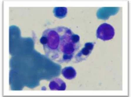
Pericardial and bilateral pleural effusion, massive hepatosplenomegaly, ascites, paraaortic lymph nodes were detected on computed tomography scan. Bone marrow examination revealed erythroid hyperplasia, elevated plasma cells and histiocytes, and erythroblasts phagocyted histiocytes (Figure 1), but no etiological factor for hemophagocytic syndrome was detected.
Figure 1. Bone marrow cells were engulfed by histiocyte cell of bone marrow.
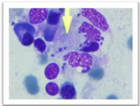
General condition and laboratory findings of the patient deteriorated gradually within two weeks. Amastigotes of Leishmania was detected in the second bone marrow examination, (Figure 2). Antibody scanning with immunofluoresence test and rK39 immunochromatic dipstick testing for Leishmania were positive. The symptoms and
Cilt/Volume 41 Yıl/Year 2016 A rare complication of visceral leishmaniasis
163
laboratory findings of the patient improved gradually within days after administration of liposomal amphotericin B.
Figure 2. Amastigotes of Leishmania were engulfed by histiocyte cell in bone marrow.
DISCUSSION
Hemophagocytic lymphohistiocytosis is a disorder characterized by nonmalignant infiltration of vital organs by activated lymphocytes and macrophages4.
Engulfment of any hematological cell type in bone marrow and reticuloendothelial system by these activated cells, called hemophagocytosis is the hall mark of the disease4. This may result in
pancytopenia, fever, organ enlargement, neurological dysfunction and disseminated intravascular coagulation4.
Visceral Leishmaniasis is a generalized protozoan infection of the reticuloendothelial system caused by the genus Leishmania and transmitted by a bite from the infected sand fly, Phlebotomus5. The disease is
endemic in the Middle East, East Africa and Southern Europe5. Visceral Leishmaniasis is
characterized by fever, hepatosplenomegaly, lymphadenopathy and various hematological abnormalities5. The most common hematologic
manifestation of Visceral Leishmaniasis is anemia, however the patient may develop leukopenia, thrombocytopenia, pancytopenia, coagulation abnormalities and hemophagocytic syndrome5-7.
Hemophagocytic syndrome is a rare life-threatening complication of Visceral Leishmaniasis6.
As discussed above, Visceral Leishmaniasis may mimic or lead to several types of hematological disorders including hemophagocytic syndrome3.
Diagnostic delay might cause severe complications and death occurs in 90% of the patients without specific anti-Leishmanial treatment4. In our patient,
we detected no etiological factors for hemophagocytic syndrome in the first bone marrow examination because of low parasitic load, so two weeks of time was lost. However we were able to detect Leishmania parasites in the second bone marrow examination.
In conclusion, the findings and symptoms of Visceral Leishmaniasis and hemophagocytic syndrome are very similar and also overlap. The early diagnosis and treatment of Visceral Leishmaniasis-related hemophagocytic syndrome is vital. Therefore, Visceral Leishmaniasis should be screened when hemophagocytic syndrome is detected in any patient.
REFERENCES
1. Fisman DN. Hemophagocytic syndromes and infection. Emerg Infect Dis. 2000;6:601-8.
2. Weitzman S. Approach to hemophagocytic syndromes. Hematology Am Soc Hematol Educ Program. 2011;2011:178-83.
3. Tapisiz A, Belet N, Ciftçi E, Ince E, Dogru U. Hemophagocytic lymphohistiocytosis associated with visceral leishmaniasis.J Trop Pediatr. 2007;53:359-61. 4. Koçak N, Eren M, Yüce A, Gümrük F.
Hemophagocytic syndrome associated with visceral leishmaniasis. Indian Pediatr. 2004;41:605-7. 5. Mokhtari M, Kumar PV. Visceral
leishmaniasis-associated hemophagocytosis: a single center experience. Arch Iran Med. 2013;16:471-3.
6. Tunç B, Ayata A. Hemophagocytic syndrome: a rare life-threatening complication of visceral leishmaniasis in a young boy. Pediatr Hematol Oncol. 2001;18:531-6.
7. Varma N, Naseem S. Hematologic changes in visceral leishmaniasis/kala azar. Indian J Hematol Blood Transfus. 2010;26:78-82.