Adıyaman Üniversitesi Sağlık Bilimleri Dergisi, 2020;6(1):13-18
doi:10.30569.adiyamansaglik.558935
Bu eser, Creative Commons Atıf-GayriTicari 4.0 Uluslararası Lisansı ile lisanslanmıştır. Telif Hakkı © 2020 Adıyaman Üniversitesi Rektörlüğü
Research Article/Özgün Araştırma
Changes in the serum of individuals exposed to extremely low frequency
electromagnetic fields for a long time
Uzun süre oldukça düşük frekanslı elektromanyetik alanlara maruz kalan
bireylerin serumundaki değişiklikler
Mehmet Cihan YAVAŞ1
1Kırşehir Ahi Evran University, Faculty of Medicine, Department of Biophysics, 40100, Kırşehir-Turkey
Atıf gösterme/Cite this article as: Yavaş MC. Changes in the serum of individuals exposed to extremely low frequency electromagnetic fields for a long time. ADYÜ Sağlık Bilimleri Derg. 2020;6(1):13-18. doi:10.30569.adiyamansaglik.558935
Abstract
Aim: Purpose of our study is to research the effect of the extremely low electromagnetic fields (ELF-EMF) generated by the hair dryer device on the hormone, biochemical and blood values of the male workers working in the hairdressing saloons.
Materials and Methods: Eight male workers were included in the study, with exclusion factors being applied, with two equal groupings as control and test group. Then the blood of the workers working in the hairdressing centers and the control group not exposed to these effects was taken. In the study, biochemistry, hormones and whole blood levels of sera were analyzed by means of full automatic analyzers.
Results: When all biochemistry and hormone parameters were examined, no significant difference was found between control and experiment group (p>0.05). In whole blood results, the red blood cell, hemoglobin and hematocrit parameters were not statistically significant (p< 0.05), and other parameters were not statistically significant (p>0.05).
Conclusion: The results suggest that ELF-EMFs over a long period of time may affect men working in hairdressers on hormone, biochemistry and whole blood parameters.
Keywords: Electromagnetic fields; 50 Hz; Individual exposure; Long time; Hair dryer device.
Öz
Amaç: Çalışmamızın amacı, saç kurutma makinesi tarafından oluşturulan oldukça düşük frekanslı elektromanyetik alanların kuaför salonlarında çalışan erkek işçilerin hormon, biyokimyasal ve kan değerleri üzerindeki etkisini araştırmaktır.
Gereç ve Yöntem: Sekiz erkek işçi çalışmaya dâhil edildi, dışlama faktörleri uygulandı, kontrol ve deney grubu olmak üzere iki eşit gruplama uygulandı. Daha sonra kuaför merkezlerinde çalışan işçilerin ve bu etkilere maruz kalmayan kontrol grubunun kanı alındı. Çalışmada, biyokimya, hormonlar ve tam kan serum seviyeleri tam otomatik analizörler ile analiz edildi. Bulgular: Tüm biyokimya ve hormon parametreleri incelendiğinde kontrol grubu ile deney grubu arasında anlamlı bir fark bulunmadı (p>0.05). Tam kan sonuçlarında, hemoglobin ve hematokrit parametresi istatistiksel olarak anlamlı (p<0.05), ve diğer parametreler ise istatistiksel olarak anlamlı bulunmadı (p>0.05).
Sonuç: Sonuçlar, kuaför salonlarında çalışan erkekler üzerinde uzun süre oldukça düşük frekanslı elektromanyetik alanların, hormon, biyokimya ve tam kan parametrelerini göstermektedir.
Anahtar Kelimeler: Elektromanyetik alanlar; 50 Hz; Bireysel maruz kalma; Uzun süre; Saç kurutma makinesi.
Yazışma Adresi/Address for Correspondence: Dr. Mehmet Cihan YAVAŞ, Department of Biophysics, Faculty of Medicine,
Kırşehir Ahi Evran University,40100, Kırşehir-Turkey, E-mail: [email protected]
Geliş Tarihi/Received:29.04.2019 Kabul Tarihi/Accepted:14.03.2020 Yayım Tarihi/Published online:23.04.2020
https://dergipark.org.tr/tr/pub/adiyamansaglik JOURNAL OF HEALTH SCIENCES OF ADIYAMAN UNIVERSITY
14
Introduction
Especially electric appliances which are used continuously at home and at work places produce the extremely low frequency
electromagnetic fields (ELF-EMF).
Increasing concentrations of this area can in turn cause oxidative stress and change the nature of the body. Both human and scientific interest have attracted attention as man-made instruments that produce electromagnetic
fields (EMF) and their increased
environmental exposure create harmful effects on biological systems and human health risks.1 Recent epidemiological studies suggest that increased exposure to ELF-EMFs in the home or business may affect depression and an increase in the proportion of certain types of cancers and health, and some studies suggest that these areas do not affect human health.2 The International Agency for Research on Cancer (IARC) has been conducting studies on the health effects of the electric field, the magnetic field and the electromagnetic field for 20 years. Particularly epidemiological studies have been carried out on whether or not there are correlations with existing childhood leukemia.3 Interaction with electrical appliances, which are more common in homes in recent years, is increasing. In relation to these, epidemiological reports on electrical and magnetic field values of sewing machines, hair dryers, television sets, shaving machines and other equipment are presented. Area measurements have been made in the x, y and z directions for hair dryers and it has been seen that the value of the magnetic field generated by the distance increases.4
The aim of this study is to investigate the effect of ELF-EMF effect of hair dryer on people working in hairdressing centers.
Materials and Methods The Experimental Protocol
This study was carried out on the employees of approximately 100 hair salon in Diyarbakır province. Exclusion factors were applied to the employed persons (such as non-smoking, no metabolic, systemic and other illnesses). Two groups were randomly divided into eight male experimental groups (mean
age, 23.12±3.56) and eight controls (mean age, 28.50±6.56). People who were taken to work were prevented from consuming any fluid until about 3 hours before blood was taken. Immediately after collection, the blood was centrifuged at 300 rpm for 10 minute and maintained at -20ºC until analysis. Serums
were analyzed with fully automated
analyzers. Biochemical serum results were analyzed using the photometric method with the Abbott Architect C16000 (Illinois, United States) instrument. Hormone results were analyzed using the electrochemiluminescence method with a Roche Cobas 601 instrument. The whole blood results were analyzed using the Abbott CELL-DYN Ruby instrument with.
This study was approved by the Dicle University Faculty of Medicine Noninvasive Clinical Ethical Research Board (4-352/13.01.2012) and continued in accordance with the Helsinki Principles Declaration. All participants signed the Informed Consent Form and their consent was obtained.
Measurement of Electrical and Magnetic Field Exposures
Specific features of standard hair dryers used in the study are; AC, 200-240 Volt, 50
Hz, 1800-2200 Watts. Reference
measurement methods are taken into account and the arithmetic mean of the measurements is calculated. Our measurements were taken at a distance of 10 cm and found an electric field of 73.5 V/m and a magnetic field of 4.61 µT, respectively. The electromagnetic field was measured with the aid of a Spectran device
NF5035 (AARONIA AG, Strickscheid,
Germany).
Evaluation of Data
The Kolmogorov-Smirnov test was used to analyze whether the data obtained in the study were normal distributions. As a result of the analysis, it was determined that the biochemical values of males were not normally distributed (p<0.05). The whole blood and hormone values provide normality assumption (p>0.05). Comparisons of biochemical values were performed using the Mann Whitney U test, a nonparametric test. Comparisons of whole blood and hormone
15 values, which provided normality hypothesis,
were made with Independent t test. Participants were asked whether they had any complaints or not, and the distribution of the obtained data according to the groups was turned into a quotient table. The results were analyzed by Fisher Exact Chi square test. Statistical analysis of the study was performed using SPSS 21.0 (Armonk, NY: IBM Corp., USA).
Results
It has been determined that gender has no significant effect on the averages of age. For this reason, the analysis did not need to be used as a covariate. The mean age of the men in the experimental group was 23.12±3.56. The men in the experimental group worked on average 8.5 years in the hairdressing salon. The men in the experiment group stated that they use hair dryer in average 395.62±343.42 minutes per day. When the distance of the hair dryers machine to the body during use was examined, It was determined that the males were 17.50±3.77 cm away.
Questionnaire questions were asked to all subjects and their complaints were asked, and
statistical analyzes of the data received were presented in Table 1. When the Fisher exact test statistics of males were examined, it was observed that the males of the experimental group were more irritable than the males of the control group (p<0.01). There were no
significant differences between the
experimental group men and the control group men in all of the other complaints (p>0.05).
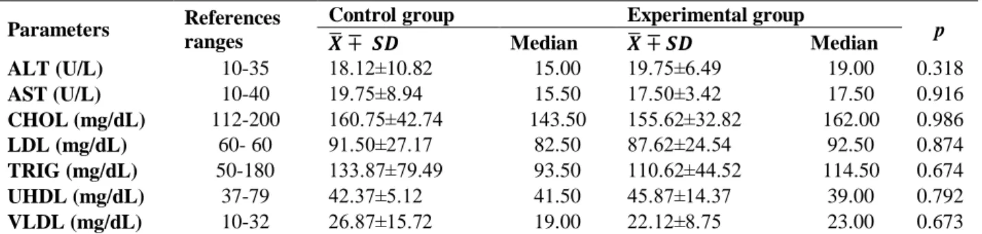
Descriptive statistics (mean±SD, median) and Mann Whitney U values for male biochemical values were also summarized. When the biochemical results of males were examined (Table 2), it was determined that the median values of the experimental group were higher than the median values of the control group for all parameters except ultra-high density lipoprotein (UHDL). In the UHDL parameter, the median value of the
control group was higher than the
experimental group. According to the Mann Whitney U test results, the differences between experiment and control groups in terms of all biochemical parameters are not statistically significant (p>0.05).
Table 1. Fisher exact chi square test results
Complaints Headache Irritability Earache Weakness Fatigue Forgetfulness Eye pain
p 0.077 0.007* 0.500 0.500 0.315 0.200 0.200
When compared with the control group of the experimental group. *p<0.05 compared to control group.
Table 2. Male’s biochemical results Parameters References
ranges
Control group Experimental group
p 𝑿̅ ∓ 𝑺𝑫 Median 𝑿̅ ∓ 𝑺𝑫 Median ALT (U/L) 10-35 18.12±10.82 15.00 19.75±6.49 19.00 0.318 AST (U/L) 10-40 19.75±8.94 15.50 17.50±3.42 17.50 0.916 CHOL (mg/dL) 112-200 160.75±42.74 143.50 155.62±32.82 162.00 0.986 LDL (mg/dL) 60- 60 91.50±27.17 82.50 87.62±24.54 92.50 0.874 TRIG (mg/dL) 50-180 133.87±79.49 93.50 110.62±44.52 114.50 0.674 UHDL (mg/dL) 37-79 42.37±5.12 41.50 45.87±14.37 39.00 0.792 VLDL (mg/dL) 10-32 26.87±15.72 19.00 22.12±8.75 23.00 0.673
Data are presented as mean±SD and median. p>0.05 compared to control (Mann Whitney U test).
ALT: alanine aminotransferase, AST:aspartate aminotransferase, CHOL:cholesterol, LDL:low-density lipoprotein, TRIG:triglyceride, UHDL: high-density lipoprotein, VLDL:very low-density lipoprotein.
Table 3. Analysis results of male hormone values Parameters References
ranges
Control group
(Mean±SD) Experimental group (Mean±SD) t p Cortizol (μg/dl) 6.2-19.4 8.97±4.15 8.91±2.86 0.032 0.975 Estrojen (pg/ml) 7.6-43 34.72±8.62 31.93±9.09 0.631 0.538 Testesteron (ng/dl) 2.8-8 4.31± .04 4.11±1.53 0.309 0.762
16
Table 4. Men's whole blood results
Parameters References ranges Control group (Mean±SD) Experimental group (Mean±SD) t p
WBC (K/µL) 3.2-12 7.42±1.20 6.83±1.60 0.845 0.413 RBC (M/uL) 3.2-6 5.23±0.45 4.37±0.17 4.999 0.000* HGB (g/dL) 10-18 15.15±2.02 13.35±0.81 2.335 0.035* HCT (%) 30-55 43.02±5.08 37.52±2.03 2.882 0.013* MCV (fL) 98-120 82.15±7.15 85.71±1.81 -1.365 0.194 RDW (%) 9-18 15.82±0.66 15.32±0.52 1.668 0.117 PLT (K/uL) 150-500 272.75± 8.35 318.50±41.38 -1.460 0.166 MPV (fL) 0-15 7.75±0.94 7.60±0.57 0.401 0.694
Data are presented as mean ± SD. p>0.05 compared to control (Independent t test).
*p< 0.05 compared to control group. HCT:hematokrit, HGB:hemoglobin, MCV:mean corpuscular volume,MPV:mean platelet volume, PLT:platelet, RBC:red blood cell, RDW:red cell distribution width, WBC: white blood cells.
Descriptive statistics (mean±SD) and independent t test results for male hormone levels are given in Table 3. According to these results, there was no statistically significant difference between experimental and control groups regarding cortisol, estrogen and testosterone values (p>0.05).
The explanatory statistics of male whole blood values and independent t test results are given in Table 4. When these results were examined, there was no statistically significant difference between control and experimental groups in white blood cells (WBC), mean corpuscular volume (MCV), red cell distribution width (RDW), platelet (PLT) and mean platelet volume (MPV) parameters (p>0.05). Red blood cell (RBC) value was lower in the experimental group and the difference between the two values was statistically significant (p<0.01). The hemoglobin (HGB) value was also found to be lower in the experimental group. A statistically significant value was found
between the control group and the
experimental group's hemoglobin results (p<0.05). The hematokrit (HCT) value of the
control group was higher than the
experimental group (p<0.05).
Discussion
Cakir and his team examined the whole blood parameters by applying a magnetic field exposure of 0.97 mT for 3 days a day for 50 and 100 days on rats. Eosinophil (EOS), HGB, and MPV levels decreased in 50-day exposure. No changes were observed in leukocyte, neutrophil, EOS, monocyte, lymphocyte and basophil counts, or PLT, HCT, RDW, MCH, MCHC, erythrocyte and PDW values at the same exposures.5 Findings
we obtained in our study are similar to the changes in the MPV, RDW levels in Çakır and his colleagues' work, but there are different results in other changes. The
complaints were determined by the
questionnaire we made. The irritability was found to be significant. But, No significant differences were found in the complaints of headache, earache, weakness, fatigue, forgetfulness and eye pain when compared with the control group (p>0.05).
Epidemiological studies have been
conducted with the electromagnetic field on human health. In his study, Johansen measured field measurements of appliances used at home and at work, measuring 17.44 at 5 cm, 0.12 at 50 cm, and 0.02 microTesla magnetic field at 100 cm.6 Dogan and colleagues reported that the 2.48 micro-magnetic field exposures on rats in the laboratory environment could alter the trace element content balance in the rat teeth, which could create a toxic effect on human health.7 Touitou and colleagues reported that long-term (1-20 years) 50 Hz and 0.3 mT larger magnetic field exposures on males may cause changes in some blood parameters.8 With the increase in mobile phones, there is
also an increase in exposure to
electromagnetic radiation. AST and ALT values were found to be significant when compared with the control group (p<0.05).9 Güler et al. constituted exposed horizontal and vertical electric field exposures on Guinea Pigs. The applied electric fields are 0.3, 0.6, 0.8, 1, 1.35, 1.5 and 1.8 kV/m. Biochemical levels such as ALT, GGT, ALP, HDL, VLDL, LDH, LDL, total cholesterol, total protein, urea, uric acid, albumin, glucose and creatin increased in the study results
17 compared to the control group.10 Coşkun and
Çömlekçi examined the effect of pulsed electric field on hematology values of rats. Compared to the control group, there was a decrease in the Ht (p<0.05), RBC (p<0.05), Hb (p<0.05), WBC (p<0.05) and PLTs (p<0.05) levels of the experimental group.11 In our study, both hormone and biochemical parameters were not statistically significant in the comparison of experimental and control groups (p<0.05). Statistical results were found for whole blood RBC (p<0.01), HGB and HCT (p<0.05), among other parameters (p>0.05). A decrease in the WBC, RBC, HGB, HCT levels of the experimental group was observed. An increase in MCV and PLT levels and no change in RDW and MPV levels. All biochemical results were found at the normal reference intervals. There was no significant difference according to the control group (p>0.05). According to the control group, there was a decrease in AST level, an increase in ALT level in the experimental group. Most of the literature reports are similar to the findings we have obtained in our study. These results indicate that extremely low frequency electromagnetic fields can cause changes in whole blood and biochemical parameters.
De Bruyn and De Jager reported that there was a significant reduction in the sperm motility and live sperm counts of randomly rotated 50 Hz 0.5 to 77 microTesla-magnetic field exposures on mice.12 Kumar et al. investigated the effect of the electromagnetic field (45 days 1 hour, 10 GHz microwave radiation, specific absorption rate (SAR): 0.014 W/kg) on the reproductive system of male rats. They reported that male rats may have a potential effect on fertility and a reduction in testosterone levels in the study.13 Al-Akhras and colleagues have applied a 50 Hz 25 micro Tesla magnetic field over sex hormones and other reproductive parameters of adult rats for 18 weeks. They reported that male rats exposed to the magnetic field would have an adverse effect on reproduction and fertilization due to decreased levels of testosterone, follicle stimulating hormone (FSH) and luteinizan hormon (LH).14 Kaur and Khera have developed cell phone
exposure on albino rats for 2 hours a day (1.25 watt/kg SAR value) and 15 cm distance for 60 days. As a result of the study, it was reported that the AST and ALT values of the experimental group were increased according to the control group and the FSH and LH levels of male rats were decreased according to the control group.15 Wang et al. examined the effect of electrical and magnetic fields (measured values: 0.1 V/m-316 kV/m and 0.1 nT-32 nT) on biomass and hormonal parameters of male workers working at the power plant. The study concluded that male workers had a reduction in T/E2 and testosterone plasma levels.16 In our study it was determined that there was no change in cortisol levels and a reduction in the test group in estrogen and testosterone levels. These changes were not statistically significant (p>0.05). Human and animal studies exposed to electromagnetic fields show that the hormonal balance has changed, with the level of testosterone decreasing. Our work is compatible with them.
There are increasing reports of negative effects of ELF-EMF areas on human health. However, some study results also indicate that extremely low frequency electromagnetic fields have not yet had any negative effects. In order for these mechanisms of action to be fully understood, it is essential to examine their work in more detail.
Limitations
The limitations of a study, the small sample size, the state of holding distances close to the body of the individuals using the
device, and the strength of the
electromagnetic field created by different brand devices may vary.
Conclusion
The results suggest that extremely low frequency electromagnetic fields over a long period of time may affect men working in hairdressers on hormone, biochemistry and whole blood parameters.
Ethics Committee Approval
This study was approved by the Dicle University Faculty of Medicine Noninvasive Clinical Ethical Research Board
(4-18 352/13.01.2012) and continued in accordance
with the Helsinki Principles Declaration.
Informed Consent
All participants signed the Informed Consent Form and their consent was obtained.
Author Contributions
In this study, it was done by the author in the creation of the method, in the acquisition and interpretation of the data.
Acknowledgments
Because of taking blood samples to Mr. Ethem YAVAŞ and Prof. Dr. Veysi AKPOLAT, Prof. Dr. M. Salih ÇELİK and Prof. Dr. Birgul ISIK for her scientific contributions.
Conflicts of Interest
The author has no conflict of interests to declare.
Financial Disclosure
No financial disclosure was declared by the author.
Statements
This study International Congress on Biological and Medical Sciences 2018, 31
October-03 November 2018, at
Nigde/TURKEY has been presented as an oral presentation.
References
1. Canseven AG, Coskun S, Seyhan N. Effects of various extremely low frequency magnetic fields on the free radical processes, natural antioxidant system and respiratory burst system activities in the heart and liver tissues. Indian J Biochem Biophys. 2008;45:326-331.
2. V Torres-Duran P, Ferreira-Hermosillo A, A Juarez-Oropeza M, Elias-Viñas D, Verdugo-Diaz L. Effect of whole exposure to extremely low frequency electromagnetic fields (ELF-EMF) on serum and liver lipid levels, in the rat. Lipids Health and Dis. 2007;6:31-6.
3. Otto M.KE, von Mühlendahl. Electromagnetic fields (EMF): Do they play a role in children’s environmental health (CEH)?. Int J Hyg Environ Health. 2007;210:635–44.
4. Kaune WT, Miller MC, Linet MS, Hatch EE, Kleinerman RA, Wacholder S. et al. Magnetic fields produced by hand held hair dryers, stereo headsets, home sewing machines, and electric clocks. Bioelectromagnetics. 2002;23:14-25.
5. Çakır DU, Yokus B, Akdag MZ, Sert C, Mete N. Alterations of hematological variations in rats exposed to extremely low frequency magnetic fields (50 Hz). Arch Med Res. 2009;40:352-6.
6. Johansen C. Electromagnetic fields and health effects-epidemiologic studies of cancer, diseases of the central nervous system and arrhythmia-related heart disease. Scand J Work Environ Health. 2004;30(1):1-80.
7. Doğan MS, Yavaş MC, Yavuz Y, Erdoğan S, Yener İ, Şimşek İ. et al. Effect of electromagnetic fields and antioxidants on the
trace element content of rat teeth. Drug Des Devel Ther. 2017;11:1393–8.
8. Touitou Y, Djeridane Y, Lambrozo J, Camus F. Long-term (up to 20 years) effects of 50-Hz magnetic field exposure on blood chemistry parameters in healthy men. Clin Biochem. 2012;45:425–8.
9. Ragy MM. Effect of exposure and withdrawal of 900-MHz-electromagnetic waves on brain, kidney and liver oxidative stress and some biochemical parameters in male rats. Electromagn Biol Med. 2015;34(4):279–84.
10. Güler G, Türközer Z, Seyhan N. Electric field effects on guinea pig serum: the role of free radicals. Electromagn Biol Med. 2007;26:207–23.
11. Coşkun O, Comlekci S. The influence of pulsed electric field on hematological parameters in rat. Toxicol Ind Health. 2012;29(9):862–6.
12. De Bruyn L, De Jager L. Effect of long - term exposure to a randomly varied 50 Hz power frequency magnetic field on the fertility of the Mouse. Electromagn Biol Med. 2010;29:52–61. 13. Kumar S, Behari J, Sisodia R. Influence of electromagnetic
fields on reproductive system of male rats. Int J Radiat Biol. 2013;89(3):147-54.
14. Al-Akhras MA, Darmani H, Elbetieha A. Influence of 50 Hz magnetic field on sex hormones and other fertility parameters of adult male rats. Bioelectromagnetics. 2006;27:127-31. 15. Kaur M Khera KS. Impact of cell phone radiations on pituitary
gland and biochemical parameters in albino rat. Octa J Biosci. 2018;6(1):1-4.
16. Wang Z, Fei Y, Liu H, Zheng S, Ding Z, Jin W. et al. Effects of electromagnetic fields exposure on plasma hormonal and inflammatory pathway biomarkers in male workers of a power plant. Int Arch Occup Environ Health. 2016;89:33–42.