EXPRESSION OF NOTCH SIGNALING PATHWAY
RECEPTORS AND LIGANDS IN
HUMAN BREAST CANCER CELL LINES AND
HUMAN BREAST TUMORS
A THESIS SUBMITTED TO
THE DEPARTMENT OF MOLECULAR BIOLOGY AND GENETICS AND THE INSTITUTE OF ENGINEERING AND SCIENCE OF
BILKENT UNIVERSITY
IN PARTIAL FULFILLMENT OF THE REQUIREMENTS FOR THE DEGREE OF
MASTER OF SCIENCE
BY
ÖZDEN YALÇIN
SEPTEMBER, 2004
I certify that I have read this thesis and that in my opinion it is fully adequate, in scope and in quality, as a thesis for the degree of Master of Science.
_______________________________________ Prof. Dr. Mehmet Öztürk
I certify that I have read this thesis and that in my opinion it is fully adequate, in scope and in quality, as a thesis for the degree of Master of Science.
________________________________________ Prof. Dr. Ay Öğüş
I certify that I have read this thesis and that in my opinion it is fully adequate, in scope and in quality, as a thesis for the degree of Master of Science.
________________________________________ Assoc. Prof. Işık Yuluğ
Approved for the Institute of Engineering and Science:
________________________________________ Prof. Dr. Mehmet Baray
ABSTRACT
Expression of Notch Signaling Pathway Receptors and Ligands in Human Breast Cancer Cell Lines and Human Breast Tumors
Özden Yalçın
M.Sc. in Molecular Biology and Genetics
Supervisor: Prof. Dr. Mehmet ÖZTÜRK
September 2004, 69 pages
Breast cancer is the most common cancer type in women. Traditional therapies targeting proliferating cells cannot be effective in all cases and recursion is observed in 40% of breast cancers within 10 years. One possible explanation is that the origin of breast cancer is ‘breast cancer stem cells’, which cannot be killed by these therapies. Cancer stem cells are thought to be formed due to deregulation of normal stem cells. Breast tissue also contains normal stem cells required for its development during puberty and pregnancy; and putative breast cancer stem cells have recently been isolated. Investigation of pathways used in stem cell regulation is the first step to understand the contribution of stem cells to tumorgenesis and design new therapeutic approaches. Notch signaling is involved in stem cell maintenance and many types of human cancers. Notch activation in mouse mammary gland development and tumorigenesis lead us to its possible role in human mammary gland tumorigenesis. The expression of Notch receptors and ligands were identified by semi-quantitative RT-PCR in human breast cancer cell lines and tumor samples. It was found that Notch3 expression was strongly upregulated in cancer cells lines and tumors compared to normal cell line, while other receptors and ligands did not have significant changes in expression. Depending on the upregulation of Notch3 expression in putative breast stem cells, we may hypothesize that its activation keeps cells in a stem cell like phenotype, inhibit differentiation and increase cancer risk.
ÖZET
İnsan Meme Kanseri Hücre Hatlarında ve Meme Tümörlerinde Notch Yolağı Reseptör ve Ligandlarının Ekspresyonu
Özden Yalçın
Yüksek Lisans Tezi, Moleküler Biyoloji ve Genetik Bölümü
Tez Yöneticisi: Prof. Dr. Mehmet ÖZTÜRK
Eylül 2004, 69 sayfa
Meme kanseri kadınlar arasında en sık görülen kanser türüdür. Çoğalan hücreleri hedefleyen geleneksel tedavi yöntemleri her durumda etkili olamamakta ve meme kanserlerinin %40’ı 10 yıl içinde tekrarlamaktadır. Meme kanserinin temelinde bu tedavi yöntemleriyle yok edilemeyen ‘meme kanseri kök hücreleri’nin olması bu duruma bir açıklama getirebilir. Kanser kök hücrelerinin, normal kök hücrelerin kontrolünün bozulması sonucunda ortaya çıktığı düşünülmektedir. Meme dokusu, ergenlik ve hamilelik sırasındaki gelişimi için gereken normal kök hücreler içermektedir, ve muhtemel meme kanseri kök hücreleri de yakın zaman önce ayrıştırılmıştır. Kök hücre kontrolünde görev yapan yolakların araştırılması, kök hücrelerin tümör oluşumuna katılımını anlamak ve yeni tedavi yöntemleri geliştirmek için gereken ilk adım olmalıdır. Notch yolağı, kök hücrelerin devamlılığında ve insanlarda bazı kanser türlerinde önemli bir role sahiptir. Farede meme dokusu gelişiminde ve tümör oluşumundaki aktivasyonu, Notch yolağının insanda da meme kanserinde bir rolü olabileceği sonucuna ulaştırmıştır. İnsan meme kanseri hücre hatları ve meme tümörlerinde Notch reseptör ve ligandlarının ekspresyonu yarı-nicel RT-PCR yöntemi ile belirlenmiştir. Diğer reseptör ve ligandların ekspresyonunda anlamlı bir değişiklik gözlenemezken, Notch3 ekspresyonunun, meme kanseri hücre hatları ve tümörlerinde normal hücre hattına göre arttığı saptanmıştır. Muhtemel meme kök höcrelerinde ekspresyonunun arttığı
göz önünde bulundurulursa, Notch3 aktivasyonunun hücreleri kök hücre fenotipinde tutup farklılaşmayı önleyerek kanser riskini artırdığı hipotezini ortaya koyabiliriz.
ACKNOWLEDGEMENT
I would like to thank Prof. Dr. Mehmet Öztürk for his supervision, valuable comments and support during my thesis. I also would like to thank him for providing me the opportunity to study molecular biology of cancer in Turkey.
I would like to express my gratitude to Assoc. Prof. Işık Yuluğ for kindly providing the cell lines and tumor samples used in this study. I also would like to thank her for the guidance that she has provided and for supporting me all the time during my undergraduate and graduate life at Bilkent.
I would like to thank our group members, especially Nuri Öztürk for sharing his valuable experience and suggestions with me and Mine Mumcuoğlu for her friendship and help in the laboratory work. I would like to thank deeply to Hilal Çelikkaya for her companionship during graduate life at Bilkent and becoming a life-long friend.
I also would like to acknowledge all MBG faculty, Sevim Baran, Abdullah Ünnü and Bilge Özbayoğlu.
Finally, my deepest gratitude is for my family and my beloved one for their love, support and trust in every step of my life.
TABLE OF CONTENTS
ABSTRACT...ii ÖZET...iii ACKNOWLEDGEMENT... v TABLE OF CONTENTS... vi LIST OF TABLES ... ix LIST OF FIGURES ... x ABBREVIATIONS ... xi CHAPTER 1 INTRODUCTION ... 1 1.1 Development of Breast ... 1 1.2 Breast Cancer... 61.2.1 Epidemiology of Breast Cancer ... 6
1.2.2 Histology of Breast Cancer ... 7
1.2.2.1 Ductal Carcinoma in Situ... 7
1.2.2.2 Lobular Carcinoma in Situ... 8
1.2.2.3 Invasive Breast Cancer... 8
1.2.3 Molecular Biology of Breast Cancer ... 10
1.2.3.1 Germ Line Mutations in Breast Cancer ... 10
1.2.3.2 Somatic Mutations in Breast Cancer... 11
1.3 Stem Cells and Cancer... 12
1.3.1 Overview of Stem Cells ... 12
1.3.2 Stem Cells and Cancer ... 14
1.4 Breast Stem Cells... 16
1.4.1 Breast Stem Cells in General ... 16
1.4.2 Evidences for Breast Stem Cells... 18
1.4.3 Breast Cancer Stem Cells... 21
1.5 Notch Pathway... 23
1.5.1 Overview of Notch Pathway ... 23
1.5.1.1 The Structure of Notch Receptors and Ligands ... 23
1.5.1.2 The Notch Signaling Pathway... 25
1.5.1.3 Regulation and Crosstalks of Notch Signaling ... 27
1.5.1.4 Function of Notch Signaling ... 28
1.5.1.5 Possible Therapeutic Applications Related to Notch Signaling .... 30
1.5.2 Notch Pathway in Diseases ... 31
1.5.3 Notch Pathway in Cancer... 31
1.5.3.1 T-cell Leukemia ... 32
1.5.3.2 Neuroblastoma ... 33
1.5.3.3 Skin Cancer ... 33
1.5.3.4 Lung Cancer... 33
1.5.3.5 Cervical Cancer... 34
1.5.4 Notch Pathway in Breast Development and Breast Cancer... 34
CHAPTER 2 AIM OF THE STUDY ... 38
2.1 Aim ... 38
2.2 Strategy... 39
CHAPTER 3 MATERIALS AND METHODS... 40
3.1 Tissue Culture... 40
3.1.1 Thawing of Cells... 41
3.1.2 Subculturing of Cells ... 42
3.1.3 Freezing of Cells ... 42
3.1.4 Preparation of Cell Pellets for RNA Isolation ... 42
3.1.5 Tumor Samples ... 43
3.2 RNA Isolation and Quantification... 43
3.3 cDNA Synthesis ... 44
3.4 Semi-Quantitative RT-PCR... 44
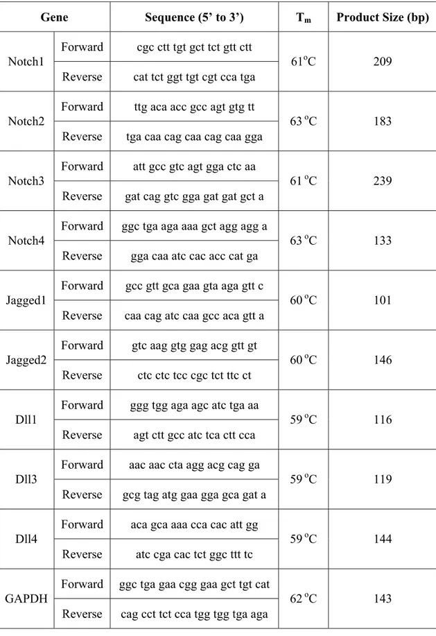
3.4.1 Reaction Conditions and Primers... 44
CHAPTER 4 RESULTS... 47
4.1 Expression of Notch Receptors and Ligands in Human Breast Cancer Cell Lines Detected by Semi-Quantitative RT-PCR... 47
4.2 Expression of Notch Receptors and Ligands in Human Breast Cancer Tumor Samples Detected by Semi-Quantitative RT-PCR ... 49
4.3 Summary of Results... 51
CHAPTER 5 DISCUSSION AND FUTURE PERSPECTIVES ... 53
LIST OF TABLES
Table 1.1 Knockout Mice of Notch Components………...……...29 Table 3.1 Human Breast Cancer Cell Lines Used in This Study.…………...……...40 Table 3.2 Human Breast Tumor Samples Used in This Study.………...43 Table 3.3 Sequences of Primers, Annealing Temperatures and Product Sizes…...45 Table 4.1 Summary of Expression Results………..………...52
LIST OF FIGURES
Figure 1.1 Structures of human breast…………..………...………...……….2
Figure 1.2 Stages of mouse mammary gland development……….………...……….4
Figure 1.3 Cyclical phase of mouse mammary gland development………...……….5
Figure 1.4 Formation of a duct in developing breast………...………...……...17
Figure 1.5 Structure of Notch Receptor………...………...……...24
Figure 1.6 Human Notch Receptors…..………...………...……...25
Figure 1.7 Notch Signaling Pathway………...………...……...26
Figure 4.1 Expression of Notch receptors in breast cancer cell lines detected by semi-quantitative RT-PCR………...……….……..48
Figure 4.2 Expression of Notch ligands in breast cancer cell lines detected by semi-quantitative RT-PCR………...………49
Figure 4.3 Expression of Notch receptors in human breast tumor samples detected by semi-quantitative RT-PCR………...………..…………50
Figure 4.4 Expression of Notch ligands in human breast tumor samples detected by semi-quantitative RT-PCR………...…………...…………51
ABBREVIATIONS
Bp Base Pairs
CADASIL Cerebral Autosomal Dominant Arteriopathy with Subcortical Infarcts and Leukoencephalopathy
DCIS Ductal Carcinoma In Situ ddH2O Double Distilled Water dH2O Distilled Water
DEPC Diethylpyrocarbonate
Dll Delta-like
DLLC Undifferentiated Large Light Cells DMEM Dulbecco’s Modified Eagle Medium ECM Extracellular Matrix
EGF Epidermal Growth Factor
EGFR Epidermal Growth Factor Receptor
ER Estrogen Receptor
ESA Epithelial specific Antigen
FACS Fluorescence Activated Cell Sorter FCS Fetal Caw Serum
FGF Fibroblast Growth Factor
GAPDH Glyceraldehyde-3-phosphate dehydrogenase HES Hairy/Enhancer of Split
HPV Human Papillomavirus HSC Haematopoietic Stem Cells
hTERT Human Telomerase Reverse Transcriptase ICM Inner Cell Mass
IDC Infiltrating (Invasive) Ductal Carcinoma ILC Infiltrating (Invasive) Lobular Carcinoma LCIS Lobular Carcinoma In Situ
LDC Large Dark Cell
LIF Leukemia Inhibitory Factor LOH Loss of Heterozygosity LRC Label Retaining Cells LTR Long Terminal Repeat MAML Mastermind-like protein MMTV Mouse Mammary Tumor Virus NICD Notch Intracellular Domain NLS Nuclear Localization Signal NSCLC Non-Small Cell Lung Cancer PBS Phosphate Buffered Saline PI3K Phosphatidylinositol 3-Kinase PR Progesterone Receptor
RT-PCR Reverse Transcriptase-Polymerase Chain Reaction SCLC Small Cell Lung Cancer
SLC Small Light Cell
SP Side Population
TACE TNF-α Converting Enzyme
T-ALL T-cell Acute Lymphoblastic Leukemia TCRβ T-cell receptor-β
TDLU Terminal Duct Lobular Unit TGF-β Transforming Growth Factor-β TEB Terminal End Buds
Tm Annealing Temperature
ULLC Undifferentiated Large Light Cells WAP Whey Acidic Protein
CHAPTER 1 INTRODUCTION
The expression patterns of Notch receptors and ligands in breast cancer cell lines and human breast tumors are presented in this study. Before introducing the project, the literature will be reviewed in order to focus on some of the basic concepts about breast cancer and Notch signaling. First, development of breast and then breast cancer will be reviewed. Next, the current studies related to stem cells and cancer will be summarized. Then, specifically breast stem cells will be presented. Finally, Notch signaling and its relation to breast cancer will be introduced.
1.1 Development of Breast
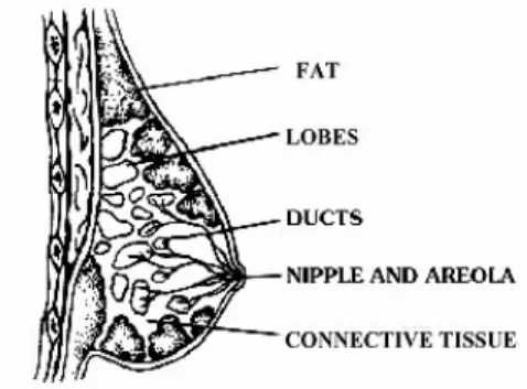
The four main structures in an adult human breast are lobules, ducts, fat and connective tissue (Figure 1.1). “Nipple” surrounded by the dark tissue “areola”, is the protruding point of the breast. “Lobes” are composed of lobules or glands, which are the milk producing parts during lactation. “Ducts” are the branching tubes that are connected to lobes, and converge to the larger “collecting ducts” towards the nipple. Ducts are responsible for carrying the milk from lobes to nipple during lactation. The branching ductal system and alveoli are supported by stroma, which is consists of fat and connective tissue, blood and lymphatic vessels (www.mammary.nih.gova).
Figure 1.1 Structures of human breast. (www.mammary.nih.gova)
Breast tissue, in contrast to most mammalian organs, is found at a rudimentary state after birth, and completes its maturation during puberty. In human embryo, breast begins to develop at around weeks 7 to 8, and visible structures are observed at around weeks 12 to 16 (www.mammary.nih.gova). The breast tissue is originated from ectoderm. First detectable structure called milk line, mammary crest or mammary band is a thickening extending across the embryo in a region, where breast bud will form. The first nipple is observed when the embryo is around 7-8mm length, it is seen as a narrow collection of ectodermal cells. Around 10mm length, the cells have a close relation with mesenchyme. Actually, the mesenchymal-epithelial interactions direct the breast development in embryo. Around day 14, following the differentiation of mesenchyme, epithelia proliferate to form a nodule that pushes mesencyme. The nodule then forms the breast bud. The bud starts to branch and form secondary buds with a clover-shape. Then secondary buds form canals and branch. The ductal morphogenesis in embryo is achieved by elongation and invasion of branches into the mesenchyme. Both female and male breast tissue shows the same development in embryo (Howard and Gusterson, 2000).
At birth, ductal system opens to the surface through a cavity on skin. The underlying mesenchyme proliferates to form nipple, and skin surrounding the nipple proliferates to form areola. The stage of breast development at birth differs from individual to individual. Some babies may have only small blunt ended tubular structures, while others may have well-developed branches. Both male and female babies may secrete some milk in response to maternal hormones. And during first
two years the lining epithelium proliferates and then involutes upon removal of the maternal hormones` effects. At the end of two years, only small ductal structures remain in a fibroblastic stroma and this state is kept until puberty (Howard and Gusterson, 2000).
Upon hormone stimulation with the beginning of the puberty, the breast structure further develops. The anatomical changes at this stage is well established, however the cellular events are not well understood. During this stage, in addition to epithelium, stroma also changes. Fibrous and fatty tissue increases that in a non-lactating breast it constitutes around 80% of the tissue. Hormones stimulate the growth of ducts, invasion into the fat pads and formation of lobular structures. The growing part of the tissue is “terminal end buds” (TEB), which consist of “body cell” mass surrounded by a layer of “cap cells”. Body cells are thought to form inner “luminal epithelial cells”, which lines the lumen. Cap cells are thought to form the outer “myoepithelial cells”, which are contractile cells found around the secretory alveoli cells and ducts; and cause squeezing the milk down to the ducts and then out of nipple upon oxytocin stimulation. Upon reaching the borders of fat pad, branching is completed and TEBs disappear (Howard and Gusterson, 2000; Smalley and Ashworth, 2003).
Human breast during pregnancy and lactation could not be studied well. In general, an increase in the number of lobules and loss of fat is observed. Lobulo-acinar structures, which have milk-secreting alveolar cells, are formed due to high proliferation followed by terminal differentiation. At weaning, removal of suckling stimulus results in involution, which means elimination of secretory epithelial cells by apoptosis and phagocytosis. At each pregnancy Terminal Duct Lobular Units (TDLUs) expand in size and then involutes. The ducts are not changed during this process. At menopause, a greater involution occurs for both lobules and ducts. The removed epithelium and interlobular connective tissue are replaced by fat (Howard and Gusterson, 2000).
It is not easy to study breast in humans. The similarities between mouse and human mammary gland development and function make mouse a good model. We have more detailed information about these processes in mice and that help us to
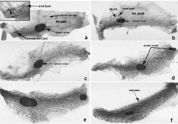
understand the mammary gland better. The figure below shows the dramatic changes of mouse mammary gland at different stages of development (Figure 1.2). Similar to development in human, mouse mammary gland development begins with thickening of epithelial cells and running from anterior to posterior direction in the embryo. The mammary bud forms and invades the mesenchyme, then encounters secondary mesenchyme, which is the precursor of mammary fat pad. The interactions with the mesenchyme induce a small amount of branching and form a rudimentary gland that remains unchanged till puberty. At puberty, hormones stimulate rapid elongation and branching by TEB similar to human. The ductal elongation ceases when the ducts reach to the periphery of the fat pad (Figure 1.2.c and 1.2.d), and the gland remain at this stage until pregnancy. The process from embryo to adult virgin animal can be called as “linear phase” of the mammary gland development (Lewis, 2000).
Figure 1.2 Stages of mouse mammary gland development. a. 3 weeks old mouse with slowly elongating and branching ducts. b. 4 weeks old virgin mouse with significantly increased growth of ducts around lymph node. c. 6 weeks old virgin mouse has ducts that reached to the periphery of mammary fat pad. d. 10 weeks old
The ductal development decreases at that time. e. Day 9 pregnant mouse with an extensive and rapid proliferation of mammary ducts. f. Day 16 pregnant mouse has an expanded mammary epithelium (www.mammary.nih.govb).
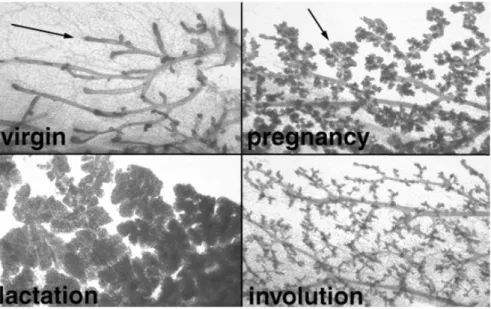
Pregnancy initiates the “cyclical phase” of the development, which is repeated in each pregnancy. The morphology of breast changes from ductal to lobuloalveolar structure (Figure 1.3). The progenitor cells proliferate and differentiate to form the alveolar buds and then alveoli. Alveolar cells gain capacity to express milk proteins at the middle of the pregnancy but secretion is inhibited until delivery. Weaning terminates the milk secretion and involution occurs by apoptosis. The gland returns to ductal morphology until next pregnancy, in which the same cycle is repeated (Lewis, 2000).
Figure 1.3 Cyclical phase of mouse mammary gland development. The predominant ductal and lobuloalveolar morphology are seen in virgin and pregnant mouse respectively. The extensively condensed milk secreting lobuloalveolar cells in lactating mouse collapsed during involution by apoptosis (www.mammary.nih.govb).
1.2 Breast Cancer
1.2.1 Epidemiology of Breast Cancer
Cancer is the second leading cause of death in U.S., and constitutes for the one fourth of all deaths. In 2004, 1,363,030 new diagnoses and 563,700 cancer deaths are expected to occur in U.S. Breast cancer is one of the important cancer types that it is the most frequently diagnosed non-skin cancer in women. 215,990 new invasive breast cancer cases, which is 32% of total, are expected to be diagnosed in women, in 2004. Breast cancer is the second for cancer deaths in women after lung cancer in U.S. 40,110 women, which is 15% of total, are expected to die because of breast cancer in 2004. In addition to invasive breast cancer, 59,390 women are expected to be diagnosed with in situ breast cancer. The number of males affected from breast cancer is not very low that the expected new diagnosis of and deaths from breast cancer is 1,450 and 470 respectively in males, in 2004. According to World Health Organization, 1.2 million people will be diagnosed with breast cancer worldwide (www.imaginis.com, American Cancer Society, 2004).
In Turkey, the facts about breast cancer are similar to U.S. According to 1999 statistics of Turkish Ministry of Health, breast cancer is the most common cancer followed by stomach cancer in Turkish women with 2,390 cases that makes 24.1% of all cancers in women.
According to statistics, breast cancer risk increases by age, 94% of new diagnosed cases and 96% of deaths occur after age 40 (American Cancer Society, 2003-2004). The menstrual history is also important that early onset and long period of menstruation, and menopause at older ages increases breast cancer risk. Although non-proliferative benign breast diseases has a very little or no effect on cancer risk, proliferative lesions without atypia and atypical hyperplasia increase breast cancer risk about two or four fold, respectively. Alcohol consumption is directly proportional to breast cancer risk that 10gr of alcohol per day increases the risk by 10% (Smith-Warner et al., 1998). Ionizing radiation and personal or familial history of breast cancer definitely increases breast cancer risk (Key et al., 2001).
Breast cancer risk increases immediately after the birth; however in long term childbearing has a protective effect. In addition, the high number of pregnancies and having the first child at an earlier age decreases risk significantly. The risk for the women with five or more children is half of the risk for the women without any child (Layde et al., 1989; Ewertz et al., 1990). The younger age at first birth has a protective effect that the women, who had first child earlier than age 20 has 30% lower risk compared to women, who had after 35 (Ewertz et al., 1990). Breastfeeding, experiencing menopause earlier, low-fat diet, a moderate physical activity and a healthy body weight are other factors decreasing the breast cancer risk (Key et al., 2001; American Cancer Society, 2004).
1.2.2 Histology of Breast Cancer
Most of tumors detected in breast are benign that they do not spread and become life threatening (American Cancer Society, 2003-2004). Breast carcinomas are originated from the malignant transformation of ductal or lobular epithelial cells. Normal epithelial cells may progressively develop ductal hyperplasia, atypical ductal hyperplasia, ductal carcinoma in situ, invasive ductal carcinoma and then metastasis, or they may directly form in situ or invasive type of breast cancer (Vogelstein and Kinzler, 1998; Russo et al., 1998).
In situ type of breast cancer indicates the early stages of breast cancer, these tumors are cancerous but they do not spread beyond the regions they originated and almost all cases can be cured.
1.2.2.1 Ductal Carcinoma in Situ
Ductal carcinoma in situ (DCIS) is the most common noninvasive breast cancer that constitutes 85% of all in situ breast cancers. The increased rate of incidence is due to early detection by mammography screening. The epithelial cells of ducts form the tumor but the tumor does not spread beyond the duct walls through the surrounding stroma (American Cancer Society, 2003-2004; www.cancer.orga).
DCIS is thought to be the precursor of invasive breast carcinoma. After removal of the DCIS lesions, 30-50% of patients developed invasive breast cancer in
6-10 years at the same site of previous DCIS (Page et al., 1982; Rosen et al., 1980). Basically DCIS is subdivided into two classes called comedo and non-comedo. Comedo type includes dead cells, and necrosis inside the tumor, and it is more malignant that precedes invasion (DeVita et al., 2001). Comedo type DCIS lesions usually lack estrogen and progesterone receptors (Albonico et al., 1998), overexpress oncogene c-erbB-2 and have mutated p53 accumulation (Poller et al., 1993; O’Malley et al., 1994). Angiogenesis is observed in the stroma surrounding the comedo DCIS lesions (Engels et al., 1997).
1.2.2.2 Lobular Carcinoma in Situ
Lobular carcinoma in situ (LCIS) is a less common type of noninvasive in situ breast cancers. It is originated from the lobules but not spread beyond them (American Cancer Society, 2003-2004; www.cancer.orga).
LCIS cannot be detected by physical examination or mammography; it is usually detected in the tissues removed for other reasons. LCIS is a homogeneous mass consist of small cells containing small and rounded-oval nuclei. The most characteristic pattern of LCIS is the loss of e-cadherin protein (Vos et al., 1997). The women, who had LCIS have a 7-10 times more risk for developing breast carcinoma. (Vogelstein and Kinzler, 1998; DeVita et al., 2001).
1.2.2.3 Invasive Breast Cancer
Infiltrating (invasive) breast cancer starts in lobules or ducts and invades the surrounding tissue. There are three stages of invasive breast cancer; in local stage tumor is fixed to the breast, in regional stage tumor spread to surrounding tissue or nearby lymph nodes, and in distant stage tumor metastasized to distant organs (American Cancer Society, 2003-2004; www.cancer.orga).
‘Infiltrating (invasive) ductal carcinoma’ (IDC) is the most common type of breast cancer, covering around 80% of all invasive breast cancers. It is originated in the ducts and spread beyond the duct wall invading the fatty tissue. ‘Infiltrating (invasive) lobular carcinoma’ (ILC) is originated from lobules and it is a less common type. Both invasive carcinomas can metastasize to other parts of body via
blood and lymphatic vessels. The other rare types of invasive breast cancer includes ‘tubular carcinoma’, in which tubules are formed; ‘medullary carcinoma’ consists of poorly differentiated and large cells, and have immune system cells around the tumor tissue; ‘mucinous (colloid) carcinoma’, which is characterized by the accumulation of extracellular mucus. The prognoses for these three types are better than IDC and ILC. Another rare type of invasive breast cancer is ‘inflammatory breast cancer’, which results in a red and pitted appearance of breast due to the blocking of lymphatic vessels or channels in the skin (Vogelstein and Kinzler, 1998; www.cancer.orga).
The prognosis and treatment strategy are directly related to the stage of the invasive breast cancer. The most commonly used staging system is the TNM system stated by American Joint Committee on Cancer. The three main criteria are the size and spreading in breast or nearby organs indicated by ‘T’, spreading to the lymph nodes indicated by ‘N’, and metastasis indicated by ‘M’. The evaluation of T, N and M stages gives the general stage of patient from stage I (the least advanced) to the stage IV (the worst case) (www.cancer.orgb) .
Several therapies including surgery, radiation therapy, chemotherapy, hormone or antibody therapy are applied to breast cancer in combination. Depending on the tumor type surgery can be applied in different ways. Lumpectomy is the local removal of tumor and a part of surrounding tissue. Mastectomy is removal of entire breast, and modified radical mastectomy is the removal of lymph node under arm in addition to entire breast. Surgery is usually followed by radiation therapy or chemotherapy together with hormone therapy, in order to destroy undetectable remaining tumor cells. Tamoxifen is one of the commonly used anti-estrogen drugs. Also some drugs interfering with estrogen synthesizing enzyme are used. These therapies can also be applied before surgery to reduce the size of tumor. (American Cancer Society, 2003-2004).
1.2.3 Molecular Biology of Breast Cancer
1.2.3.1 Germ Line Mutations in Breast Cancer
BRCA-1 and BRCA-2 are breast cancer susceptibility genes, which function as tumor suppressors and involved in the repair of damaged DNA in normal cells. BRCA1 and BRCA2 mutations constitute 5% of all breast cancer cases and they are associated with hereditary breast cancer (American Cancer Society, 2004). Female carriers of BRCA1 mutation have 87% life time risk for developing breast cancer and 40-60% for ovarian cancer. Almost all mutations of this gene are germ line mutations, and depending on the rare somatic mutations we can conclude that BRCA-1 mutations do not play a major role in sporadic breast cancers in contrast to hereditary cases. However, in sporadic cancer cell lines abnormal cytoplasmic localization that it is found in nucleus in normal cells and decreased expression during progression of sporadic breast cancers are observed (Chen et al. 1995; Thompson et al., 1995). Similar to BRCA-1, BRCA-2 is not thought to have a role in sporadic breast cancer and female carriers of BRCA-2 mutations have 85% life time risk for developing breast cancer but the risk for ovarian cancer is lower (10-20%).(Vogelstein and Kinzler, 1998; DeVita et al., 2001).
Other germ line mutations causing breast cancer are also associated with other tumor types and diseases. Li-Fraumeni syndrome is a result of germ line mutation in TP53 gene, which in addition to breast cancer may give rise to other types of tumor like soft-tissue sarcomas, brain tumor, and leukemia. Cowden disease, increases the risk of breast cancer in addition to several different features, and carries PTEN mutation. MLH1, MSH2 are mutated in Muir-Torre syndrome, which increases the breast cancer risk and associated with skin and upper and lower gastrointestinal tumors. Ataxia-telangiectasia is another disease associated to breast cancer risk but lower than other diseases, with non-Hodgkin Lymphoma, ovarian cancer, stomach, pancreas and bladder malignancies. Mutations in ATM gene are found in Ataxia-telangiectasia (Vogelstein and Kinzler, 1998; DeVita et al., 2001).
1.2.3.2 Somatic Mutations in Breast Cancer
Loss of heterozygosity (LOH) in cancers is usually involved in tumor suppressor genes. In breast cancer LOH is observed in some important locus including the genes RB-1, CDKN2, CDH1, and TP53. RB-1 and CDKN2 encode retinioblastoma and p16 proteins respectively, which are involved in cell cycle regulation. CDH1 encodes E-cadherin, which regulates differentiation and tissue compartmentalization. Point mutations for TP53 and RB-1 are more common, while methylation is mostly observed in CDKN2 and CDH1. TP53 mutations are analyzed in several groups of breast cancer samples and results showed that TP53 mutations are associated with poor prognosis. Cyclin D1, another important protein involved in cell cycle regulation. It was overexpressed in some of the breast cancer cell lines and in 45% of breast tumors in a study (Buckley et al., 1993). Increase in cyclin D1 expression is thought to be an early event in breast cancer development. One of the most common gene amplifications in breast cancer occur in c-MYC gene, which is a protooncogene, involved in cell proliferation or apoptosis depending on the cellular context. However the detection of c-Myc at protein level is harder because of short half-life of protein (Vogelstein and Kinzler, 1998; DeVita et al., 2001).
Epidermal growth factor receptors (EGFR), including EGFR, erbB-2 or HER-2/neu, erbB-3, and erbB-4, behave like oncogenes in breast cancer due to gene amplifications or overexpression, and results in uncontrolled cell proliferation (Bacus
et al.,1994; Vogelstein and Kinzler, 1998).
Fibroblast growth factors (FGF) are another group of growth factors involved in breast cancer. FGF-3 (mouse int-2), is one of the genes activated by the insertion of mouse mammary tumor virus (MMTV), and become oncogenic (Coleman-Krnacik and Rosen, 1994; DeVita et al., 2001).
Estrogen and progesterone are important in mammary gland development by regulating epithelial growth, differentiation and survival. In addition, these hormones are involved in the development of breast cancer. Estrogen receptor (ER) and progesterone receptor (PR), through which the hormones act, have an association with the prognosis and response to the antihormonal therapy of the patient. Estrogen
and progesterone induces the expression of protooncogenes c-MYC, c-FOS, c-JUN. In normal tissue, ER and PR are expressed in luminal epithelial cells and dividing cells are negative for both receptor. However, most of the breast tumors are steroid receptors positive (DeVita et al., 2001; Clarke et al., 2003).
1.3 Stem Cells and Cancer
1.3.1 Overview of Stem Cells
The cells, which have the ability to self-renew and to produce daughter cells that will differentiate into different cell types, are called ‘stem cells’. Stem cells are classified according to their capacities. The fertilized oocyte and its first daughter cells have the capacity to generate whole embryo and the surrounding tissue of placenta, and called totipotent stem cells. Throughout the embryonic development blastocyst and inner cell mass (ICM) is formed. The ICM cells are pluripotent stem cells that can from the all cell types of three germ layers, but not supporting tissue and a complete embryo. Adult tissues are thought to have multipotential stem cells (Alison et al., 2002). Repair and regeneration are essential in adult tissues, and require a stock of cells, the multipotential stem cells, which have an increased lifespan, and capacity to generate necessary tissue specific cells. It is easier to detect stem cells in tissues with high cell turn over like skin, intestine and bone-marrow. In some tissues stem cells are required for regeneration in response to tissue injury like in liver, and it is harder to identify these stem cells because they are hidden and activated only after an injury or pathologic condition (Nagy, 1995; Presnell et al., 2002). The last group is unipotential stem cells, which can only generate one type of differentiated cell, and sometimes considered as committed progenitor cells, because of their limited proliferation and differentiation capacity (Alison et al., 2002).
The intermediate cells between the stem cells and the terminally differentiated cells are called “progenitor cells”, “transit cells” or “transit amplifying cells”. A stem cell can generate one stem cell (self-renewal property) and one progenitor cell (differentiating daughter cell producing property) by asymmetric division, or can generate two identical types of cells by symmetric division. The
division generating two stem cells cause expansion or symmetric division generating two progenitor cells cause depletion of the stem cell compartment (Smalley and Ashworth, 2003).
Stem cells are undifferentiated and usually do not function like their progeny. They are thought to be found in very small numbers in the tissues, like 1 in 10,000 in haematopoietic tissue, and have a slow rate of proliferation compared to progenitor cells (Alison et al., 2002).
The stem cells are thought to be found in a specific part of the tissue, ‘stem cell niche’, which include extracellular matrix (ECM) and supporting cells with specific signaling functions. The base of the small intestine is an example for the stem cell niche. It was shown that in the case of stem cell depletion, the stem cell niche have the capacity to induce the formation of stem cells or can induce the progenitor cells to behave like stem cells (Smalley and Ashworth, 2003).
Stem cells should keep a balance between self-renewal and differentiation. Uncontrolled self-renewal will cause problems like tumor formation. It is crucial to understand pathways behind the decision of two fates in order to understand the stem cell derived tumor formation. The most detailed studies in stem cells are done in haematopoietic stem cells (HSC), which are isolated both from mice and human. The knowledge about stem cells mostly comes from HSC studies. Activation of notch signaling pathway by Jagged-1 ligand promoted HSC self-renewal or maintenance of multipotentiality (Varnum-Finney et al., 2000; Karanu F.N., et al., 2000). Sonic hedgehog signaling is another pathway that can increase self-renewal capacity of human HSC in vitro (Bhardwaj et al., 2001). Overexpression of activated β-catenin, which functions at the downstream of Wnt signaling, increased the cell population that can reconstitute haematopoietic system in vivo and have stem cell markers in isolated HSC. In addition, inhibition of Wnt signaling due to Axin overexpression resulted in inhibition of HSC proliferation and reduction in in vivo reconstitution. Activation of Wnt signaling results in increased self-renewal and decreased differentiation in epidermal (Zhu and Watt, 1999) and intestinal stem cells (Sancho et
al., 2003). We may conclude that Wnt signaling plays a role in self-renewal of stem
cells.
1.3.2 Stem Cells and Cancer
Tumors are also tissues that consist of heterogeneous cell populations, they can form the required structures like blood vessels to survive, they can metastasize to new tissues and form similar tumor tissues, and they can regenerate after surgical removal. All these features require a group of cells that have capacity to survive for a longer time and differentiate into several distinct cell types. As explained in section 1.3.1, stem cells have a high capacity to replicate, and live for a longer time compared to differentiated cells. These properties give them a greater potential to accumulate mutations, which is the main requirement for cancer formation. Trott expanded the hypothesis that only a small part, around 1%, of tumor cells behave like tumor stem cells, which can proliferate to form both tumor stem cells and progenitor cells to differentiate and form heterogeneous cell types of a tumor (Trott, 1994; Presnell et al., 2002; Smalley and Ashworth, 2003).
The dedifferentiation of differentiated adult cells was first thought to be the origin of cancer, they are thought to gain the ability to proliferate rapidly and give rise to different types of cells due to dedifferentiation. However cancer formation occurs within several months or years, and requires more than one ‘hit’ that changes the cellular behavior. Even when mature cells get the first ‘hit’ they will probably die before secondary hits occur. However; stem cells can live long enough to accumulate mutations (Sell and Pierce, 1994). Skin is one of the best examples for the stem cell origin of cancer. Skin cancer is mostly observed in the middle or late life time; however the initial promoting events occur early in life. The mature skin cells have a high turn over that, a fully differentiated keratinocyte derived from daughters of stem cells, removed from the skin in a day. Since the differentiated skin cells do not have enough time to accumulate mutations, there should be long-lived skin stem cells that begin to accumulate mutations early at life and give rise to cancer several years later (Presnell et al., 2002).
Another advantage of stem cells is that they already have self-renewal capacity, in contrast to differentiated cells. And uncontrolled activation of self-renewal will cause tumor formation due to proliferating stem cells. Stem cells require fewer mutations to maintain self-renewal capacity compared to differentiated cells, which should gain this capacity de novo. So, stem cells are more susceptible for being the origin of cancer (Reya et al., 2001).
Normal stem cells and cancer cells have many similar properties. First of all, both have ability to self-renew that stem cells use tightly controlled proliferation capacity for organogenesis, adult tissue maintenance, repair, and regeneration; while uncontrolled self-renewal of cancer cells cause tumor formation. Second, both can differentiate to form different types of tissue specific cells, or organogenesis in the case of stem cells and tumor heterogeneity in the case of cancer cells. Third, both have active antiapoptotic pathways and telomerase activity, which results in long life and high risk of mutation accumulation. Fourth, they have resistance to damaging agents. Stem cells have an increased transporter activity to exclude toxic agents, and cancer cells can remove chemotherapeutics and become resistant. Last, both can survive independent of anchorage and migrate, which gives cancer cells ability to metastasize (Dontu et al., 2003a).
Tu and colleagues hypothesize that heterogeneity of the tumors depends on the origin stem cell. If the origin is an early stem cell the tumor will be more heterogeneous than the tumors derived from a late stem cell with a narrower capacity. The early stem cells will generate tumors containing all cell types of germ layers, like in germ cell tumors; intermediate stem cells will generate tumors with restricted cell phenotypes, like in respiratory and gastrointestinal cancers; and a late stem cell will generate tumors containing only one type of cell, like in basal cell skin carcinoma (Tu et al., 2002).
The signaling of stem cell niche may contribute to tumor formation too. Because an abnormality in the signaling can cause accumulation of stem cells and that will increase the risk of tumor formation. The repopulation of stem cells by progenitor cells in response to stem cell niche signals will also increase the tumor formation risk according to the Cairns hypothesis. Cairns hypothesis states that after
asymmetric divisions, always stem cells take the original DNA strand, which will serve as template for the following divisions. In that case daughter progenitor cells always carry the newly synthesized DNA strand, which have more error due to the nature of the replication and accumulate mutations (Smalley and Ashworth, 2003).
Current therapeutic approaches target proliferating cells. However; stem cells have a relatively low proliferation rate and will not be affected by traditional therapies. Reducing the tumor size will not cure the cancer because it will recur as long as the stem cells exist in the tissue. However; a novel therapy targeting the stem cells will be effective against the origin of tumorigeneis and prevent recursion. A therapy targeting the stem cells may use the advantage of common phenotypic properties of stem cells. Instead of designing different therapies for different genotypic aberration in each cancer case, removing the stem cells directly may be easier. Some cases, like in breast cancer, a preventive treatment can be applied to the women in risk group. Since breast is not a vital organ, eliminating cancer prone stem cells at early ages will prevent tumor formation. However, these kinds of applications should be discussed deeply because depleting whole stem cells may generate serious side effects. For example repopulation of stem cells, will result in the accumulation of more mutations according to Cairns hypothesis, and this will increase the risk of tumorigenesis (Smalley and Ashworth, 2003).
1.4 Breast Stem Cells
1.4.1 Breast Stem Cells in General
Stem cells are mostly studied in detail in blood tissue, because studying with blood cells is easier than studying with the solid tissues. As a result, proliferation and differentiation process of haematopoietic stem cells are explained in detail, but the stem cells of solid tissues are not well identified and studied (Smalley and Ashworth, 2003).
The idea of breast stem cells first comes from a study done in 1959, by DeOme. He did the first transplantation experiment in breast, and showed that
epithelium isolated from different regions of postnatal mammary gland, can generate functional mammary epithelial structures. (Welm et al., 2003)
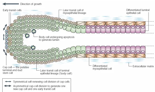
Figure 1.4 Formation of a duct in developing breast. The inner luminal epithelial cell layer and outer myoepithelial cell layer are formed from TEB. Cap cells at the tip of the TEB generate two different types of transit cells; first type on the outer site differentiate into myoepithelial cells, and second type at the center, called “body cells” differentiate into luminal epithelial cells. Central body cells undergo apoptosis while outer layer differentiate into the luminal epithelial cells in order to form the ductal lumen (Smalley and Ashworth, 2003).
A stem cell population cannot be identified in breast tissue yet; however it is thought that there are cell populations that behave like stem cells in specific microenvironments. In developing mammary gland, depending on the structure and function of TEB, we can say that the TEB are most probably the structures that contain the stem cell activity in breast tissue (Figure 1.4). In terminal end buds, the cap cells are the candidates for stem cells because it was shown that they migrated into the body cell mass (Smalley and Ashworth, 2003).
In addition to developing mammary gland, breast stem cells should be found in adult mammary gland for at least three processes. During pregnancy, side
branches are developed and secretory alveoli are generated to produce milk. Upon weaning involution occurs and these structures are removed by apoptosis. In each pregnancy this mammary cycle is repeated and dramatic changes occur in breast tissue (Figure 1.3). Only stem cells have the capacity to proliferate and differentiate into different structures for many times during the life of a female animal. In addition, non-pregnant animals have a similar alveolar bud expansion and regression process during each menstrual cycle. The other process is the replacement of epithelial cells, which are shed into the lumen during cell turnover. The cells may be lost during lactation, since they may be found in the milk, or they may be lost in resting gland (Smalley and Ashworth, 2003).
1.4.2 Evidences for Breast Stem Cells
The experimental techniques like cleared fat pad transplantation, retroviral tagging and x-chromosome inactivation patterning gave some evidences about breast stem cells. Cleared fat pad transplantation depends on the growth of the epithelia after birth, and enables removal of all epithelial cells of host breast and transplantation of new epithelial cells. The primary epithelial cells transplanted into the cleared fat pads resulted in mostly complete ductal system generation that can respond to hormones during pregnancy and generate alveolar cells, less commonly only alveoli without ducts, and rarely only ducts without alveoli generation. These results show that in the transplanted population there are some cells, which are able to form all or a few parts of an adult breast. Retroviral tagging system, in which retroviral sequence is incorporated into the host genome and enables marking of the cells, and monitoring the progeny of cells transplanted to the cleared fat pads. This system showed that entire mammary epithelial growth could be clonal in origin (Kordon and Smith, 1998). X chromosome inactivation patterns also showed that terminal duct lobular units (TDLU) are clonal, so there may be adult stem cells distributed to the mammary gland (Diallo et al., 2001; Welm et al., 2003; Smalley and Ashworth, 2003).
The histological and microscopic studies in mouse and rat identified a cell population, which morphologically is a candidate for a stem cell population. The
myoepithelial cells, and small in size. SLCs have a pale cytoplasm and do not have specialized organelles. They are found together as homogeneous or heterogeneous pairs, probably representing symmetric and asymmetric divisions respectively. SLC can differentiate into undifferentiated large light cells (ULLC), differentiated large light cells (DLLC) and large dark cells (LDC). It is thought that ULLC and DLLC are the progenitor/transit cells. Cells similar to SLC are identified in cattle and human mammary glands too (Smith and Chepko, 2001).
Another candidate cell population is identified by bromodeoxyuridine (BrdU) labeling experiment, in which the dye is incorporated into DNA and diluted by each cell proliferation. Some cells were shown to retain the label for a longer time, and that indicates a slow rate of proliferation, which is a property of stem cells. Some of these label-retaining cells (LRC) did not express neither luminal nor myoepithelial markers, while some express both. So it is thought that undifferentiated LRC stem cells can form differentiated transit cells (Welm et al., 2002; Smalley and Ashworth, 2003).
As mentioned before, luminal cells are thought to be the candidate for stem cell population. Freshly isolated human luminal and myoepithelial cells can be grown in specific mediums. When luminal cells are switched to the myoepithelial medium, after a certain time some cells with myoepithelial markers, and also some other cells with both myoepithelial and luminal markers are observed, indicating the intermediate transit cells. However, myoepithelial cells do not give this result (Pechoux et al., 1999). In addition, some cells positive for ESA (epithelial specific antigen, a marker for luminal cells), negative for MUC1 (luminal epithelial marker) and positive for cytokeratin 19, can generate ESA+/MUC1+ cells and myoepithelial cells. And when these ESA+/MUC1-/Cytokeratin 19+ cells are grown on three-dimensional cultures and xenografts, they can form TDLU like structures, and are thought to be TDLU progenitors (Gudjonsson et al., 2002; Smalley and Ashworth, 2003).
A common property of stem cells is their ability to efflux chemicals like chemotherapeutics or Hoechst dye out of their cytoplasm. Side population (SP), is accepted as a universal stem cell marker, because it is found in putative stem cells of
many tissues. SP cell population also pumps chemicals out of cell. This phenomenon is thought to be due to ABC transporter protein ABCG. SP cells are also found in breast epithelium of human and mouse, in which they are undifferentiated and express none of the cytoskeletal markers of myoepithelial and luminal epithelial cells. Depending on the efflux of Hoechst dye SP cells can be isolated from mouse mammary gland by FACS. The isolated SP cells were shown to reconstitute mammary gland and form ductal and alveolar cell populations. Breast SP cells are rich for expression of Sca-1, a stem cell marker. The Sca-1 enriched cells were able to reconstitute mammary gland. However since 20-30% of breast cells are positive for Sca-1, we can say that this is mixed population containing stem cells. (Welm et
al., 2003; Smalley and Ashworth, 2003).
Mammospheres, which is based on the principle that normal epithelial cells cannot survive in the absence of a substratum to attach; but in contrast stem cells are anchorage independent that they can survive and proliferate in this condition, was used to enrich the candidate breast stem cells. Cultured mammospheres lost the normal epithelial cells and enriched in the undifferentiated cells. These cells were able to differentiate and form myoepithelial, ductal epithelial and alveolar epithelial cells. In addition, they had self-renewal capacity that they could form new mammospheres containing cells with the same differentiation capacity. These studies should be expanded for in vivo experimental conditions as well. The expression profiles of mammospheres gave some clues about the pathways important in breast stem cells that TGF-β, and growth hormone signaling were activated, and Wnt-1 and Notch3 were among the signaling proteins upregulated (Dontu et al., 2003b).
Depending on these experimental results, we can conclude that there are breast stem cells, which are most probably luminal origin, can differentiate into both luminal epithelial and myoepithelial cells, and can generate TDLU like structures. The probable stem cells are located at the base of luminal epithelial layer, next to myoepithelial layer, and do not have a contact with lumen or basement membrane. These cells do not express all markers of differentiated luminal epithelial or myoepithelial cells, but may express some of them together. They generate transit
epithelial or myoepithelial cells. And these cells also express some common markers for stem cells (Smalley and Ashworth, 2003).
In spite of these experimental evidences, we still cannot say that there were breast stem cells in adult breast. The self-renewal capacity of the candidate stem cells is not shown yet. In order to prove the presence of breast stem cells exactly, the breast cells with multipotent differentiation and self-renewal capacity should be isolated.
1.4.3 Breast Cancer Stem Cells
Breast stem cells are strong candidates for the origin of breast cancer. In addition to the general ideas explained in section 1.3.2., some observations also supports this idea. For example, women exposed to ionizing irradiation as teenagers are more susceptible to breast cancer compared to women exposed as adults. In 40% of breast cancer cases, recursion is observed in 10 years after the diagnosis and removal of the tumor. So, the origin should have an extremely long life and should be found in young women more, with a higher proliferative capacity (Welm, 2003). These observations direct us to the breast stem cells as origin of breast caner.
Al-Hajj et al identified a group of candidate “breast cancer stem cells” in human breast tissue. In solid tumors only a small proportion of cells are able to from colonies in vitro, and large number of cells are required in transplantation to form tumor in animals. So, they think that in a tumor there is small number of cells, which have capacity to proliferate and from new tumors. They isolated different group of cells depending on the heterogeneous expression of cell surface markers from human breast tumors. A specific group of cell, which is positive for cell adhesion molecule CD44, negative or has low expression for CD24, and negative for lineage markers (CD2, CD3, CD10, CD16, CD18, CD31, CD64, CD140b, which are associated to normal cell type and not expressed in cancer cells) was isolated. It is interesting that the transplantation of 103 CD44+/CD24-/low/Lineage- cells generated tumor in all cases, however transplantation of 104 mixed tumor cells generated tumors in minority of cases. In ESA+ subpopulation, even 200 cells give rise to tumor. The complexity of the new tumor with respect to ESA, CD24, and CD44 expression
patterns is similar to the original tumor, from which CD44+/CD24-/low/Lineage- cells were isolated. The newly formed tumor contained tumorigenic CD44+/CD24 -/low/Lineage- cells, representing the self-renewal property. And also the tumor contained non-tumorigenic cells with different cell surface marker expression patterns, representing the differentiation property. The experiments strongly states that CD44+/CD24-/low/Lineage- group of cells have stem cell properties, however in order to prove definitely a single cell transplantation should be able to generate tumor (Al-Hajj et al., 2003). A possibility is that this group of cells is enriched in stem cells, but may also contain non-tumorigenic cells. So, further analysis is required for the isolation of exact stem cells within CD44+/CD24-/low/Lineage- group.
Do the tumor stem cells originated from normal adult stem cells? The multipotent cells of breast are also known to express ESA and CD44, which are positive in putative tumor stem cells as well (Al-Hajj et al., 2003). In addition, depending on the experimental results we can say that they are probably originated from normal stem cells. When the epithelial cells of a Wap-TGFβ1 transgenic mouse are transplanted to a cleared fat pad, reduced repopulation is observed, and this is thought to be due the premature stem cell senescence. And these transplanted animals are resistant to MMTV induced tumorigenesis. Another evidence for the normal adult stem cell origin of breast cancer stem cells is the protective role of early pregnancy against breast cancer. Pregnancy causes terminal differentiation, and decreases the proliferative capacity. Early pregnancy results in the increase in number of terminally differentiated cells depleting the stem cell compartments before they accumulate mutations and begin tumorigenesis (Smalley and Ashworth, 2003).
1.4.4 Regulation of Breast Stem Cells
The regulation of stem cell self-renewal and differentiation is based on the balance between signaling pathways and many factors like environment. Understanding the pathways involved in regulation of self-renewal and differentiation will be important to establish the relation between breast stem cells and tumorigenesis, and will direct us to novel therapeutic approaches.
Wnt signaling is involved in the self-renewal of haematopoietic, epidermal and intestinal stem cells as explained in section 1.3.1. Mammosphere experiments showed that Wnt pathway components are differentially expressed in putative breast stem cells. In addition overexpression of Wnt in the mouse mammary gland by MMTV promoter increased tumor formation (Schroeder et al., 2002). So, abnormal Wnt signaling may be involved in tumor formation due to stem cell deregulation in mammary gland (Dontu et al., 2003a).
Leukemia Inhibitory Factor (LIF), which stimulates self-renewal of neural stem cells and maintenance of embryonic stem cells in undifferentiated state, was shown to be involved in mammary gland tumorigenesis. Overexpression of LIF in transgenic mouse caused mammary tumor development and its overexpression is observed in human breast cancers (Dhingra et al., 1998). PTEN, is another protein shown to be involved in self-renewal of neural stem cell self-renewal, and formation of neurospeheres, have mutation in breast cancer (Dontu et al., 2003a).
Notch signaling pathway is an emerging topic in breast stem cells and tumorigenesis and will be discussed in the next section.
1.5 Notch Pathway
1.5.1 Overview of Notch Pathway
1.5.1.1 The Structure of Notch Receptors and Ligands
A Drosophila strain with notches at the end of its wings was first described in 1917. Later it was identified that this phenotype is related to a partial loss of function of Notch gene. Notch gene is evolutionary conserved from flies to mammals (Nam et
al., 2002; Radtke and Raj, 2003).
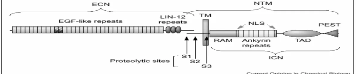
Notch receptor is single-pass transmembrane protein, which is synthesized as a single precursor protein but cleaved during transportation to the membrane by a furin-like protease in trans-Golgi. This first cleavage of Notch occurs at around 70 amino acids external to the transmembrane domain and generates two subunits, extracellular and transmembrane. The subunits are not dissociated but transported to the membrane as a heterodimeric receptor, which is held together by non-covalent
interactions and calcium. Notch consists of highly conserved structural motifs (Figure 1.5). Extracellular subunit of Notch receptor contains 29-36 N-terminal epidermal growth factor (EGF)-like repeats, which are involved in ligand binding. EGF-like repeats are followed by three tandemly repeated LIN-12 motifs, which are required to inhibit activation in the absence of ligand binding. First cleavage site is around 100 residues after LIN-12 repeats. The extracellular part of transmembrane subunit is very short and consists of conserved cysteine residues. The intracellular part of transmembrane subunit contains RAM domain followed by ankyrin/CDC10 repeats, both of which are involved in binding to the transcription factors in the nucleus. Ankyrin/CDC10 repeats are flanked by two nuclear localization signals (NLS) and followed by transactivation domain and PEST (PEST for proline, glutamate, serine and threonine) sequence (Kadesch, 2000; Nam et al., 2002).
Figure 1.5 Structure of Notch Receptor. ECN and NTM show the extracellular and transmembrane subunits of Notch receptor, respectively. S1, S2 and S3 represent the sites of three cleavages in order (Nam et al., 2002).
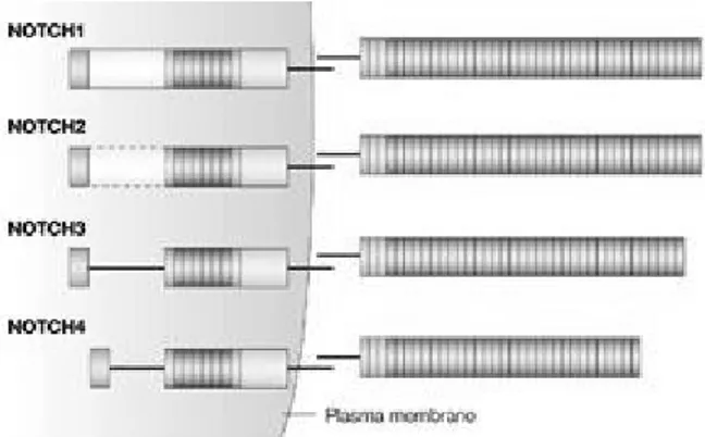
Drosophila has only one Notch gene, while C.elegans have two (glp-1 and
lin-12) and mammals have four (Notch1-4), which are thought to be evolved by gene duplication. Mammalian Notch receptors are highly homologues, but have some differences in extracellular and cytoplasmic domains (Figure 1.6). Notch1 and Notch2 have 36 EGF-like repeats while Notch3 has 34 and the shortest one Notch4 has 29 repeats. Notch1 has the strongest transactivation domain, Notch2 has a weaker one, but Notch3 and Notch4 do not have. Ankyrin/CDC10 repeats are the least conserved part of the receptors and affects transactivating activity (Nam et al., 2002; Radtke and Raj, 2003).
Figure 1.6 Human Notch Receptors. Human Notch receptors are highly conserved but have differences in the number of extracellular EGF repeats and cytoplasmic Ankyrin repeats and transactivating domain (Radtke and Raj, 2003).
Notch ligands, DSL, are also single-pass transmembrane proteins. Drosophila has two ligands Delta and Serrate, while in human, two Serrate homologues Jagged1 and Jagged2, and three Delta homologues Delta-like1, Delta-like3, and Delta-like4, are found. Each of the ligands contain N-terminal DSL motif, which is required for receptor interaction. DSL is followed by EGF-like repeats, and then transmembrane domain. The cytoplasmic tail of ligands is shorter and consists of 70-215 amino acids. The number and spacing of EGF-like repeats are different in ligands. Jagged1 and Jagged2 have additional cysteine rich domain below EGF-like repeats (Nam et
al., 2002; Radtke and Raj, 2003).
1.5.1.2 The Notch Signaling Pathway
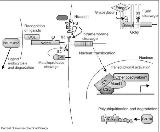
In contrast to usual signaling pathways, Notch signaling does not contain an enzymatic amplification step, and Notch receptor itself directly involved in the transduction of the signal to the nucleus (Figure 1.7).
Notch signaling is activated upon interaction with ligand represented by adjacent cells. Receptor-ligand interaction disrupts the heterodimeric structure of Notch and induces the second proteolytic cleavage. A member of ADAM family of proteases, TNF-α converting enzyme (TACE) in human and Kuzbanian in
Drosophila, cleaves the transmembrane subunit of receptor from a site adjacent to
Figure 1.7 Notch Signaling Pathway. S1, S2, and S3 represents the three cleavages in order. ICN represents the released intracellular domain of Notch (NICD) (Nam et
al., 2002).
which is catalyzed by a complex of Presenilin, nicastrin, Aph1, and Pen2 protein complex and releases functional cytoplasmic part, Notch intracellular domain (NICD). NICD translocates to the nucleus and interacts with CSL (CSL stands for CBF1 in mammals, Su(H) in Drosophila, and Lag-1 in C.Elegans) transcription factors. In the absence of Notch signaling CSL binds to the target genes’ promoters in a complex with several corepressors and histone deacetylases to inhibit the transcription. NICD competes with the inhibitory proteins, like CIR, KyoT2, and SMRT. Then displace them and recruit coactivators and histone acetylases like CBP/p300, pCAF, GCN5 and Mastemind like protein (MAML) converting CSL from repressor to activator, and initiate transcription. The signaling is terminated by
an E3 ubiquitin ligase (Kadesch, 2000; Nam et al., 2002; Radtke and Raj, 2003; Wu and Griffin, 2004).
The first identified target gene of Notch signaling is HES (Hairy/enhancer of split) basic helix-loop-helix transcription factors, which inhibits expression or function of Neurogenin, Mash, and MyoD proteins. Notch signaling is thought to activate different targets at different tissues. For example in keratinocytes Notch1 was shown to induce p21Waf1, which has CSL binding site, inhibiting proliferation and inducing terminal differentiation (Rangarajan et al., 2001a). CSL binding site is also described in cyclin D1 gene, which is upregulated by Notch signaling in rat kidney epithelial cells (Ronchini and Capobianco, 2001). Notch1 was shown to activate phosphatidylinositol 3-kinase (PI3K), which confers resistance to anoikis and p53 mediated apoptosis. ERBB2 and NF-κB2 promoters have CSL binding sites, and induced by Notch1 (Chen et al., 1997; Oswald et al., 1998). ERBB2 stimulates proliferation, while NF-κB2 is mainly involved in development of lymphoid organs and induces expression of many genes, some of which have anti-apoptotic roles (Radtke and Raj, 2003; Hansson et al., 2004).
There are also evidences for CSL-independent signaling of Notch. NICD lacking CSL-interacting domain can still function in blocking myogenesis. Deltex protein is involved in CSL-independent signaling (Radtke and Raj, 2003; Hansson et
al., 2004).
1.5.1.3 Regulation and Crosstalks of Notch Signaling
Notch receptors are glycosylated by addition of fucose to serine and threonine residues of EGF-like repeats by O-fucosyl transferase. The glycsoylation is essential for the regulation of Notch activity that loss of function or overexpression of the particular enzyme may inhibit Notch activity. Fringe is another enzyme involved in modification of O-linked fucose on EGF-like repeats, which are important in ligand binding. Modification by Fringe increases the affinity of Notch for Delta while decreasing the affinity for Serrate in Drosophila (Schweisguth, 2004). The orthologues of Fringe in human are Radical, Manic and Lunatic Fringe, which perform gycosylation in Golgi. Similar to Drosophila, modification of Notch inhibits
activation by Jagged1 and Jagged2 but not by Delta-like (Dll) ligands in mammals (Mumm and Kopan, 2000; Nam et al., 2002).
The internalization of receptors by endocytosis, and degradation by ubiqitination is another mechanism of Notch regulation. Numb is one of the proteins that are thought to be involved in endocytosis. The exact mechanism of Numb function is not clear but it segregates differentially to daughter cells and affect the cell fate by inhibiting Notch (Hansson et al., 2004; Schweisguth, 2004).
The crosstalk with several pathways may alter the result of Notch signaling depending on the cellular context. For example, in C.elegans Ras activation downregulates Notch. However, in cultured human cell lines Ras activates Notch signaling and Notch is required to maintain Ras induced neoplastic phenotype (Weijzen et al., 2002). Wnt signaling has an inhibitory effect on Notch signaling, and NICD has a week interaction with LEF-1, and GSK-3β, which is thought to phosphorylate and stabilize NICD (Hansson et al., 2004).
1.5.1.4 Function of Notch Signaling
Notch is one of the pathways involved in development in coordination with the pathways Wnt, Hedgehog, Transforming Growth Factor-β (TGF-β), and Receptor Tyrosine Kinase/Phosphatase. Notch signaling is important in several cellular processes like maintenance of stem cells, specification of cell fate, differentiation, and proliferation depending on the cell type (Mumm and Kopan, 2000; Radtke and Raj, 2003).
Maintenance of an undifferentiated state is one of the major roles of the signaling. Activated Notch signaling prevents differentiation neuronal progenitor cells, while inhibition results in excessive neurogenesis and depletion of progenitor cells. In a hematopoietic cell population, induction of Notch signaling by Jagged1, increases the stem cell ratio. So, Notch signaling keeps progenitor cells in a stem cell like character (Radtke and Raj, 2003).
Table 1.1 Knockout Mice of Notch Components
Gene Knockout Phenotype Reference
Notch1 Embryonic lethality Swiatek et al., 1994; Conlon et al., 1995 Notch2 Embryonic lethality Hamada et al., 1999
Notch3 No apparent phenotype Krebs et al., 2003 Notch4 No apparent phenotype Krebs et al., 2000
Notch1 and Notch4 More severe phenotype than
Notch1 null mice Krebs et al., 2000
Notch1 and Notch3 Embryonic lethality similar to
Notch1 null mice Krebs et al., 2003 Jagged1 Embryonic lethality Xue et al., 1999
Delta like1 Embryonic lethality Hrabe et al., 1997
Delta like3 Skeletal defects Kusumi et al., 1998; Dunwoodie et al., 2002 CBF-1 Embryonic lethality Oka et al., 1995
Notch signaling is also involved in binary cell fate decision, which means the determination of a cell fate between two possible options. During development in
Drosophila, the fate of precursor cells, which can differentiate into
neuronal-precursor or epidermal cells, are determined by Notch signaling. In lateral inhibition, Notch receptor and ligands are expressed equally in all cells at the beginning, but the concentrations changes over time and cells begin to express either receptors or ligands dominantly. The cells expressing ligand differentiate into neuronal cells,