IMAgENs EM REUMATOLOgIA
ÓRgÃO OFICIAL DA sOCIEDADE PORTUgUEsA DE REUMATOLOgIA 349
A rare cause of chronic hip pain:
intraarticular synovial chondromatosis
ACTA REUMATOL PORT. 2014;39:349-350
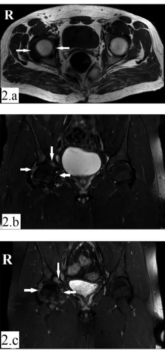
A 23-year-old male patient presented to our clinic with pain and limited motion in the right hip. The pain start -ed about 3 years previously and increas-ed over time, spreading to the trochanteric region of the right hip and the right groin. The characteristic of the pain was me-chanical. He did not feel pain while sleeping. Prolon-ged sitting or standing caused the hip to lock. Pre-viously, he had received physical therapy and analge-sic medications but there had been no significant im-provement. There was no pain in any other joint, and there was no history of disease or trauma associated with the hip. Physical examination revealed an antalgic gait. Palpation of the iliopsoas muscle caused pain. The motion of the right hip joint was limited and painful in all directions, whereas lumbar and left hip joint mo-tions were unrestricted and painless. There were no neurological deficits of the lower extremities. Radio-graphy of the pelvis indicated a narrowing joint space, and there were erosions on acetabular side of the joint (Figure 1). Magnetic resonance imaging (MRI) of the right hip revealed findings consistent with synovial chondromatosis, which filled the joint space comple-tely (Figure 2 a, b, c). Serological and biochemical mar-kers and superficial ultrasound of the inguinal region were normal. Orthopedic surgical procedures inclu-ding arthroscopic debridement were planned.
Synovial chondromatosis is defined as benign proli-feration of hyaline cartilage at the joint, bursa, or tendon sheath. It is relatively rare and its etiology is un known1. It most commonly occurs at the knee joint, but may also affect hip, shoulder, elbow, ankle, and wrist joints2,3. It is observed more often in males of 30–50 years of age, and usually involves single rather than multiple joints3. However, symptoms are nonspecific, often delaying
Kose MM1, Durmus O1, Ayhan MY1, Batmaz AG2
diagnosis. The most common symptoms are insidiousonset mechanical pain around the affected joint, swell -ing, joint lock-ing, and/or stiffness. The most common finding is a limited range of mo tion1,4. If there is no cal-cification at cartilaginous nodu les, it is difficult to iden-tify in radiographs. MRI can be used for synovial and soft tissue pathologies4, but may be negative in the initial period of synovial chondromatosis. In advanced stages, the synovium may form cartilaginous nodules called loose bodies. These may grow, calcify, and even ossify. This, in turn, can cau se mechanical damage to articular cartilage, leading to osteoarthritis. Therefore, loose bo-dies must be removed surgically1.
Hip pain is a frequent disorder of the musculoskeletal system. Young patients, in particular, have non -specific symptoms. Medical history review and physi-cal examination often cannot provide enough clues for diagnosis5. Because synovial chondromatosis starts with nonspecific symptoms and early diagnosis is difficult, it is associated with significant disability. In this res-pect, we recommend keeping synovial chondromato-sis in mind during differential diagnochondromato-sis of chronic and stubborn hip pain.
1. Department of Physical Medicine and Rehabilitation, Istanbul Medipol University, Faculty of Medicine, Istanbul
2. Department of Orthopedics and Traumatology, Istanbul Medipol University, Faculty of Medicine, Istanbul
FIGURE 1.Radiography of the pelvis: narrowing of the right hip joint space and erosions on acetabular side of the joint. Nodule formation is not observed
CoRREspondEnCE to
Oguz Durmus
Department of Physical Medicine and Rehabilitation, Istanbul Medipol University, Faculty of Medicine. TEM Avrupa Otoyolu Goztepe Cikisi, No 1, Bagcilar Istanbul - TURKEY. 34214 E-mail: [email protected]
REFEREnCEs
1. Griesser MJ, Harris JD, Likes RL, Jones GL. Synovial Chon-dromatosis of the Elbow Causing a Mechanical Block to Range of Motion: A Case Report and Review of the Literature. Am J Orthop (Belle Mead NJ) 2011;40:253-256.
2. de Carvalho JF. Knee synovial osteochondromatosis. Acta Reu-matol Port 2010; 35:107-108.
3. Tutun S, Ozgonenel L, Cetin E, Aytekin E. Two rare involvement sites: synovial chondromatosis. Rheumatol Int 2011;31:687--689.
4. Tibor LM, Sekiya JK. Differential Diagnosis of Pain Around the Hip Joint. Arthroscopy 2008;24:1407-1421.
5. Poultsides LA, Bedi A, Kelly BT. An Algorithmic Approach to Mechanical Hip Pain. HSS J 2012;8:213–224.
ÓRgÃO OFICIAL DA sOCIEDADE PORTUgUEsA DE REUMATOLOgIA 350
A rAre cAuse of chronic hip pAin: intrAArticulAr synoviAl chondromAtosis
FIGURE 2.MRI of the right hip: Millimeter-sized, multiple intra-articular cartilaginous fragments, not all of which are ossified (white-arrows) can be seen on the axial-T1-sequence (a) and coronal-T2-sequence (b).They are heterogeneous--hypointense and fill the entire right coxofemoral joint space. Contrast enhancement can be seen in coronal-T1 post-contrast sequence (c)