International Journal of Surgery 83 (2020) 89–97
Available online 16 September 2020
1743-9191/© 2020 IJS Publishing Group Ltd. Published by Elsevier Ltd. All rights reserved.
Experimental Research
Effects of endothelin receptor blockade and COX inhibition on intestinal I/R
injury in a rat model: Experimental research
Bercis Imge Ucar
a,*,1, Acelya Erikci
b,2, Kemal Kosemehmetoglu
c, Ceren Ozkul
d, Alper
Bektas Iskit
e, Gulberk Ucar
b, Sezgin Zeren
aaDepartment of General Surgery, Faculty of Medicine, Kutahya Health Sciences University, Kutahya, Turkey bDepartment of Biochemistry, Faculty of Pharmacy, Hacettepe University, Ankara, Turkey
cDepartment of Pathology, Faculty of Medicine, Hacettepe University, Ankara, Turkey
dDepartment of Pharmaceutical Microbiology, Faculty of Pharmacy, Hacettepe University, Ankara, Turkey eDepartment of Medical Pharmacology, Faculty of Medicine, Hacettepe University, Ankara, Turkey
A R T I C L E I N F O Keywords:
Intestinal ischemia reperfusion injury Rat
Cyclooxygenase inhibition Endothelin receptor blockers Nitric oxide
A B S T R A C T
Background: Intestinal ischemia is a highly morbid and mortal condition with no specific treatment. The present study aimed to investigate the effects of cyclooxygenase (COX) inhibition synchronized with nitric oxide (NO) release and endothelin (ET) receptor blockade on oxidative stress, inflammation, vasoconstriction, and bacterial translocation which occur during ischemia-reperfusion (I/R) injury in in-vivo rat intestinal I/R model. Materials and methods: 36 male Wistar rats were randomly divided into six groups (n = 6). Superior mesenteric artery blood flow (SMABF) was recorded; SMA was occluded for 30 min; SMABF was re-recorded at the beginning of the reperfusion phase. Rats were sacrificed after the reperfusion period of 60 min. Blood and tissue samples were obtained. Acetylsalicylic acid (ASA), NO-ASA, flurbiprofen (FLUR), and Tezosentan (TS) were administered 15 min after ischemia. Histopathological examination, bacterial translocation, and biochemical analysis were performed in plasma and tissue samples.
Results: SMABF difference, mean Chiu’s score and bacterial translocation were increased in the I/R group and decreased in the treatment groups. Plasma LDH, transaminases, intestinal fatty acid-binding protein (I-FABP), TNF-α, ICAM-1, interferon-gamma (IFN-Ɣ) and proinflammatory cytokine panel; tissue lipid peroxidation, MPO, xanthine oxidase (XO), NO, NF-kB levels and the expression of TNF-α were significantly elevated in the I/R group and markedly decreased in the treatment groups. The tissue antioxidant status was decreased in the I/R group and increased in the treatment groups.
Conclusion: It is suggested that NO-ASA, TS, and FLUR can be introduced as promising therapeutics to improve intestinal I/R injury.
Institutional protocol no: 2018-29-05 (Animal Experimentations Ethics Committee, Hacettepe University).
1. Introduction
Intestinal ischemia/reperfusion (I/R) injury is a life-threatening perfusion problem of the gastrointestinal (GI) system associated with the superior mesenteric artery (SMA) which mostly occur following abdominal or thoracic vascular surgery, cardiopulmonary by-pass; hemorrhagic, traumatic or septic shock, severe burns, strangulated hernias, small bowel transplantation, necrotizing enterocolitis, and acute mesenteric ischemia (AMI). The protection of organs from I/R
injury still remains a challenge for surgeons since there has been a limited number of therapeutic possibilities for I/R injury occurred dur-ing surgery [1–6].
Pathophysiology of AMI includes intestinal barrier injury due to perfusion deficit, local and systemic inflammation, and I/R injury, which leads to excessive production of reactive oxygen species (ROS) and bacterial translocation. A critical decrease in the intestinal blood flow leads to intestinal necrosis, which is the main reason for the high mortality rate. Medical treatment includes administering antibiotics, * Corresponding author. Yozgat City Hospital, Department of General Surgery, 66100, Yozgat, Turkey.
E-mail address: [email protected] (B.I. Ucar).
1 Yozgat City Hospital, Department of General Surgery, Yozgat, TURKEY.
2 Lokman Hekim University, Faculty of Pharmacy, Department of Biochemistry, Ankara, TURKEY. Contents lists available at ScienceDirect
International Journal of Surgery
journal homepage: www.elsevier.com/locate/ijsu
https://doi.org/10.1016/j.ijsu.2020.08.061
anticoagulants, and vasodilator agents, while surgical treatment aims to provide reperfusion with recanalization of the occluded structure via conventional surgery or with the help of interventional radiology [7]. However, there is no ideal treatment until now and mortality has not decreased yet.
Nitric oxide is shown to modulate ROS, decrease leukocyte adhesion to mesenteric endothelium, and sustain normal vascular permeability. NO-releasing anti-inflammatory drugs (NO-NSAIDs) were reported as having immuno-modulatory properties with reduced GI toxicity compared to their parent compounds. NO-releasing acetylsalicylic acid (NO-ASA) causes local vasodilation, decreases oxidative stress, prevents inflammation, and modulates vascular cell proliferation [4,8–10].
Endothelins (ET) are the most potent vasoconstrictor substances vasoconstricting peptides, and ET-1 mediates its biologic effects via the activation of ETA and ETB receptors. ETA and ETB receptor antagonists are involved in the pathophysiology of I/R [6,7] Tezosentan (TS), a non-selective ETA and ETB receptor antagonist was suggested to improve intestinal and hepatic microcirculation in sepsis, pulmonary and renal dysfunction [11] and in endotoxemic shock [9,12,13]. TS was suggested to increase NO signaling via enhanced hydrogen peroxide (H2O2) generation, decreases IL-6 and tumor necrotizing factor-alpha (TNF-α) levels during I/R injury [4,12,13].
In the present study, the effects of NO release plus cyclooxygenase (COX) inhibition and also ET receptor blockade on oxidative stress, inflammation, vasoconstriction, and bacterial translocation occur dur-ing I/R injury were investigated in an experimental in vivo intestinal I/R model. For his purpose, flurbiprofen (FLUR), as the COX enzyme in-hibitor; acetylsalicylic acid (ASA), as the non-specific irreversible COX inhibitor; NO-ASA as NO-releasing NSAID; and TS, as dual ETA/ETB receptor antagonist, were used in the study.
2. Materials and methods
ASA, FLUR, NO-ASA (NCX 4016), and other chemicals were pur-chased from SIGMA (Sigma-Aldrich, Germany). TS kindly provided by Dr. Martine Clozel and Dr. Marc Iglarz from Actelion Pharmaceuticals Ltd., Allschwil, Switzerland to Prof. Alper Bektas Iskit.
The details of the experimental design, study groups and the per-formed tests including rat intestinal I/R model, SMA blood flow (SMABF) measurement, blood and tissue sampling, the assessment of edema, histopathological examination, analysis of bacterial trans-location, biochemical tests and statistical analysis which have been performed can be found in supplementary data section (Online-Only). This study is reported in accordance with the ARRIVE guidelines (Ani-mals in Research: Reporting In Vivo Experiments) [14].
3. Results
3.1. Rat intestinal I/R model
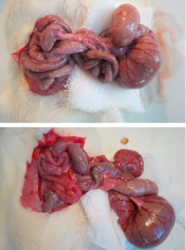
The intestine’s pale color verified the success of the intestinal I/R model in all groups after 30 min of ischemia. After 60 min of reperfusion period, the colour of the intestines was still found to be pale in I/R, I/R +ASA, I/R+(NO-ASA) groups; the colour of the intestines was slightly pink in I/R + FLUR group, whereas the colour of the intestines of I/R + FLUR group was similar to Sham group (Fig. 1).
3.1.1. SMABF values
SMABF values before and after ischemia were found to be signifi-cantly different in I/R group (p < 0.001). The said difference was reduced in the groups of I/R + NO-ASA and I/R + TS (p < 0.01 and p < 0.001, respectively). Reduction of the difference between the SMABF values of before ischemia and after reperfusion was highly distinctive in I/R group, indicating that treatment with TS ameliorates the impaired SMABF during intestinal I/R. However, treatment with FLUR and ASA had no significant effect on SMABF during the intestinal I/R (Fig. 2).
3.2. Histopathological examination
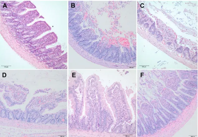
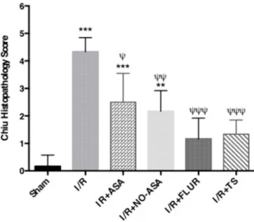
The intestinal mucosal injury was evaluated using Chiu’s scores. In Sham group, normal intestinal villous architecture was observed without any mucosal erosion. In I/R group, loss of epithelium, digestion, and disintegration of lamina propria, hemorrhage, and ulceration were observed. In I/R-ASA and I/R + NO-ASA groups, loss of epithelium, the disintegration of lamina propria, and ulceration were observed. In I/R + FLUR and I/R + TS groups, villi were intact, yet the development of a subepithelial space at the tips of the villi was observed (Fig. 3). Ac-cording to the Chiu scoring system, the injury in the I/R group was found to be markedly increased when compared with Sham group (p < 0.0001). Chiu scores of all treatment groups were lower than those of I/ R group’s Chiu score. The I/R + NO-ASA and I/R + TS groups showed a significant decrease in intestinal injury score when compared with the I/ R group, indicating a remarkable histopathological improvement via the treatment with these drugs (Fig. 4).
3.3. Bacterial translocation
Microbiological evaluation showed that the counts of microorgan-isms cultured in the liver, spleen, and mesenteric lymph nodes (MLN) in I/R group were significantly higher than in the treatment groups of I/R +ASA, I/R + NO-ASA, I/R + FLUR, and I/R + TS. Data demonstrated that the treatment with ASA, NO-ASA, FLUR, and TS provided a sig-nificant reduction in bacteria count in liver, spleen, and MLN of rats, while administration of TS after I/R appeared as the most effective treatment leads to significantly reduced bacterial translocation partic-ularly in spleen tissue and MLN after intestinal I/R.
Fig. 1. Pale-looking intestinal segments of I/R group (a) and pink-looking
Fig. 2. Superior mesenteric arterial blood flow (SMABF) values before and after I/R in the study groups (a) mL/min (b) mL/min/body weight (kg). Values are
presented as mean ± SEM (n = 6) (***p < 0.001 compared to I/R + SF group; **p < 0.01 compared to I/R + SF group).
Fig. 3. Histopathological examination of intestinal tissue samples. A: All villi are intact in negative control (sham) group. There is neither erosion nor subepithelial
space formation (grade 0). B: Complete disruption of villi with total loss of intestinal epithelium and disintegration of lamina propria (grade 5) in IR group. C: Erosion of epithelium causing denudation of villi in ASA group (grade 4). D: Massive epithelial lifting leading to exposition of some parts of the lamina propria to the lumen in NO-ASA group (grade 3). E: Development of subtle subepithelial spaces at the apex of intact villi in FLUR group (grade 1). F: Expanded subepithelial spaces without denudation of villi in TS group (grade 2).
Since quantification of bacterial DNA encoding the ribosomal 16S rRNA gene, which is a gene with well-conserved regions shared by most bacteria, gene expression of 16S rRNA was detected by real-time poly-merase chain reaction (RT-PCR) in tissue samples and demonstrated as a copy number per gram of tissue. 16S rRNA copy number was signifi-cantly increased in the samples of liver, spleen, and MLN. In liver tissue samples, no statistically significant difference was found between the gene copy numbers of I/R group and the treatment groups. However, in the spleen and MLN samples, a significant difference was found between the gene copy numbers of I/R and the treatment groups. Administration of TS following I/R caused a highly remarkable reduction in the
bacterial translocation occurred during intestinal I/R in spleen and MLN (p < 0.001) (Fig. 5).
3.4. The assessment of edema
Since the most detectable sign of intestinal mucosal injury in ischemia is the increased capillary permeability resulting in intestinal edema, this parameter was estimated as wet-to-dry tissue weight (W/D) ratio in ileum, liver, spleen and MLN samples of the study groups. Rats in I/R group exhibited significantly higher intestinal edema compared to the Sham-operated animals. W/D ratio decreased in tissue samples of all study groups compared with those of I/R group. W/D ratio in tissue samples of I/R + TS group was remarkably lowered and approached the values of Sham group, suggesting that treatment with TS contributes to the improvement of intestinal I/R injury (Table 1).
3.5. Plasma lactate dehydrogenase (LDH) and transaminase activities As the non-specific markers of cell damage, plasma LDH, alanine aminotransferase (ALT), and aspartate aminotransferase (AST) activities were determined. Enzyme levels were found to be markedly elevated in I/R group compared with those of Sham group and decreased in the
Fig. 4. I/R injury evaluated by Chiu scoring system in the study groups.
Grading as (0 = normal mucosa, 1 = slight-, 2 = moderate-, 3 = massive subepithelial detachments, 4 = denudes villi, 5 = ulceration). Data are expressed as mean ± SEM (one way ANOVA followed by Tukey’s post-test) (n =6) (****p < 0.0001 vs Sham group; ***p < 0.001 vs Sham group; **p < 0.01 vs Sham group; **p < 0.05 vs Sham group; ψψψ p < 0.001 vs IR group; ψψ p < 0.01 vs IR group; ψ p < 0.05 vs IR group).
Fig. 5. Bacterial translocation in liver, spleen and mesenteric lymph node (MLN) tissue samples (a) colony counts in tissue samples (CFU/gr tissue) (***p < 0.001;
**p < 0.01). (b) Copy numbers of bacterial 16SrRNA in tissue samples (n = 6) (***p < 0.001 compared to control group; **p < 0.01 compared to control group; *p < 0.05 compared to control group; ψψψ p < 0.001 compared to IR + SF group; ψ p < 0.05 compared to IR + SF group).
Table 1
W/D weight ratios in tissue samples of study groups. Results were presented as mean ± SEM (n = 6).
Study groups Liver Spleen Ileum MLN
Sham 1.80 ± 0.01 1.95 ± 0.05 1.98 ± 0.08 1.89 ± 0.01 I/R 8.49 ± 0.52 *** 6.44 ± 2.27 *** 7.11 ± 0.11 *** 7.46 ± 2.25 *** I/R + FLUR 3.16 ± 0.25 * ††† 4.84 ± 1.16 ** † 4.79 ± 1.01 *** †† 3.86 ± 2.14 ** ††† I/R + ASA 5.40 ± 0.39 ** ††† 4.57 ± 0.37 * †† 3.90 ± 0.10 ** ††† 4.45 ± 0.45 ** †† I/R + NO-ASA 4.0 ± 0.01 ** ††† 3.35 ± 0.05 * †† 3.04 ± 0.04 * ††† 4.04 ± 0.04 ** †† I/R + TS 1.90 ± 0.01 ††† 2.05 ± 0.05 ††† 1.75 ± 0.35 ††† 1.89 ± 0.10 ††† ***p < 0.001 vs. Sham; **p < 0.01 vs. Sham; *p < 0.05 vs. Sham; †††p < 0.001 vs. I/R; ††p < 0.01 vs. I/R; †p < 0.05 vs. I/R.
treatment groups. Treatment with TS dramatically reduced the enzyme levels nearly towards the levels of Sham group, which demonstrates that TS administration after intestinal I/R injury may be helpful to reverse the tissue damage that occurred in I/R (Table 2).
3.6. Oxidative stress parameters
Since oxidative stress has been proposed as an underlying mecha-nism of cell damage induced by ischemia, oxidative stress parameters were estimated in study groups. Malondialdehyde (MDA) contents of the tissues in I/R group were higher than those of Sham group, whereas this parameter was significantly decreased in tissue samples of all treatment groups. The most significant reduction in tissue MDA level was detected by the administration of TS after I/R; thus, it was suggested that treat-ment with TS might be beneficial to the recovery of intestinal I/R injury. Tissue reduced glutathione (GSH) level was decreased; oxidized gluta-thione (GGSG) level was increased, and GSH/GSSG ratio was decreased in I/R group while GSH level and GSH/GSSG ratio were increased and GSSG level was decreased in the treatment groups. TS appeared as the most effective drug for improving the impaired oxidative stress status during intestinal I/R injury. Antioxidant enzyme activities were signif-icantly diminished in the intestinal tissue of I/R group and elevated in the tissues of treatment groups, particularly in NO-ASA and TS groups. TS may be introduced as a promising drug for the recovery of deterio-rated antioxidant capacity during intestinal I/R injury (Table 2). 3.7. Tissue myeloperoxidase (MPO) and xanthine oxidase (XO) activities
The MPO activity was determined to evaluate the amount of neutrophil accumulation in the tissues. Tissue MPO activity in the I/R group significantly increased compared to the one of Sham group while a significant reduction was found in the treatment groups, mostly in TS ±I/R group (Table 2). Tissue XO activity was markedly elevated in I/R group compared to that of Sham group and prominently diminished in the treatment groups. Treatment with TS caused a dramatic reduction in XO activity by approximating almost to the level of Sham group (Table 2).
3.8. NO level in intestinal tissue
Although the role of NO in intestinal I/R injury is controversial, it has been reported that NO causes tissue dysfunction in I/R injury. In the present study, NO contents of tissue samples were increased in I/R group compared to that of Sham group while decreased in the treatment groups but not fully recovered. However, the most significant reduction was found in TS + I/R group, and the NO level of TS + I/R group significantly approximated that of Sham group (Table 2).
3.9. Nuclear factor kappa-B (NF-κB) level in intestinal tissue
Tissue NF-κB level in I/R group was found to be higher than that of Sham group. Tissue NF-κB level was significantly lowered in the treat-ment groups compared with that of I/R group. Treattreat-ment with TS caused the most decrease in tissue NF-κB level (Table 2).
3.10. Tumor necrosis factor-α (TNF-α) expression in intestinal tissue Since TNF-α plays an essential role in I/R injury, the expression of
TNF-α was determined in tissue samples. TNF-α expression is
signifi-cantly elevated in the tissue samples of I/R group when compared with that of Sham group and markedly diminished in the treatment groups. The most reduction was observed in TS + I/R group (Fig. 6).
3.11. Plasma cytokine and chemokine levels
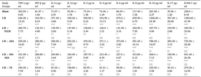
Plasma levels of TNF-α, interferon-gamma (IFN-Ɣ), interleukin 1
Table 2
Biochemical parameters in plasma and intestinal tissues of the study groups. Values are presented as mean ± SEM.
Sham I/R I/R +
FLUR I/R +ASA I/R +NO-ASA I/R +TS Plasma LDH (U/ L) 240.20 ±12.48 916.30 ±70.16 *** 676.50 ±36.26 *** †† 548.00 ±30.07 *** ††† 499.90 ±24.40 *** ††† 349.10 ±21.27 * ††† Plasma AST (U/ L) 27.30 ±1.30 84.25 ±8.97 *** 87.91 ±5.98 *** † 71.13 ±7.93 *** †† 70.10 ±6.59 *** †† 33.18 ±3.40 *** ††† Plasma ALT (U/ L) 133.20 ±3.02 202.40 ±8.43 *** 181.90 ±8.74 *** † 171.4 ±6.89 ** †† 151.60 ±3.23 ††† 136.00 ±6.29 ††† Tissue MDA (nmol/ mg protein) 7.43 ± 0.42 38.49 ±3.20 *** 12.19 ±1.23 ††† 22.16 ±0.93 *** ††† 16.68 ±1.67 ** ††† 8.62 ± 0.72 ††† Tissue GSH (nmol/ mg protein) 24.12 ±0.53 4.69 ± 0.25 *** 11.86 ±0.82 *** ††† 12.55 ±0.62 *** ††† 15.69 ±1.26 *** ††† 20.54 ±0.95 ††† Tissue GSSG (nmol/ mg protein) 0.63 ± 0.026 1.68 ±0.04 *** 1.40 ± 0.05 *** 1.29 ± 0.05 *** † 0.78 ± 0.04 ††† 0.65 ± 0.04 ††† Tissue GSH/ GSSG 38.34 ±0.76 2.79 ± 0.13 *** 8.46 ± 0.57 *** ††† 9.63 ± 0.56 *** ††† 20.44 ±2.31 *** ††† 32.51 ±2.80 ††† Tissue SOD (nmol/ mg protein) 26.71 ±0.63 6.932 ±0.22 *** 22.63 ±1.50 *††† 15.44 ±0.86 *** ††† 16.86 ±1.23 *** ††† 24.7 ± 0.92 ††† Tissue CAT (nmol/ mg protein) 8.62 ± 0.19 3.22 ±0.30 *** 5.954 ±0.54 ** ††† 4.37 ± 0.25 *** † 5.86 ± 0.36 ** †† 6.54 ± 0.51 ** ††† Tissue GR (nmol/ mg protein) 49.39 ±2.98 12.16 ±1.18 *** 40.91 ±1.20 * ††† 21.84 ±1.63 *** † 35.69 ±1.76 ** ††† 47.88 ±1.95 ††† Tissue GST (U/ mg protein) 5.36 ± 0.42 2.12 ±0.08 *** 3.79 ± 0.27 ** †† 3.37 ± 0.39 ** † 4.74 ± 0.44 ††† 4.95 ± 0.15 ††† Tissue GPx (U/ mg protein) 4.95 ± 0.13 1.64 ±0.24 *** 3.435 ±0.43 ** †† 2.612 ±0.30 *** † 3.715 ±0.13 * ††† 4.31 ± 0.33 ††† Tissue MPO activity (U/mg protein) 0.38 ± 0.032 2.78 ±0.08 *** 1.62 ± 0.11 *** ††† 1.47 ± 0.08 *** ††† 1.26 ± 0.04 *** ††† 0.44 ± 0.06 ††† Tissue XO activity (nmol/ mg protein) 0.64 ± 0.04 1.73 ±0.04 *** 1.40 ± 0.08 *** † 1.51 ± 0.03 *** 1.32 ± 0.03 *** †† 0.83 ± 0.05 *††† Tissue NO level (μM) 0.64 ± 0.04 1.73 ±0.04 *** 1.40 ± 0.08 *** † 1.41 ± 0.03 *** † 1.52 ± 0.03 *** † 0.83 ± 0.05 *††† Tissue NF- κB level (ng/mg protein) 0.36 ± 0.02 1.38 ±0.04 *** 1.22 ± 0.05 *** 1.17 ± 0.03 *** † 1.32 ± 0.12 *** 0.44 ± 0.04 †††
***p < 0.001 vs Sham; **p < 0.01 vs. Sham; *p < 0.05 vs Sham; †††p < 0.001 vs I/R; ††p < 0.01 vs I/R; †p < 0.05 vs I/R.
alpha (IL-1-α), IL-1β, IL-2, IL-4, IL-6, IL-8, IL-10, IL-17 and intercellular
adhesion molecule-1 (ICAM-1) were markedly increased in I/R group when compared with that of Sham group and diminished in the treat-ment groups. The most reduction was observed in TS + I/R group (Table 3).
3.12. Plasma intestinal fatty acid-binding protein (I-FABP) level Plasma I-FABP level in I/R group was found to be significantly higher than that of Sham group. This level was significantly lowered in the treatment groups when compared with that of I/R group. Treatment with TS caused the most decrease in plasma I-FABP levels Fig. 7.
4. Discussion
ROS formation during I/R leads to impairment of the microvascular structure of enterocytes, edema, release of inflammatory cytokines into the circulation, complement system activation, and PMNs infiltration to the affected site [8,11]. Loss of integrity of the intestinal functional barrier generates bacterial translocation, systemic inflammatory response, sepsis, shock, and multiple organ failure (MOF) [5,6,8,9]. COX-1 and COX-2 expressions are shown to be altered in I/R injury, and COX inhibitors are suggested to exhibit marked improvement in I/R [6, 15–18]. On the other hand, endothelium adjusts vascular tonus and platelet functions via secreting vasoactive mediators or autacoids, and recent studies showed that NO and ET releases from endothelium are crucial mediators in systemic inflammatory response syndrome pro-gressing to MOF [17].
The present study was designed to investigate and compare the beneficial effects of ASA (irreversible COX inhibitor), FLUR (reversible COX inhibitor), NO-ASA (NO-releasing NSAID) and TS (non-specific ERA) to overcome the consequences of intestinal I/R injury such as vasoconstriction, oxidative stress, inflammatory response, PMNs acti-vation and continuous cytokine release due to this response, loss of
integrity of the intestinal wall and eventually the bacterial translocation in the light of the information above.
SMABF difference recorded before and after the I/R was found to be higher in I/R group and significantly lowered in the treatment groups of I/R + NO-ASA and I/R + TS in accordance with the previous reports suggesting that altered mesenteric circulation and diminished blood flow decrease the oxygen delivery to visceral organs. Vasodilators and ET receptor antagonists may be useful in combating the persistent vasospasm that occurs in acute mesenteric ischemia and leads to microvascular flow improvement, sinusoidal dilatation, and reduced intravascular cell adhesion [19]. Thus, TS was found to be the most effective one among the drugs.
W/D ratio was increased in tissue samples of I/R group and decreased in the treatment groups. TS administration markedly reduced the W/D ratio. Histopathological examination in intestinal tissue sam-ples showed that Chiu score was higher in I/R group compared to Sham group. I/R + TS and I/R + FLUR groups had the most significant his-topathological improvement. The present study is the first one sug-gesting that TS administration remarkably recovers the impaired histological scores following intestinal I/R injury, possibly due to the improvements in vascular congestion, edema, and leukocyte infiltration. The previous studies showed that the integrity of cell walls lost during ischemia leads to epithelial dysfunction, increases intestinal permeability, and provokes translocation of bacteria through MLN and intra-abdominal organs. It is also reported that endotoxins, which are transported to circulation due to the intestinal permeability change, trigger cytokine production during I/R and lead to MOF eventually [4, 20].
In the present study, counts of microorganisms in tissues were significantly higher in I/R group. Gene expression of 16S rRNA in tissues was also increased in I/R group. These parameters were decreased in the treatment groups, whereas TS was found to be the most effective drug to reverse the bacterial translocation caused by intestinal I/R. A previous report postulating that ET-1 protects the cellular integrity of intestinal mucosa [21] supports our finding.
Elevated plasma LDH, AST and ALT activities in I/R group, which were previously reported as an indication of the contribution of remote organ damage to intestinal I/R injury [22], were found to be decreased in the treatment groups; TS appeared as the most potent drug in this view.
Tissue MDA level was significantly increased in I/R group and decreased in treatment groups. It has been postulated that COX-2 se-lective inhibitors may have protective effects in I/R by reducing oxidative damage [23,24]. Many NSAIDs are suggested to act as ROS scavengers. NO-ASA decreased lipid peroxidation better than ASA in our study, which is coherent with the previous studies suggesting that NO-ASA is more beneficial than ASA in terms of diminishing oxidative stress in peripheral I/R [25,26]. TS administration remarkably decreased lipid peroxidation in agreement with the earlier reports sug-gested that ET-1 induces oxidative stress by producing prostacyclin and NO via ETB and promotes vasodilation [27].
Tissue GSH content, GSH/GSSG ratio, and antioxidant enzyme levels were reduced; GSSG content was increased in I/R group, and all these parameters were recovered in the treatment groups. TS appeared as the most effective drug for the improvement of the redox balance during intestinal I/R. COX inhibitors decrease oxidative stress via inhibiting the triggering effects of COX-2 on ROS production, which leads to vascular inflammation and endothelial dysfunction [27] while ET-1 has been shown to disrupt the vasomotor functions and causes endothelial dysfunction through ROS production [27].
XO activity of I/R group was found as significantly higher than that of Sham group; and lower in the treatment groups. The most reduction was detected in I/R + TS group. It was demonstrated that ATP catabo-lizes into hypoxanthine, and NAD-reducing XOD transforms into XO during I/R. XO produces oxygen radicals leading to lipid peroxidation, facilitates granulocytes to adhere to microvascular endothelium,
Fig. 6. Gene expression of TNF-α in intestinal tissue samples. Values are pre-sented as mean ± SEM (n = 6). (***p < 0.001 vs Sham; **p < 0.01 vs Sham; *p <0.05 vs Sham; ψψψp<0.001 vs I/R; ψψp<0.01 vs I/R; ψp < 0.05 vs I/R).
enhances cell injury, and induces cytokine cascades and expressions of adhesion proteins, such as P-selectin and ICAM-1 and increases the production of NO [11,29].
It is known that activation of ETA receptor evokes vasoconstriction, whereas activation of ETB receptor leads to vasodilation via the release of NO or prostacyclin. Treatment with an ETA receptor antagonist has been demonstrated to improve glomerular dysfunction in ischemia [12]. It was also suggested that elevation of ET-1 during I/R activates NADPH oxidase/XO and NOS, resulting in ROS production, and increased vasoconstriction [30]. Our findings demonstrate that TS treatment significantly improved the intestinal I/R injury, suggesting that the ET receptor blockade maintains endothelial NO production, which may contribute to tissue protection.
Tissue MPO activity was elevated in I/R group and decreased in the treatment groups. TS was found to be the most effective drug in accor-dance with the earlier studies indicating that MPO released from the infiltrating neutrophils during I/R increases the ETB expression and contributes to ET-mediated vasoconstriction [31].
The role of NO in I/R is still a controversy. It is suggested that proinflammatory cytokines induce iNOS in many pathological condi-tions, and imbalanced NO production is involved particularly in in-flammatory diseases due to its role in oxidative stress and tissue damage [32]. On the other hand, it was suggested that NO decreases mucosal damage in intestinal I/R by reducing ROS production and destructive enzyme release, and preventing PMNs to reach the ischemic region by inhibiting the expression of endothelial adhesion molecules, such as ICAM and VCAM [6,8]. In the present study, NO levels were significantly increased in I/R group and decreased in the treatment groups. Inter-estingly, tissue NO level in I/R + NO-ASA group did not differ from that of the other treatment groups indicating that NO released from NO-ASA has no influence on the tissue NO level. This level was markedly diminished in I/R + TS group, even approaching the levels of Sham group, which is in accordance with some previous reports suggesting that increased NO and peroxynitrite levels in intestinal I/R were decreased by using ET receptor blockers [33].
Inflammation is the major response to I/R, and it is reported that intestinal I/R induces disruption of the intestinal mucosal barrier which allows bacterial translocation which initiates a systemic inflammatory response and activation of inflammatory mediators including cytokines, chemokines, TNF-α, NO and complement factors [11,28]. TNF-α,
pro-duced by macrophage-monocytes during acute inflammation in I/R injury, is shown to be responsible for the generation of ROS, disruption of microcirculation, and production of other inflammatory mediators [34]. In the present study, intestinal tissue expression and the plasma level of TNF-α were significantly increased in I/R group than those of
Sham group while these parameters were decreased in the treatment groups. Along with the increased expression and plasma levels of TNF-α,
Plasma levels of IFN-Ɣ, IL-1α, IL-1β, IL-2, IL-4, IL-6, IL-8, IL-10, IL-17,
NF-κB and ICAM-1 are also elevated in I/R group and decreased in the treatment groups. The most diminishing effect in proinflammatory mediators was observed in I/R + TS group. Data was in accordance with the previous reports showed that in intestinal I/R, tissue proin-flammatory cytokine expressions were markedly increased with facili-tating the influx of neutrophils into I/R-damaged villus tips causing severe I/R damage [35]. The significant increase in plasma level of IL-17 is also in agreement with the recent reports demonstrating that bacterial translocation is linked to the over-expression of IFN-γ, IL-4, IL-17, which might facilitate the intestinal permeability and leads to organ damage [36–38]. Besides, NF-κB signaling is crucial for inflammatory responses to microorganisms by activating innate and adaptive immune cells and
Table 3
Plasma proinflammatory cytokines and chemokines in study groups. Data are presented as mean ± SEM. Study
Groups TNF-α pg/
ml IFN-Ɣ pg/ ml IL-1 α pg/
ml IL-1β pg/ ml IL-2 pg/m IL-4 pg/ml IL-6 pg/ml IL-8 pg/ml IL-10 pg/ml IL-17 pg/ ml ICAM-1 ng/ ml
Sham 203.50 ± 6.83 81.97 ±1.66 71.07 ±2.91 93.96 ±1.78 73.34 ±1.48 1.29 74.46 ± 86.59 ±1.70 117.40 ±1.58 201.90 ±2.37 35.96 ±1.20 238.0 ±8.44 I/R 646.40 ± 15.22 *** 410.30 ± 8.23 *** 371.40 ± 4.86 *** 449.20 ± 5.29 *** 309.90 ± 6.23 *** 316.90 ± 13.13 *** 679.0 ± 3.112 *** 659.90 ± 6.75 *** 1206.00 ± 24.38 *** 357.20 ± 20.68 *** 1384.00 ± 41.99 *** I/R + FLUR 282.70 ±7.73 * ††† 132.10 ± 4.80 * ††† 145.90 ± 2.06 ** ††† 147.80 ± 5.18 ** ††† 138.60 ± 2.44 ** 126.0 ± 1.51 * ††† 141.40 ± 3.16 * ††† 322.90 ± 7.50 *** ††† 357.70 ± 4.90 * ††† 104.90 ± 2.80 * ††† 429.30 ± 35.66 ** ††† I/R + ASA 529.20 ± 16.81 *** † 280.50 ± 7.47 *** †† 300.90 ± 7.59 *** † 255.20 ± 3.83 *** ††† 270.30 ± 9.73 *** †† 271.1 ± 3.52 *** 373.90 ± 3.82 *** ††† 491.30 ± 10.14 *** †† 788.30 ± 10.07 *** †† 284.30 ± 3.15 *** †† 735.50 ± 35.68 *** ††† I/R + NO- ASA 431.90 ±19.17 *** †† 200.80 ± 1.72 *** ††† 204.80 ± 4.64 *** ††† 184.40 ± 2.07 *** ††† 187.70 ± 5.09 *** ††† 215.40 ± 8.39 *** † 257.0 ± 6.07 *** ††† 415.80 ± 6.15 *** †† 568.4 ± 7.13 *** ††† 160.90 ± 2.71 *** ††† 651.40 ± 26.38 *** ††† I/R + TS 240.30 ± 7.94 ††† 89.69 ± 1.64 ††† 81.50 ± 0.96 ††† 108.80 ± 1.84 ††† 82.95 ± 2.26 ††† 81.51 ± 1.17 ††† 98.00 ± 1.86 ††† 135.80 ± 1.81 ††† 225.50 ± 4.98 ††† 45.33 ± 0.86 ††† 279.30 ± 12.05 ††† ***p < 0.001 vs. Sham; **p < 0.01 vs. Sham; *p < 0.05 vs. Sham; †††p < 0.001 vs. I/R; ††p < 0.01 vs. I/R; †p < 0.05 vs. I/R.
Fig. 7. Plasma I-FABP level. Values are presented as mean ± SEM (n = 6).
suggesting that NF-κB is activated in I/R [39,40]. Reports suggesting that ET-1 induces proinflammatory mechanisms by increasing super-oxide anion production, activating NF-κB and increasing the expression of proinflammatory cytokines, especially through NO production and TNF-α release from macrophages [41], TS prevents the transcriptional
activity of NF-κB [42]; COX-2 inhibition suppresses the inflammatory cascade in I/R injury [17], and NO-releasing NSAIDs reduces NF-kB and TNF-α levels [43] support our data.
Plasma I-FABP level of I/R group was found six times higher than the level of Sham group, reduced in the treatment groups. TS was the most effective drug.
5. Conclusion
Our cumulative results encourage us to design further studies with NO-releasing NSAIDs and ETA/ETB antagonists to clarify the efficacy of these drugs in I/R injury, and to target a combination of them to the damaged tissue via a nanocarrier system.
Provenance and peer review
Not commissioned, externally peer-reviewed.
Data statement
We state here that all data concerning to the present study are reproducable and transparent. However, it is not possible to present all of the raw data in the article since all of our the raw data are kept for sharing with the researches upon request.
CRediT authorship contribution statement
Bercis Imge Ucar: Writing - original draft, Conceptualization,
Formal analysis, Investigation, Visualization. Acelya Erikci: Data curation, Methodology, Formal analysis, Investigation. Kemal
Kose-mehmetoglu: Data curation, Methodology, Formal analysis,
Investiga-tion. Ceren Ozkul: Data curation, Methodology, Formal analysis, Investigation. Alper Bektas Iskit: Conceptualization, Writing - review & editing, Methodology, Formal analysis, Investigation. Gulberk Ucar: Writing - original draft, Conceptualization, Methodology, Project administration, Funding acquisition, Data curation, Investigation.
Sez-gin Zeren: Supervision, Writing - review & editing, Visualization. Declaration of competing interest
The authors declare that they have no known competing financial interests or personal relationships that could have appeared to influence the work reported in this paper.
Appendix A. Supplementary data
Supplementary data to this article can be found online at https://doi. org/10.1016/j.ijsu.2020.08.061.
References
[1] D. Alexandropoulos, G.V. Bazigos, I.P. Doulamis, A. Tzani, P. Konstantopoulos, N. Tragotsalou, A. Kondi-Pafiti, T. Kotsis, N. Arkadopoulos, V. Smyrniotis, D. N. Perrea, Protective effects of N-acetylcysteine and atorvastatin against renal and hepatic injury in a rat model of intestinal ischemia-reperfusion, Biomed. Pharmacother. 89 (2017) 673–680, https://doi.org/10.1016/j. biopha.2017.02.086.
[2] P. Stringa, N. Lausada, D. Romanin, E. Portiansky, C. Zanuzzi, M. Machuca, G. Gondolesi, M. Rumbo, Pretreatment combination reduces remote organ damage secondary to intestinal reperfusion injury in mice: Follow-up study, Transplant. Proc. 48 (2016) 210–216, https://doi.org/10.1016/j.transproceed.2015.12.002. [3] S.H. Wen, Y.H. Ling, Y. Li, C. Li, J.X. Liu, Y.S. Li, X. Yao, Z.Q. Xia, K.X. Liu,
Ischemic postconditioning during reperfusion attenuates oxidative stress and intestinal mucosal apoptosis induced by intestinal ischemia/reperfusion via aldose
reductase, Surgery 153 (2013) 555–564, https://doi.org/10.1016/j. surg.2012.09.017.
[4] H. Nakagawa, N. Tsunooka, Y. Yamamoto, M. Yoshida, T. Nakata, K. Kawachi, Pitavastatin prevents intestinal ischemia/reperfusion-induced bacterial translocation and lung injury in atherosclerotic rats with hypoadiponectinemia, Surgery 145 (2009) 542–549, https://doi.org/10.1016/j.surg.2009.01.002. [5] A. de P.L. Oliveira, J. da P. Piccoli-Rangel, B.S. Monteiro, Pathophysiology of the
intestinal ischemic reperfusion injury, Glob. J. Anim. Sci. Res. 2 (2014) 351–356.
http://archives.gjasr.com/index.php/GJASR/article/view/83.
[6] ´I.M. Azevedo, I. Araújo-Filho, A.C.M. Rˆego, V.B. Medeiros, M.D.F. Carvalho, E.S. T. Egito, A.C. Medeiros, Bacterial translocation in rats treated with simvastatin undergoing intestinal ischemia and reperfusion, J. Surg. Clin. Res. 1 (2011),
https://doi.org/10.20398/jscr.v1i1.930, 54.
[7] W. Berland, Andrew Oldenburg, acute mesenteric ischemia, Curr. Treat. Options Gastroenterol. 11 (2008) 3–10, https://doi.org/10.1007/s11938-008-0001-2. [8] Y. Takizawa, H. Kishimoto, T. Kitazato, M. Tomita, M. Hayashi, Effects of nitric
oxide on mucosal barrier dysfunction during early phase of intestinal ischemia/ reperfusion, Eur. J. Pharmaceut. Sci. 42 (2011) 246–252, https://doi.org/10.1016/ j.ejps.2010.11.016.
[9] N. Ulu, A.B. ˙Iskit, C. S¨okmensüer, M.O. Güç, The effects of aspirin, flurbiprofen, and NO-donating acetylsalicylic acid (NCX 4016) on mice models of endotoxic and septic shock, Turk. J. Med. Sci. 45 (2015) 812–819, https://doi.org/10.3906/sag- 1402-82.
[10] K. Sinha, P.C. Sil, Targeting oxidative stress and inflammation in NSAIDs induced gastropathy: a plausible therapeutic approach, Inflamm. Cell Signal. (2015),
https://doi.org/10.14800/ics.763.
[11] R.O.S. Soares, D.M. Losada, M.C. Jordani, P. ´Evora, O. Castro-E-Silva, Ischemia/ reperfusion injury revisited: an overview of the latest pharmacological strategies, Int. J. Mol. Sci. 20 (2019), https://doi.org/10.3390/ijms20205034.
[12] S.M. Wilhelm, N.T. Stowe, A. V Robinson, J.A. Schulak, The use of the endothelin receptor antagonist, Tezosentan, before or after renal ischemia protects renal function, Transplantation 71 (2001) 211–216, https://doi.org/10.1097/ 00007890-200101270-00007.
[13] J. Dingemanse, M. Clozel, P.L.M. Van Giersbergen, Pharmacokinetics and pharmacodynamics of tezosentan, an intravenous dual endothelin receptor antagonist, following chronic infusion in healthy subjects, Br. J. Clin. Pharmacol. 53 (2002) 355–362, https://doi.org/10.1046/j.1365-2125.2002.01158.x. [14] C. Kilkenny, W.J. Browne, I.C. Cuthill, M. Emerson, D.G. Altman, Improving
bioscience Research reporting: the ARRIVE guidelines for reporting animal Research, PLoS Biol. 8 (6) (2010), e1000412.
[15] H. Fu, H. Chen, C. Wang, H. Xu, F. Liu, M. Guo, Q. Wang, X. Shi, Flurbiprofen, a cyclooxygenase inhibitor, protects mice from hepatic ischemia/reperfusion injury by inhibiting GSK-3β signaling and mitochondrial permeability transition, Mol. Med. 18 (2012) 1128–1135, https://doi.org/10.2119/molmed.2012.00088. [16] M. Karthikeyan, K. Deepa, Therapeutic applications of nitric oxide releasing non
steroidal anti-inflammatory drugs, J. Chem. Pharmaceut. Res. 1 (2009) 134–147.
www.jocpr.com.
[17] R.H. Tolba, N. Fet, K. Yonezawa, K. Taura, A. Nakajima, K. Hata, Y. Okamura, H. Uchinami, U. Klinge, T. Minor, Y. Yamaoka, Y. Yamamoto, Role of preferential cyclooxygenase-2 inhibition by meloxicam in ischemia/reperfusion injury of the rat liver, Eur. Surg. Res. 53 (2014) 11–24, https://doi.org/10.1159/000362411. [18] S.L. Bourque, S.T. Davidge, M.A. Adams, The interaction between endothelin-1 and
nitric oxide in the vasculature: new perspectives, Am. J. Physiol. Regul. Integr. Comp. Physiol. 300 (2011) 1288–1295, https://doi.org/10.1152/
ajpregu.00397.2010.
[19] D.G. Clair, J.M. Beach, Mesenteric ischemia, N. Engl. J. Med. 374 (2016) 959–968,
https://doi.org/10.1056/NEJMra1503884.
[20] C. Vaishnavi, Translocation of gut flora and its role in sepsis, Indian J. Med. Microbiol. 31 (2013) 334–342, https://doi.org/10.4103/0255-0857.118870. [21] Y.S. Li, Z.X. Wang, C. Li, M. Xu, Y. Li, W.Q. Huang, Z. Xia, K.X. Liu, Proteomics of
ischemia/reperfusion injury in rat intestine with and without ischemic postconditioning, J. Surg. Res. 164 (2010), https://doi.org/10.1016/j. jss.2009.10.003.
[22] S. Bertoni, V. Ballabeni, E. Barocelli, M. Tognolini, Mesenteric ischemia- reperfusion: an overview of preclinical drug strategies, Drug Discov. Today 23 (2018) 1416–1425, https://doi.org/10.1016/j.drudis.2018.05.034.
[23] E. Candelario-Jalil, A. Gonz´alez-Falc´on, M. García-Cabrera, D. ´Alvarez, S. Al- Dalain, G. Martínez, O.S. Le´on, J.E. Springer, Assessment of the relative contribution of COX-1 and COX-2 isoforms to ischemia-induced oxidative damage and neurodegeneration following transient global cerebral ischemia,
J. Neurochem. 86 (2003) 545–555, https://doi.org/10.1046/j.1471- 4159.2003.01812.x.
[24] B. Zarrouki, A.F. Soares, M. Guichardant, M. Lagarde, A. G´elo¨en, The lipid peroxidation end-product 4-HNE induces COX-2 expression through p38MAPK activation in 3T3-L1 adipose cell, FEBS Lett. 581 (2007) 2394–2400, https://doi. org/10.1016/j.febslet.2007.04.048.
[25] A.M.L. Mouithys-Mickalad, S.X. Zheng, G.P. Deby-Dupont, C.M.T. Deby, M. M. Lamy, J.Y.Y. Reginster, Y.E. Henrotin, In vitro study of the antioxidant properties of non steroidal anti-inflammatory drugs by chemiluminescence and electron spin resonance (ESR), Free Radic. Res. 33 (2000) 607–621, https://doi. org/10.1080/10715760000301131.
[26] C. Emanueli, S. Van Linthout, M.B. Salis, A. Monopoli, P. Del Soldato, E. Ongini, P. Madeddu, Nitric oxide-releasing aspirin derivative, NCX 4016, promotes reparative angiogenesis and prevents apoptosis and oxidative stress in a mouse model of peripheral ischemia, Arterioscler. Thromb. Vasc. Biol. 24 (2004) 2082–2087, https://doi.org/10.1161/01.ATV.0000144030.39087.3b.
[27] F. Dong, X. Zhang, L.E. Wold, Q. Ren, Z. Zhang, J. Ren, Endothelin-1 enhances oxidative stress, cell proliferation and reduces apoptosis in human umbilical vein endothelial cells: role of ET B receptor, NADPH oxidase and caveolin-1, Br. J. Pharmacol. 145 (2005) 323–333, https://doi.org/10.1038/sj.bjp.0706193. [28] M. Mu˜noz, A. S´anchez, M. Pilar Martínez, S. Benedito, M.E. L´opez-Oliva, A. García-
Sacrist´an, M. Hern´andez, D. Prieto, COX-2 is involved in vascular oxidative stress and endothelial dysfunction of renal interlobar arteries from obese Zucker rats, Free Radic. Biol. Med. 84 (2015) 77–90, https://doi.org/10.1016/j. freeradbiomed.2015.03.024.
[29] M.Y. Wu, G.T. Yiang, W.T. Liao, A.P.Y. Tsai, Y.L. Cheng, P.W. Cheng, C.Y. Li, C. J. Li, Current mechanistic concepts in ischemia and reperfusion injury, Cell. Physiol. Biochem. 46 (2018) 1650–1667, https://doi.org/10.1159/000489241. [30] E. Dabbs Loomis, J.C. Sullivan, D.A. Osmond, D.M. Pollock, J.S. Pollock,
Endothelin mediates superoxide production and vasoconstriction through activation of NADPH oxidase and uncoupled nitric-oxide synthase in the rat aorta, J. Pharmacol. Exp. Therapeut. 315 (2005) 1058–1064, https://doi.org/10.1124/ jpet.105.091728.
[31] D. Lau, K. Sz¨ocs, A. Klinke, S. Baldus, Myeloperoxidase modulates endothelin receptor type B expression, Free Radic. Biol. Med. 49 (2010), https://doi.org/ 10.1016/j.freeradbiomed.2010.10.404. S145.
[32] E. Barocelli, V. Ballabeni, P. Ghizzardi, F. Cattaruzza, S. Bertoni, C.A.M. Lagrasta, M. Impicciatore, The selective inhibition of inducible nitric oxide synthase prevents intestinal ischemia-reperfusion injury in mice, Nitric Oxide - Biol. Chem. 14 (2006) 212–218, https://doi.org/10.1016/j.niox.2005.11.006.
[33] S¸.K. ¨Ozel, M. Yüksel, G. Haklar, Ç.U. Durakbas¸a, T.E. Dagli, A.¨O. Aktan, Nitric oxide and endothelin relationship in intestinal ischemia/reperfusion injury (II), Prostaglandins Leukot. Essent. Fat. Acids. 64 (2001) 253–257, https://doi.org/ 10.1054/plef.2001.0268.
[34] X. Gao, H. Zhang, S. Belmadani, J. Wu, X. Xu, H. Elford, B.J. Potter, C. Zhang, Role of TNF-α-induced reactive oxygen species in endothelial dysfunction during reperfusion injury, Am. J. Physiol. Heart Circ. Physiol. 295 (2008), https://doi. org/10.1152/ajpheart.00587.2008.
[35] J. Grootjans, K. Lenaerts, J.P.M. Derikx, R.A. Matthijsen, A.P. De Bruïne, A.A. Van Bijnen, R.M. Van Dam, C.H.C. Dejong, W.A. Buurman, Human intestinal ischemia-
reperfusion-induced inflammation characterized: Experiences from a new translational model, Am. J. Pathol. 176 (2010) 2283–2291, https://doi.org/ 10.2353/ajpath.2010.091069.
[36] M.A.R.C. Daemen, T.G.A.M. Wolfs, W.A. Buurman, Ischemia/reperfusion-induced IFN-γ up-regulation: involvement of IL-12 and IL-18, J. Immunol, 162 (n.d.) 5506–5510, https://www.jimmunol.org/content/162/9/5506.short. [37] A.S. Farivar, B. Krishnadasan, B.V. Naidu, S.M. Woolley, E.D. Verrier, M.
S. Mulligan, Endogenous interleukin-4 and interleukin-10 regulate experimental lung ischemia reperfusion injury, Ann. Thorac. Surg. 76 (2003) 253–259, https:// doi.org/10.1016/S0003-4975(03)00335-7.
[38] N.C. Nüssler, A.R. Müller, H. Weidenbach, A. Vergopoulos, K.P. Platz, H.D. Volk, P. Neuhaus, A.K. Nussler, IL-10 increases tissue injury after selective intestinal ischemia/reperfusion, Ann. Surg. 238 (2003) 49–58, https://doi.org/10.1097/ 00000658-200307000-00007.
[39] M. Geha, M.G. Tsokos, R.E. Bosse, T. Sannikova, Y. Iwakura, J.J. Dalle Lucca, R. De Waal Malefyt, G.C. Tsokos, IL-17A produced by innate lymphoid cells is essential for intestinal ischemia-reperfusion injury, J. Immunol. 199 (2017) 2921–2929,
https://doi.org/10.4049/jimmunol.1700655.
[40] N. Vega-Maga˜na, V. Delgado-Rizo, L. García-Benavides, S. Del Toro-Arreola, J. Segura-Ortega, A.S.M.Z. Morales, J.S. Zepeda-Nu˜no, M. Escarra-Senmarti, J. Guti´errez-Franco, J. Haramati, M.R. Bueno-Topete, Bacterial translocation is linked to increased intestinal IFN-γ, IL-4, IL-17, and mucin-2 in cholestatic rats, Ann. Hepatol. 17 (2018) 318–329, https://doi.org/10.5604/01.3001.0010.8662. [41] A. Kowalczyk, P. Kleniewska, M. Kolodziejczyk, B. Skibska, A. Goraca, The role of
endothelin-1 and endothelin receptor antagonists in inflammatory response and sepsis, Arch. Immunol. Ther. Exp. 63 (2015) 41–52, https://doi.org/10.1007/ s00005-014-0310-1.
[42] M. Iribarne, V. Torbidoni, M. Casta˜neda, Blockade of endothelinergic receptors reduces stress pathway signaling in endotoxin-induced uveitis, Invest. Ophthalmol. Vis. Sci, 48 (n.d. https://iovs.arvojournals.org/article.aspx?articleid=2387837. [43] M. Chattopadhyay, S. Goswami, D.B. Rodes, R. Kodela, C.A. Velazquez, D. Boring,
J.A. Crowell, K. Kashfi, NO-releasing NSAIDs suppress NF-κB signaling in vitro and in vivo through S-nitrosylation, Canc. Lett. 298 (2010) 204–211, https://doi.org/ 10.1016/j.canlet.2010.07.006.