http://journals.tubitak.gov.tr/medical/ © TÜBİTAK
doi:10.3906/sag-1411-77
Evaluation of brain perfusion in Alzheimer disease with perfusion computed
tomography and comparison to elderly patient without dementia
Tülin YILDIRIM1, Başak KARAKURUM GÖKSEL2,*, Şenay DEMİR3, Naime TOKMAK3, Meliha TAN2
1Department of Radiology, Faculty of Medicine, Başkent University, Ankara, Turkey
2Department of Neurology, Başkent University Adana Medical and Teaching Research Center, Adana, Turkey 3Department of Radiology, Başkent University Adana Medical and Teaching Research Center, Adana, Turkey
1. Introduction
Alzheimer disease (AD) occurs in more than half of patients with dementia and is a progressive, degenerative neurological disorder (1). The aging population is increasing and AD is becoming an increasingly important public health problem. Diagnosis of AD is made via a combination of clinical history, the results of physical and neurological examinations, and neuroimaging and laboratory evaluations (2). Neuropathological studies suggest that evidence of AD may be present in the brain years before the onset of clinical symptoms. Identification of AD at the earliest possible time is crucial for optimal care and treatment. Therefore, recent studies have focused on prodromal AD (3).
Neuroimaging studies are used in the diagnosis of dementia. Structural techniques such as computed tomography (CT) and magnetic resonance imaging (MRI) allow for evaluation of the degree of cerebral atrophy and detection of organic causes of dementia. Newer imaging techniques such as single photon-emission CT (SPECT),
positron emission tomography (PET), and perfusion MRI are useful to evaluate blood perfusion in AD dementia and other dementia groups (4–6). Although these techniques have been shown to be valuable, they are often not readily available. Perfusion CT (PCT) is another perfusion technique that provides rapid and minimally invasive assessment of brain perfusion. There are a few studies concerning PCT in which dementia has been reported (6–8). The goal of the present study was to evaluate the usefulness of PCT in assessing brain perfusion in patients with AD and to compare it with that of patients without dementia.
2. Materials and methods
This study was approved by the Institutional Review Board of our university hospital and written informed consent was obtained from all subjects. Twenty patients with AD were recruited among outpatients visiting the Neurology Department of our university. Clinical diagnosis of dementia was made according to criteria in the
DSM-Background/aim: The aim of this study was to evaluate perfusion computed tomography (PCT) findings in patients with Alzheimer
disease and to compare them with those of patients without dementia.
Materials and methods: PCT was performed in 35 patients: 20 with Alzheimer disease (mean age, 69.7 ± 5.5 years) and 15 control
subjects (mean age, 67.5 ± 3.5 years). Control subjects were elderly individuals with no cognitive problems who were admitted with headaches. All PCT examinations were performed on a 4-slice CT unit. The PCT analysis software program was used to calculate regional cerebral blood flow (rCBF), regional cerebral blood volume (rCBV), regional time-to-peak (rTTP) values in the bilateral frontal, temporal, and occipital cortices, and bilateral lentiform nucleus.
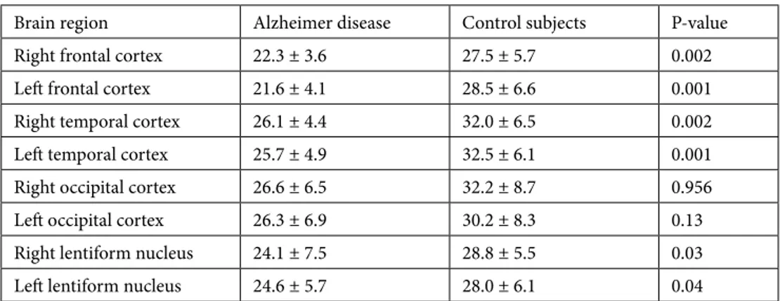
Results: rCBF values in the bilateral frontal and temporal cortices and bilateral lentiform nucleus were significantly lower in the patients
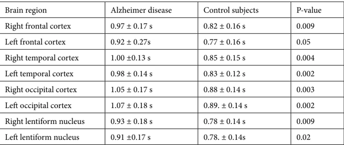
with Alzheimer disease than in the control subjects. There were no significant differences in rCBV values between Alzheimer disease and the control group. rTTP values in all cortical areas and bilateral lentiform nucleus were significantly higher in the patients with Alzheimer disease than in the control subjects.
Conclusion: PCT is a rapid and reliable imaging modality for evaluating brain perfusion in Alzheimer disease. Key words: Alzheimer disease, brain perfusion, perfusion computed tomography
Received: 14.11.2014 Accepted/Published Online: 06.07.2015 Final Version: 19.04.2016
IV and probable AD was determined by excluding other dementias in accordance with criteria from the National Institute of Neurological and Communicative Disorders and Stroke and the AD and Related Disorders Association (NINCDS-ADRDA). Patients showed mini-mental state examination (MMSE) scores ranging from 14 to 26. The MMSE is a standardized, 30-point scale that encompasses the following items: orientation to time (5 points), orientation to place (5 points), word registration (3 points), attention (5 points), word recall (3 points), naming (2 points), repetition (1 point), following commands (3 points), reading (1 point), writing (1 point), and pentagon copying (1 point).
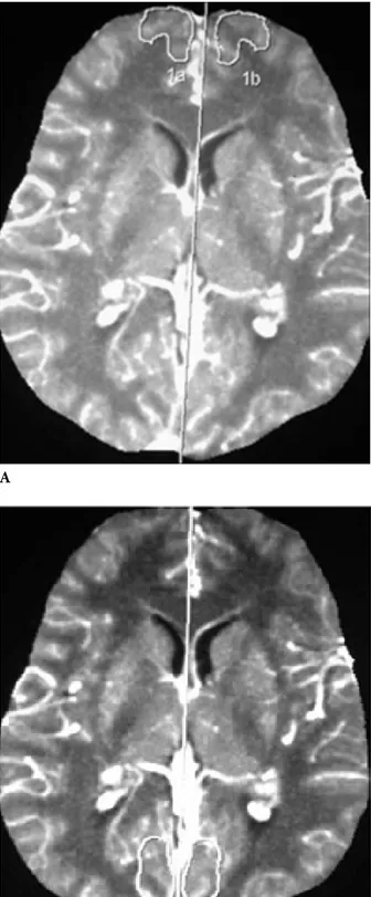
The 15 control subjects were healthy individuals with no cognitive problems who visited our outpatient clinic complaining of headaches and who showed normal results on cranial CT. All PCT examinations were done on the same 4-detector CT scanner (Sensation 4; Siemens). Routine whole-brain CT scans were done before and after PCT. A volume of 40 mL of iodinated contrast medium (370 mg iodine/mL) was injected via a 14-gauge cannula into the antecubital fossa with an automatic injector. The injection rate was 4 mL/s and the delay time was 5 s. Forty images were obtained from the level of the basal ganglia. Slice thickness was 10 mm. All perfusion images were postprocessed with PCT analysis software on a workstation (Wizard; Siemens). We used the standard convolution method for CT perfusion processing. Maximum intensity projection perfusion, cerebral blood flow (CBF), cerebral blood volume (CBV), and time to peak (TTP) maps were created. The region of interest (ROI) was placed superior to the sagittal sinus for the assessment of perfusion parameters. Relative CBF (rCBF), relative CBV (rCBV), and relative TTP (rTTP) values were measured symmetrically on maps. All symmetric ROIs were drawn by hand on the maximum intensity projection perfusion image by a single radiologist (T.Y.) in the bilateral frontal, temporal, and occipital cortices, and bilateral lentiform nucleus (Figures 1a, 1b, 1c, and 1d).
We investigated differences in sociodemographic features between the groups with the chi-square test for categorical variables. Quantitative data obtained from the frontal, temporal, and occipital cortices and the lentiform nucleus were compared by independent t-test. P < 0.05 was considered significant.
3. Results
Demographic features of the AD patients and control subjects are listed in Table 1. There were no significant differences between the two groups. Values for rCBF
significantly lower in the AD group than in the control group.
Values for rCBV are listed in Table 3. Values for the bilateral temporal cortex were lower in the AD group than in the control group, but these differences were not significant. Values obtained from bilateral frontal and occipital cortices and lentiform nucleus were not significantly different between the AD patients and the control group.
Values for rTTP are listed in Table 4. Values for the bilateral frontal, temporal, and occipital lobes, and the bilateral lentiform nucleus were significantly higher in the AD group than they were in the control group.
4. Discussion
CT is the most widely used and oldest imaging modality used to evaluate dementia patients. This technique has been used to determine reversible causes of dementia such as normal pressure hydrocephaly, chronic subdural hematoma, mass lesion (i.e. large meningioma), and infection. MRI is preferred to identify specific pathologies such as atrophy of the medial temporal lobe, hippocampal formation, or parahippocampal gyrus, each of which is highly predictive of AD (5).
Magnetic resonance spectroscopy is useful in the differential diagnosis and evaluation of progression of dementia. Findings of increased myo-inositol to creatine levels, increased choline to creatine levels, and decreased N-acetylaspartate to creatine levels in the medial temporal lobe, and to a lesser degree in the parietal lobe, are markers of early stage AD (9).
Volumetric studies evaluate volume loss in limbic structures such as the entorhinal cortex, hippocampus, amygdala, and hypothalamus. Magnetic resonance volumetric analysis may help in distinguishing patients with mild cognitive impairments (at risk for proceeding to AD) from normal elderly subjects (10).
Several studies showed cerebral perfusion in AD, other dementia groups, and mild cognitive impairment patients. Caroli et al (3) showed that parahippocampal and inferior temporal hypoperfusion in amnestic mild cognitive impairment patients correlated with conversion to AD. Some studies have also shown that decreased blood flow in the temporoparietal association area is common in early stage AD, before cerebral atrophy is detectable. As dementia progresses, blood flow decreases in the frontal lobe and is relatively well preserved in the pons, primary motor cortex, and primary visual cortex of the occipital lobe, basal ganglia, and thalamus (7,8,11–20).
contrast-Figure 1. MIP perfusion of pCT at the basal ganglia. A-Frontal, B-Temporal, C-Occipital, D-Lentiform nucleus with freehand drawn ROI. A C B D
decreased regional CBF observed by PET and SPECT. The specificity/sensitivity of 18F-fluorodeoxyglucose PET is limited; however, 15O H2O PET or SPECT can be used to show perfusion deficits (decreased regional CBF) in the hippocampus and temporal and parietal regions (6,8,18–20). Although volumetric studies and perfusion MRI, PET,
still limited due to availability and cost (16). PCT represents an inexpensive, noninvasive, and widely available tool that can be applied to measure brain perfusion quantitatively. This technique is usually used for evaluating decreased perfusion in patients with acute stroke. In addition, it can be used to detect brain tumor perfusion and brain perfusion
Table 1. Demographic features of the subjects included in the study.
Number Sex Age (years)
Alzheimer disease 20 12 F, 8 M 69.7 ± 5.5
Control subjects 15 11 F, 4 M 67.5 ± 3.5
P-value 0.350 0.091
M: male, F: female.
Table 2. Regional cerebral blood flow values determined by perfusion computed tomography in patients
with Alzheimer disease and control subjects.
Brain region Alzheimer disease Control subjects P-value
Right frontal cortex 22.3 ± 3.6 27.5 ± 5.7 0.002
Left frontal cortex 21.6 ± 4.1 28.5 ± 6.6 0.001
Right temporal cortex 26.1 ± 4.4 32.0 ± 6.5 0.002
Left temporal cortex 25.7 ± 4.9 32.5 ± 6.1 0.001
Right occipital cortex 26.6 ± 6.5 32.2 ± 8.7 0.956
Left occipital cortex 26.3 ± 6.9 30.2 ± 8.3 0.13
Right lentiform nucleus 24.1 ± 7.5 28.8 ± 5.5 0.03
Left lentiform nucleus 24.6 ± 5.7 28.0 ± 6.1 0.04
Table 3. Regional cerebral blood volume values determined by perfusion computed tomography in
patients with Alzheimer disease and control subjects.
Brain region Alzheimer disease Control subjects P-value
Right frontal cortex 36.2 ± 8.0 36.4 ± 5.8 0.93
Left frontal cortex 36.3 ± 9.5 37.5 ± 7.1 0.66
Right temporal cortex 40.7 ± 7.7 42.3 ± 6.4 0.49
Left temporal cortex 40.0 ± 9.5 42.6 ± 7.2 0.35
Right occipital cortex 42.1 ± 10.7 41.9 ± 8.5 0.95
Left occipital cortex 42.6 ± 8.4 39.5 ± 8.8 0.29
Right lentiform nucleus 35.2 ± 10.9 37.1 ± 4.2 0.49
Zimny et al. (19) performed PCT in 41 patients with AD, vascular dementia, or mixed dementia. They showed that rCBF and rCBV values in the grey and white matter of the bilateral frontal and temporal lobes were lower in patients with AD than in patients with vascular dementia; however, the authors found no significant difference in rCBF, rCBV, or rMTT between patients with AD and patients with mixed dementia. However, the authors did not include control subjects in that study. Streitparth et al. (18) found that the occipital and temporal blood volume decreased significantly with an increasing degree of dementia. They showed regional hypoperfusion in the frontal, basal ganglia, temporal, and occipital region in the group with AD when compared with the group of cognitively normal subjects. They suggested that PCT may be useful in differentiating severity and types of dementia. Zimny et al. (19) suggest that analysis of CBF and CBV parameters assessed in PCT can be useful in evaluating cognitive impairment in patients with dementia and may thus have a role in monitoring disease progression or therapeutic response.
In the present study, we aimed to show usefulness of PCT in evaluation of brain perfusion in AD. We included 15 cognitively normal control subjects and compared their perfusion findings with those of the AD group. Our study demonstrated cerebral cortical perfusion failure in AD by using PCT. We observed decreased rCBF in the frontal and temporal cortex and bilateral lentiform nucleus. It should be noted that perfusion parameters are relative values that depend on software programs and that they are lower than physiological values in patient and control groups.
It is known that CBF is the most important parameter for ischemia of PCT. It is measured in milliliters of blood per 100 g brain tissue/min. CBF values obtained from areas of the brain with high energy requirements such as the cortical surface or basal ganglia were higher than those from the white matter. In the AD group, diffuse
vascular disturbances affected large brain regions as compared with vascular dementia caused by focal vascular pathology (20). This result may be related to decreased rCBF values. CBF is controlled by changes in the vessel diameter (autoregulation) and local calculated rCBF can change depending on software program, injection speed, and cardiac function. rCBV is defined as the percentage of blood vessels in a specific volume of tissue. It is a functional parameter and alters with vessel size changes in the context of vascular autoregulation. In our study, there was no significant difference in rCBV values between the two groups. This finding may be explained with vessel size changes. We also observed increasing rTTP values in bilateral frontal, temporal, and occipital cortex, and lentiform nucleus. It is known that mean transit time (MTT) and TTP values indicate retarded perfusion, but these values are not generally specific. It is also known that rTTP values are affected by cardiac problems, but our perfusion program did not contain MTT maps. A metabolic decrease and hypoperfusion have been reported to occur in the posterior parietal cortex, temporal cortex, and limbic system in patients with AD. We also found hypoperfusion of the temporal lobes in patients with AD, as well as a decrease in regional CBF in the frontal lobes and bilateral lentiform nucleus, when compared with control subjects. Previous studies have shown that mild AD is localized more in the cingulate gyrus and that a more advanced disease is localized in the temporoparietal and frontal cortices (2,14). Tang et al. (22) showed that the CBV and CBF of the frontal and temporal lobe, hippocampus, and basal ganglia in patients with senile dementia were much lower, and the MTT and TTP of these areas were higher than those values in the healthy group at a low-dose perfusion program. We did not use this program in a 4-slice scanner, but our dose values were within safe limits.
Table 4. Time-to-peak values determined by perfusion computed tomography in patients with
Alzheimer disease and control subjects.
Brain region Alzheimer disease Control subjects P-value
Right frontal cortex 0.97 ± 0.17 s 0.82 ± 0.16 s 0.009
Left frontal cortex 0.92 ± 0.27s 0.77 ± 0.16 s 0.05
Right temporal cortex 1.00 ±0.13 s 0.85 ± 0.15 s 0.004
Left temporal cortex 0.98 ± 0.14 s 0.83 ± 0.12 s 0.002
Right occipital cortex 1.05 ± 0.17 s 0.88 ± 0.14 s 0.003
Left occipital cortex 1.07 ± 0.18 s 0.89. ± 0.14 s 0.002
Right lentiform nucleus 0.93 ± 0.18 s 0.78 ± 0.14 s 0.009
The patients in the present study had moderate to advanced AD; hypoperfusion of the frontal, temporal, and basal ganglia regions was observed. One limitation of the present study is its limited coverage, which is a disadvantage of perfusion CT. Perfusion evaluation at the hippocampal level and parietal lobes was not possible in this technique in our 4-slice CT scanner. Second, we could not evaluate the relationship between cognitive impairment according to
the MMSE and according to perfusion parameters. Further studies are needed to evaluate the role of PCT in monitoring disease progression and therapy response in patients with AD.
In conclusion, PCT is readily available and cost effective compared with other perfusion techniques for evaluation of brain perfusion in AD patients. rCBF values can be more useful in assessing hypoperfusion in the bilateral frontal and temporal lobes and the lentiform nucleus.
References
1. Golden R. Dementia and Alzheimer’s disease. Indications, diagnosis, and treatment. Minn Med 1995; 78: 25-29.
2. Bennett DA, Evans DA. Alzheimer’s disease. DM-Dis Mon 1992; 38: 1-64.
3. Caroli A, Testa C, Geroldi C, Nobili F, Barnden LR, Guerra UP, Bonetti M, Frisoni GB. Cerebral perfusion correlates of conversion to Alzheimer’s disease in amnestic mild cognitive impairment. J Neurol 2007; 254: 1698-1707.
4. Matsuda H. The role of neuroimaging in mild cognitive impairment. Neuropathology 2007; 27: 570-577.
5. Scheltens P, Korf ES. Contribution of neuroimaging in the diagnosis of Alzheimer’s disease and other dementias. Curr Opin Neurol 2000; 13: 391-396.
6. Zimny A, Sasiadek M, Leszek J, Czarnecka A, Trypka E, Kiejna A. Does perfusion CT enable differentiating Alzheimer’s disease from vascular dementia and mixed dementia? A preliminary report. J Neurol Sci 2007; 257: 114-120.
7. McKhann G, Drachman D, Folstein M, Katzman R, Price D, Stadlan EM. Clinical diagnosis of Alzheimer’s disease: report of the NINCDS-ADADRDA Work Group under the auspices of Department of Health and Human Services Task Force on Alzheimer’s Disease. Neurology 1984; 34: 939-944.
8. Diagnostic patterns of regional atrophy on MRI and regional cerebral blood flow change on SPECT on SPECT in young onset patients with Alzheimer’s disease, frontotemporal dementia and vascular dementia. Acta Neurol Scand 2002; 105: 261-269.
9. Kantarci K. H magnetic resonance spectroscopy in dementia. Brit J Radiol 2007; 80: 146-152.
10. Callen DJ, Black SE, Gao F, Caldwell CB, Szalai JP. Beyond the hippocampus: MRI volumetry confirms widespread limbic atrophy in Alzheimer disease. Neurology 2001; 57: 1669-1674.
11. Yoshikawa T, Murase K, Oku N, Imaizumi M, Takasawa M, Rishu P, Kimura Y, Ikejiri Y, Kitagawa K, Hori M, et al. Heterogeneity of cerebral blood flow in Alzheimer disease and vascular dementia. Am J Neuroradiol 2003; 24: 1341-1347.
12. Hanyu H, Shimizu S, Hirao K, Kanetaka H, Sakurai H, Iwamoto T, Koizumi K, Abe K. Differentiation of dementia with Lewy bodies from Alzheimer’s disease using Mini Mental State Examination and brain perfusion SPECT. J Neurol Sci
13. Du AT, Jahng GH, Hayasaka S, Kramer JH, Rosen HJ, Gorno-Tempini ML, Rankin KP, Miller BL, Weiner MW, Schuff N. Hypoperfusion in frontotemporal dementia and Alzheimer disease by arterial spin labeling MRI. Neurology 2006; 67: 1215-1220.
14. Nagga K, Radberg C, Marcusson J. CT brain findings in clinical dementia investigation -underestimation of mixed dementia. Dement Geriatr Cogn 2004; 18: 59-66.
15. Hoeffner EG, Case I, Jain R, Gujar SK, Shah GV, Deveikis JP, Carlos RC, Thompson BG, Harrigan MR, Mukherji SK. Cerebral perfusion CT: technique and clinical applications. Radiology 2004; 231: 632-644.
16. Missler U, Schulte-Altedorneburg G, Brueckmann HJ. Dynamic CT perfusion imaging of acute stroke. Am J Neuroradiol 2000; 21: 1441-1449.
17. Cianfoni A, Colosimo C, Basile M, Wintermark M, Bonomo L. Brain perfusion CT: principles, technique and clinical applications. Radiol Med 2007; 112: 1225-1243.
18. Streitparth F, Wieners G, Kämena A, Schröder RJ, Stiepani H, Kokocinski T, Röttgen R, Steinhagen-Thiessen E, Lenzen-Grossimlimghaus R, Hidajat N. Diagnostic value of multislice perfusion CT in dementia patients. Radiologe 2008; 48: 175-184.
19. Zimny A, Leszek J, Kiejna A, Sasiadek M. Analysis of correlation between the degree of cognitive impairment and the results of perfusion CT in patients with dementia. Med Sci Monitor 2007; 13: 23-30.
20. Benson DF, Kuhl DE, Hawkins RA, Phelps ME, Cummings JL, Tsai SY. The fluorodeoxyglucose 18F scan in Alzheimer’s disease and multi-infarct dementia. Arch Neurol-Chicago 1983; 40: 711-714.
21. Minoshima S, Giordani B, Berent S, Frey KA, Foster NL, Kuhl DE. Metabolic reduction in the posterior cingulate cortex in very early Alzheimer’s disease. Ann Neurol 1997; 42: 85-94. 22. Tang Z, Chen F, Huang J, Shi L, Gong HT, Fu H, Qu Z, Pi X.
Low-dose cerebral CT perfusion imaging (CTPI) of senile dementia: diagnostic performance Arch Gerontol Geriat 2013; 56: 61-67.