CARTAS AL DIRECTOR / EDITOR
ARCHIVOS ESPAÑOLES DE UROLOGÍA
LEIOMYOMA IN A VERY RARE LOCATION: BLADDER
Yelda Dere
1, Serkan Yasar Çelik
1, Özgür Ilhan Çelik
1, Hasan Deliktas
2and Hayrettin Sahin
2.
1Department of Pathology and 2Department of Urology. Mugla Sitki Kocman University Faculty
of Medicine, Mugla, Turkey. INTRODUCTION
Leiomyomas are generally seen in the uterus in 4th decades, however different
localizatio-ns were known for this tumor (1). Forming 35% of mesenchymal bladder tumors and only 0.4% of all bladder tumors, leiomyomas remain as extremely rare tumors although they can easily be recognized by radiologic methods. Only ~200 cases of bladder leiomyoma were reported in the literature.
CASE DESCRIPTION
A 41 year old male who has been under follow up for appendiceal neuroendocrine tumor since 2011 was directed to our Urology Department because of a well-defined, 0.7 cm lesion with soft tissue density located in left anterolateral part of the bladder and protruding to peritoneal fat detected by lower abdominal tomography in 2012. The patient had no urinary symptoms and the physical examination was normal. The lesion was decided for being followed up. Because of the significant rise in the size of the lesion (0.7 cm to 1.8 cm) cystoscopy was planned in July 2014. No endovesical lesion was detected and partial excision of the bladder was performed. After the operation, 2.5x2x2 cm sized, gray-white coloured excisional biopsy with fatty areas around was sent to our laboratory. The cut surface revealed a solid, white coloured, well defined, 2 cm sized mass.
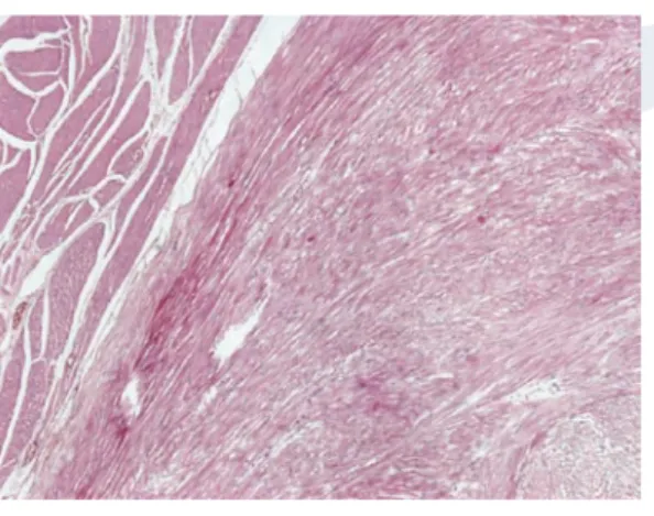
Microscopically the lesion was characterized by bundles of spindle shaped cells similar to smooth muscle cells with small nuclei, without atypia and mitotic figures arranged with hapha-zard intersections (Figure 1) The tumor cells showed diffuse expression of smooth muscle actin and desmin and Ki 67 proliferation index was 1% (Figure 2). Final diagnosis was given as bladder leiomyoma. The patient is under follow up as disease-free after 3 months from the operation with no additional treatment.
DISCUSSION
Leiomyomas are benign soft tissue tumors with smooth muscle transformation, predominan-tly seen in the uterus of premenopausal women (2). Leiomyomas occur only 0.4% of all bladder tumors however it is the most common type of mesenchymal tumors seen in the bladder (2). The rate of bladder leiomyomas was similar in females and males however there are some studies showing
FUNDADOS EN 1944 POR E. PÉREZ CASTRO, A. PUIGVERT GORRO Y L. CIFUENTES DELATTE Director / Editor: E. Pérez-Castro Ellendt Editor Asociado: L. Martínez-Piñeiro Lorenzo Editor Asociado Internacional: J. de la Rosette Editor Asociado para Latinoamérica: F. Secín
Arch. Esp. Urol. 2015; 68 (7): 585-586 585
Figure 1: Tumor composed of bundles of spindle shaped cells located in the muscular
layer of the bladder, H&E, X100
Jain SK, Tanwar R, Mitra A. Rev Urol. 2014; 16(1): 50–54.
Kabalin JN, Freiha FS, Niebel JD. Leiomyoma of bladder. Report of 2 cases and demonstration of ultrasonic appearance. Urology. 1990 Mar;35(3):210-212.
Knoll LD, Segura JW, Scheithauer BW. Leiomyoma of the bladder. J Urol. 1986 Oct;136(4):906-908. Goluboff ET, O’Toole K, Sawczuk IS. Leiomyoma of bladder:report of case and review of literature. Urology 1994;43:238–41.
Jiang XZ, Chao XU, Zhang NZ, Zhi-shunChin XU. Med J 2012;125(14):2436-2439.
Broessner C, Klingler CH, Bayer G, et al. A 3,500-gram leiomyoma of the bladder: case report on a 3-year follow-up after surgical enucleation. Urol Int. 1998;61:175–7.
Goktug GH, Ozturk U, Imamoglu AM. Transurethral resection of a bladder leiomyoma: A case report. Can Urol Assoc J. 2014 Jan-Feb; 8(1-2): 111–113.
Lyons TL, Lee T, Winer WK. Laparoscopic removal of a bladder leiomyoma. J Am Assoc Gynecol Laparosc. 1998 Nov;5(4):423-6.
Kim IY, Sadeghi F, Slawin KM. Dyspareunia: An unusual presentation of leiomyoma of the bladder. Rev Urol. 2001;3:152–4. **1. *2. *3. **4. *5. *6. **7. **8. **9. BIBLIOGRAFÍA 586
female predominance (2,3). Although the etiology of the tumor remains a mystery, hormonal irre-gularities, recurrent infections and chromosomal alterations are being accused (2,4). Patients with bladder leiomyomas usually apply with obstructive and irritative symptoms. Despite that, 20-30% of the cases were asymptomatic and the tumor was detected incidentally (4,5).
Leiomyomas are classified as endovesical, intramural and extravesical according to their anatomic locations (3). Endovesical type is the most common form comprising 63-86% of the cases. Intramural leiomyomas, like our case, and extravesical localizations forms 3-7% and 11-30% of the cases respectively (3,4).
Cystoscopical evaluation is an effective method for diagnosing, subtyping and treating these tumors. Small sized endovesical tumors can be diagnosed and excised by cystoscopy in the same session (6). Cystoscopy is recommended for small tumors however, Göktug et al. (7) were reported a 7 cm endovesical bladder leiomyoma treated by cystoscopic resection. In general endo-vesical tumors over 3 cm, intramural and extraendo-vesical tumors are suggested for partial cystectomy and tumor enucleation (6,8,9). In our case,because of the anatomical location the tumor was ex-cised by partial cystectomy with 1 cm surgical margins. In addition to the favorable outcome, no malign transformation was reported in the literature.
Remembering this rare location of leiomyomas will increase the importance of cystoscopic examination in order to diagnose and even treat these lesions. In addition, the optimal macroscopic sampling especially in cases without preliminary diagnosis will also help the pathologists in diffe-rential diagnosis of many different entities.
Figure 2: Diffuse positivity for Smooth Muscle Actin, DAB, X200.
View publication stats View publication stats